. 2012;7(8):e44177.
doi: 10.1371/journal.pone.0044177. Epub 2012 Aug 31.

Improper tagging of the non-essential small capsid protein VP26 impairs nuclear capsid egress of herpes simplex virus

Affiliations

Improper tagging of the non-essential small capsid protein VP26 impairs nuclear capsid egress of herpes simplex virus

Claus-Henning Nagel et al. PLoS One. 2012.

Abstract

To analyze the subcellular trafficking of herpesvirus capsids, the small capsid protein has been labeled with different fluorescent proteins. Here, we analyzed the infectivity of several HSV1(17(+)) strains in which the N-terminal region of the non-essential small capsid protein VP26 had been tagged at different positions. While some variants replicated with similar kinetics as their parental wild type strain, others were not infectious at all. Improper tagging resulted in the aggregation of VP26 in the nucleus, prevented efficient nuclear egress of viral capsids, and thus virion formation. Correlative fluorescence and electron microscopy showed that these aggregates had sequestered several other viral proteins, but often did not contain viral capsids. The propensity for aggregate formation was influenced by the type of the fluorescent protein domain, the position of the inserted tag, the cell type, and the progression of infection. Among the tags that we have tested, mRFPVP26 had the lowest tendency to induce nuclear aggregates, and showed the least reduction in replication when compared to wild type. Our data suggest that bona fide monomeric fluorescent protein tags have less impact on proper assembly of HSV1 capsids and nuclear capsid egress than tags that tend to dimerize. Small chemical compounds capable of inducing aggregate formation of VP26 may lead to new antiviral drugs against HSV infections.


Conflict of interest statement

Competing Interests: The authors have declared that no competing interests exist.

Figures

Figure 1
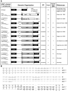
Figure 1. HSV1-VP26 constructs.
(A) 1st column: HSV1 constructs in which the SCP VP26 has been tagged with different fluorescent protein domains. 2nd column: Genomic organization of the UL35 region approximately drawn to scale. The gene UL35 coding for VP26 has been disrupted by replacing it with lacZ or an rpsLneo cassette out of frame. Some constructs lack a 65 bp region upstream of UL35 (D65 bp) including the first seven N-terminal codons of VP26 (Daa1–7), while others lack only four (Δaa1–4) or just one (Daa1) codon. For the present study, the fluorescent protein tag was inserted between VP26 residues 4 and 8 (Daa5–7). Due to the mutagenesis, some strains contain additional linkers (*, AW; **, NSS; ***, HST). 3rd column: Propensity of the fluorescent protein (FP) to dimerize –. 4th column: Ability of the construct to replicate and to form plaques (+++, similar to wild type; ++ attenuated, but robust growth; − strongly attenuated, tiny plaques; −−, single fluorescent cells, no plaques). 5th column: Propensity of the construct to induce nuclear aggregates (+++, large irregular shaped aggregates; ++, large aggregates early after infection or transfection; +, aggregates late in infection; − aggregates in less than 2% of cells even late in infection). 6th column: References. (B) Nucleotide (upper lines) and amino acid (lower lines) sequences of the UL34/UL35 (pUL34/VP26) intergenic region. The 3¢ end of the UL34 ORF until the 5¢ start of the UL35 ORF are shown for wild type HSV-1, the GFPVP26Äaa5–7 (Äaa5–7) and GFPVP26Äaa1–7 (Äaa1–7) mutants. Additional nucleotides inserted during mutagenesis are shown in bold capitals, and the GFP amino acids are shown in italics. Putative Inr late promoter elements are underlined with the element perfectly matching the consensus sequence being underlined and in italics. The original amino acids encoded by UL35 are shown in bold capitals, the inserted GFP residues in italic capitals and the additional linker residues in normal script capitals.
Figure 2
Figure 2. Characterization of HSV1(17+)blueLox-VP26 strains.
(A) Restriction digestion analysis of different BAC clones with NotI and EcoRI. Band shifts resulting from modifications in the UL35 ORF (arrowheads) and the NotI joint fragments containing the viral a-sequences (asterisks) are indicated. DNA sizes in kb. (B) For single-step growth kinetics, Vero cells were infected in duplicates with 10 PFU/cell, and the amount of secreted infectious virus at a given time point was determined by duplicate plaque assays. (C) Vero cells were infected at an MOI of 10 PFU/cell with HSV1(17+) or HSV1(17+)blueLox and its derivatives as indicated. After 48 h, the cells and virions secreted into the culture medium were harvested, and analyzed by SDS-PAGE and immunoblotting for expression of VP5 (α-NC-1) and VP26 (α-VP26). The signals in the molecular weight range below 25 kDa were further contrast enhanced.
Figure 3
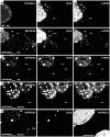
Figure 3. Nuclear aggregates induced by HSV1-XFPVP26 impair nuclear capsid egress.
Vero cells were infected (inf.) with 10 PFU/cell of HSV1(17+)blueLox (A, wild type), HSV1(17+)blueLox-mRFPVP26Δaa1–7 (B), or HSV1(17+)blueLox-GFPVP26Δaa1–7(C), and fixed at 9 h with PFA. Alternatively, cells were transfected (transf.) with pHSV1(17+)blueLox-GFPVP26Δaa5–7 (D) or pHSV1(17+)blueLox-GFPVP26Δaa1–7 (E), and fixed at 24 h. In addition to the intrinsic fluorescence of the XFPVP26 constructs (mRFPVP26 or GFPVP26), the subcellular localization of VP26 (α-VP26) and VP5 (MAb 5C10) were analyzed after permeabilization with TX-100 and immunolabeling by confocal fluorescence microscopy. The nuclei were stained with TO-PRO-3 (A, E). Arrows highlight cytoplasmic capsids (A–D) or incoming capsids at the nuclear rim of a neighboring cell (Ei). Scale bar: 10 µm.
Figure 4
Figure 4. Quantification of nuclear aggregate formation.
Vero cells were infected with 10 PFU/cell of HSV1(17+)blueLox (B) or HSV1(17+)blueLox-GFPVP26Δaa1–7-gDmRFP (C), and the cells were fixed at 4, 6, 8, 10, or 12 h. After permeabilization, HSV1(17+)blueLox infected cells were labeled with an antibody directed against VP26. According to their intranuclear VP26 phenotype, the cells were classified into “none”, “single”, “grainy” and “aggregate” (A). The numbers above the columns describe the number of nuclei analyzed for each time point.
Figure 5
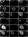
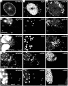
Figure 5. Nuclear aggregates contain capsid proteins.
Vero cells were infected (inf.) with 10 PFU/cell of HSV1(17+)blueLox-GFPVP26Δaa1–7 for 9 h (A–D) or transfected (transf.) with pHSV1(17+)blueLox-GFPVP26Δaa5–7 (E) or pHSV1(17+)blueLox-GFPVP26Δaa1–7 (F) for 24 h. The cells were fixed with PFA and permeabilized with TX-100. GFPVP26 was detected by its intrinsic fluorescence (left column). Furthermore, the cells were labeled with different VP5 antibodies (MAb 8F5 or 5C10) and with α-VP26, α-pUL25 or α-pUL17, and analyzed by confocal fluorescence microscopy. For row D, the specimen was scanned at higher magnification and lower photomultiplier settings. The arrows point to cytoplasmic capsids. Bars: 5 µm (D), 10 µm (F).
Figure 6
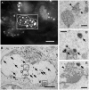
Figure 6. Nuclear aggregates do not contain capsids.
RPE cells were infected with HSV1(KOS)-GFPVP26Δaa1–7 at an MOI of 10 PFU/cell. At 19.5 h, the cells were analyzed by fluorescence microscopy to identify nuclei with bright fluorescence spots (A). After fixation, embedding and sectioning these cells were further analyzed by electron microscopy (B–E). The nuclei contain nuclear capsids (arrowheads) as well as the amorphous electron dense material representing the aggregates (arrows) identified by fluorescence microscopy. The aggregates do not contain capsids (arrowheads). Bars: 10 µm (A), 5 µm (B), and 500 nm (C–E).
Figure 7
Figure 7. Nuclear GFPVP26 aggregates do not sequester pUL36 and are not DNA replication compartments.
Vero cells were infected (inf.) for 9 h with 10 PFU/cell of HSV1(17+)blueLox (A) or HSV1(17+)blueLox-GFPVP26Δaa1–7 (B, D), or transfected (transf.) with the pHSV1(17+)blueLox-GFPVP26Δaa5–7 for 24 h(C, E). The cells were fixed with PFA and permeabilized with TX-100. GFPVP26 was detected by its intrinsic fluorescence. Furthermore, the cells were labeled with anti-pUL36aa1408–2112 and with MAb 5C10 against VP5 (A–C), against the single-strand DNA binding protein ICP8 and VP26 (D, E), or with TO-PRO-3 to stain the DNA (A). The arrows point to cytoplasmic capsids. Scale bar: 10 µm.

Similar articles

Cited by

References

    1. Bosse JB, Bauerfeind R, Popilka L, Marcinowski L, Taeglich M, et al. (2012) A Beta-herpesvirus with fluorescent capsids to study transport in living cells. PLoS One 7: e40585. - PMC - PubMed
    1. Antinone SE, Zaichick SV, Smith GA (2010) Resolving the assembly state of herpes simplex virus during axon transport by live-cell imaging. J Virol 84: 13019–30. - PMC - PubMed
    1. Cheng SB, Ferland P, Webster P, Bearer EL (2011) Herpes Simplex Virus Dances with Amyloid Precursor Protein while Exiting the Cell. PLoS One 6: e17966. - PMC - PubMed
    1. de Oliveira AP, Glauser DL, Laimbacher AS, Strasser R, Schraner EM, et al. (2008) Live visualization of herpes simplex virus type 1 compartment dynamics. J Virol 82: 4974–90. - PMC - PubMed
    1. Döhner K, Radtke K, Schmidt S, Sodeik B (2006) Eclipse phase of herpes simplex virus type 1 infection: Efficient dynein-mediated capsid transport without the small capsid protein VP26. J Virol 80: 8211–24. - PMC - PubMed

Publication types

MeSH terms

Substances