. 2012;7(8):e44244.
doi: 10.1371/journal.pone.0044244. Epub 2012 Aug 31.

SHP-1 phosphatase is a critical regulator in preventing natural killer cell self-killing

Affiliations

SHP-1 phosphatase is a critical regulator in preventing natural killer cell self-killing

Sajid Mahmood et al. PLoS One. 2012.

Abstract

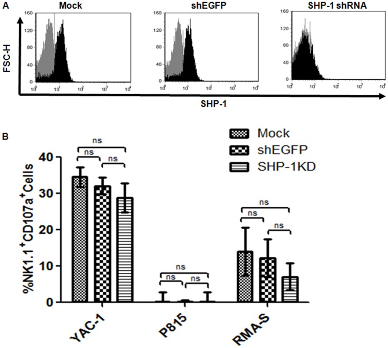
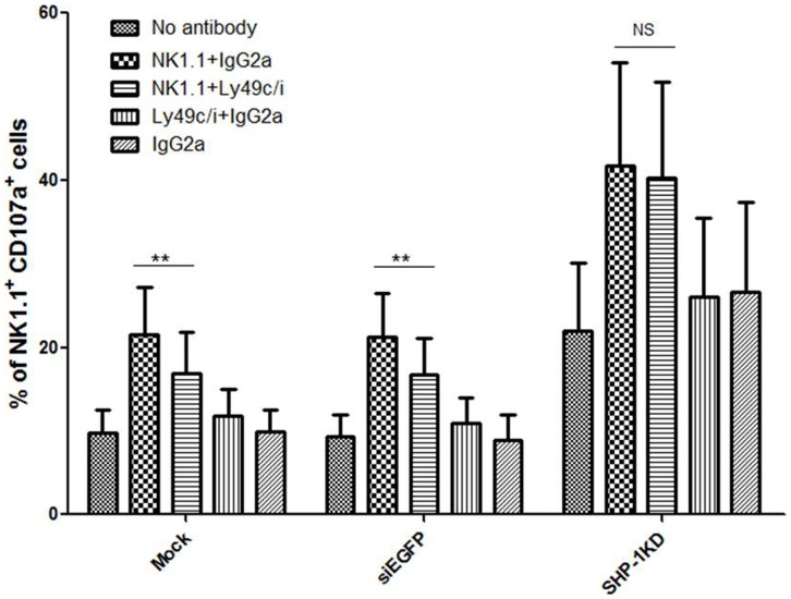
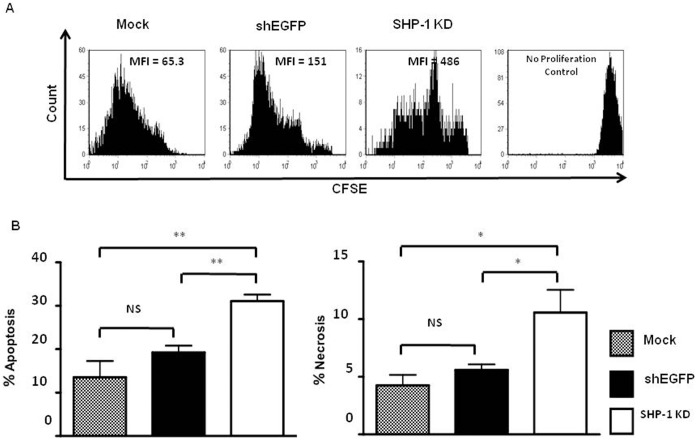
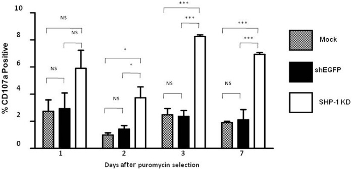
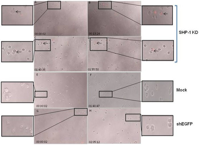
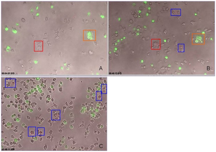
Balance of signals generated from the engaged activating and inhibitory surface receptors regulates mature NK cell activities. The inhibitory receptors signal through immunoreceptor tyrosine based inhibitory motifs (ITIM), and recruit phosphatases such as SHP-1 to inhibit NK cell activation. To directly examine the importance of SHP-1 in regulating activities and cell fate of mature NK cells, we used our established lentiviral-based engineering protocol to knock down the SHP-1 protein expression in primary C57BL/6NCrl cells. Gene silencing of the SHP-1 in primary NK cells abrogated the ability of ITIM-containing NK inhibitory receptors to suppress the activation signals induced by NK1.1 activating receptors. We followed the fates of stably transduced SHP-1 silenced primary NK cells over a longer period of time in IL-2 containing cultures. We observed an impaired IL-2 induced proliferation in the SHP-1 knockdown NK cells. More interestingly, these "de-regulated" SHP-1 knockdown NK cells mediated specific self-killing in a real-time live cell microscopic imaging system we developed to study NK cell cytotoxicity in vitro. Selective target recognition of the SHP-1 knockdown NK cells revealed also possible involvement of the SHP-1 phosphatase in regulating other NK functions in mature NK cells.


Conflict of interest statement

Competing Interests: The authors have declared that no competing interests exist.

Figures

Figure 1
Figure 1. SHP-1 gene knockdown NK cells showed normal cytotoxicity towards prototypic tumor target cells.
A. Efficient SHP-1 gene silencing in primary murine IL-2-activated NK cells. Purified C57BL/6NCrl IL-2 activated NK cells were transduced on two consecutive days with TRC lentiviral vectors and incubated for 3 days post-transduction. Transduced cells were puromycin selected for 48 hours followed by 3 days incubation. Cells were assayed for SHP-1 expression by intracellular staining with primary rabbit anti-SHP-1 and secondary anti-rabbit Alexa Fluor 488 antibodies in flow cytometry (Black). Secondary antibodies alone (Grey) was used as negative control. Data is representative of 3 experiments. B. IL-2 activated NK cells were transduced on two consecutive days with mock, shEGFP and SHP-1 shRNA and incubated for 3 more days. Transduced cells were puromycin-selected and used as effectors against Yac-1, P815 and RMA-S targets at E:T ratio 1∶1 in CD107a cytotoxicity assay. Background degranulation responses of the NK effectors (in the absence of target cells) were subtracted from the degranulation responses observed in the presence of the target cells. Data of three independent experiments were pooled together for statistical analyses. N.S., (p>0.05), non significant.
Figure 2
Figure 2. SHP-1 knockdown NK cells showed loss of inhibition in the antibody induced redirected lysis assay.
Mock, shEGFP and SHP-1 shRNA transduced and puromycin selected LAK cells incubated with purified antibodies for 20-minutes. Effector:target (P815) cells (1∶1 ratio) in V-shaped 96-well plate incubated for 5 & 4 hours in the presence of CD107a antibody and monensin respectively. Percentages of NK1.1+CD107+ cells were calculated and statistically analyzed using graph pad prism software. A p-value of <0.05 was considered statistically significant. Data is representative of 5 experiments.
Figure 3
Figure 3. Stable SHP-1 gene knockdown in primary NK cells resulted in impaired IL-2 induced NK cell proliferation.
A. Mock, shEGFP and SHP-1 shRNA transduced and puromycin selected LAK cells were labeled with carboxyfluorescein succinimidyl ester (CFSE) and cultured them again in IL-2 supplemented media. On day 7 post CFSE staining, cells were surface stained with NK1.1 antibody. Samples were acquired and gated on NK1.1+ population for CFSE dilution analysis. B. Loss of cell viability in the SHP-1 knockdown NK cells. Mock, shEGFP and SHP-1 shRNA transduced and puromycin selected LAK cells were for apoptosis and necrosis by stained with Annexin V and 7-amino actinomycin D (7AAD) respectively in flow cytometery. Data is representative of 3 experiments. NS, non-significant; *, p<0.05; **, p<0.01.
Figure 4
Figure 4. SHP-1 gene knockdown NK cells showed increased spontaneous degranulation.
Mock, shEGFP and SHP-1 shRNA transduced and puromycin selected LAK cells were incubated in IL-2 supplemented media for 1,2,3 or 7 days. Cells were then used in a CD107a degranulation assay. Briefly, cells were incubated with CD107a antibody and monensin in 5 ml centrifuge tubes for 5 hours at 37°C before analysis in flow cytometry. Data is representative of 3 experiments. NS, non-significant; *, p<0.05; ***, p<0.0001.
Figure 5
Figure 5. Real-time imaging of spontaneous killing of the SHP-1-knockdown NK cells in vitro.
Mock, shEGFP transduced and SHP-1 shRNA transduced and puromycin selected purified NK cells resuspended in Hanks Buffered Salt Solution (HBSS) with 10% FCS and 50 U/ml IL-2 containing 7AAD. Cells were imaged every 25 seconds for up to 5 hours using 10X magnification objective on a Zeiss Observer 710 station. The images in the figures were excised from live cell imaging movies (Videos S4, S5, S6) at different time points. Dying cells appeared red due to 7AAD staining. Frames A–B, C–D: events at different time points of the real-time live cell imaging. Frames B, D represented late time points of the same cells after 10–20 minutes of interactions (as noted in A and C respectively, indicated by the arrows). The inset images showed the zoom-in regions of interest for clarity.
Figure 6
Figure 6. Specific recognition of the SHP-1 gene knockdown NK cells as targets.
SHP-1 shRNA transduced and puromycin selected purified NK cells mixed with green CMFDA-labelled normal C57BL/6NCrl NK cells at 1∶1 ratio, resuspended in Hanks Buffered Salt Solution (HBSS) with 10% FCS and 50 U/ml IL-2 containing 7AAD. Cells were imaged every 25 seconds for up to 5 hours using 10X magnification objective on a Zeiss Observer 710 station. The images in the figure were taken from the live cell imaging videos (Video S7) at different time points. Red box in insert A indicated real cell conjugate formation between two unlabelled NK cells. These conjugated cells can be tracked 46 minutes later (red box, insert B) to show subsequent cytolysis as the dying cells took up the 7-AAD dye. Orange box indicated transient aggregates that disintegrated at later time points, and showed no evidence of cytolysis. At late time-points (such as 150 minutes of imaging), more apoptotic cells (red) were observed in the unlabelled SHP-1 knockdown NK cells (Blue box).

Similar articles

Cited by

References

    1. Sentman CL, Barber MA, Barber A, Zhang T (2006) NK cell receptors as tools in cancer immunotherapy. Adv Cancer Res 95: 249–292. - PubMed
    1. Biassoni R, Ugolotti E, De Maria A (2010) Comparative analysis of NK-cell receptor expression and function across primate species: Perspective on antiviral defenses. Self Nonself 1: 103–113. - PMC - PubMed
    1. Lanier LL (1998) NK cell receptors. Annu Rev Immunol 16: 359–393. - PubMed
    1. Moretta L, Bottino C, Cantoni C, Mingari MC, Moretta A (2001) Human natural killer cell function and receptors. Curr Opin Pharmacol 1: 387–391. - PubMed
    1. Yokoyama WM, Plougastel BF (2003) Immune functions encoded by the natural killer gene complex. Nat Rev Immunol 3: 304–316. - PubMed

Publication types

MeSH terms

Substances