. 2011:1.
doi: 10.3402/pba.v1i0.7202. Epub 2011 Jun 1.

Practical pathology of aging mice

Affiliations

Practical pathology of aging mice

Christina Pettan-Brewer et al. Pathobiol Aging Age Relat Dis. 2011.

Abstract

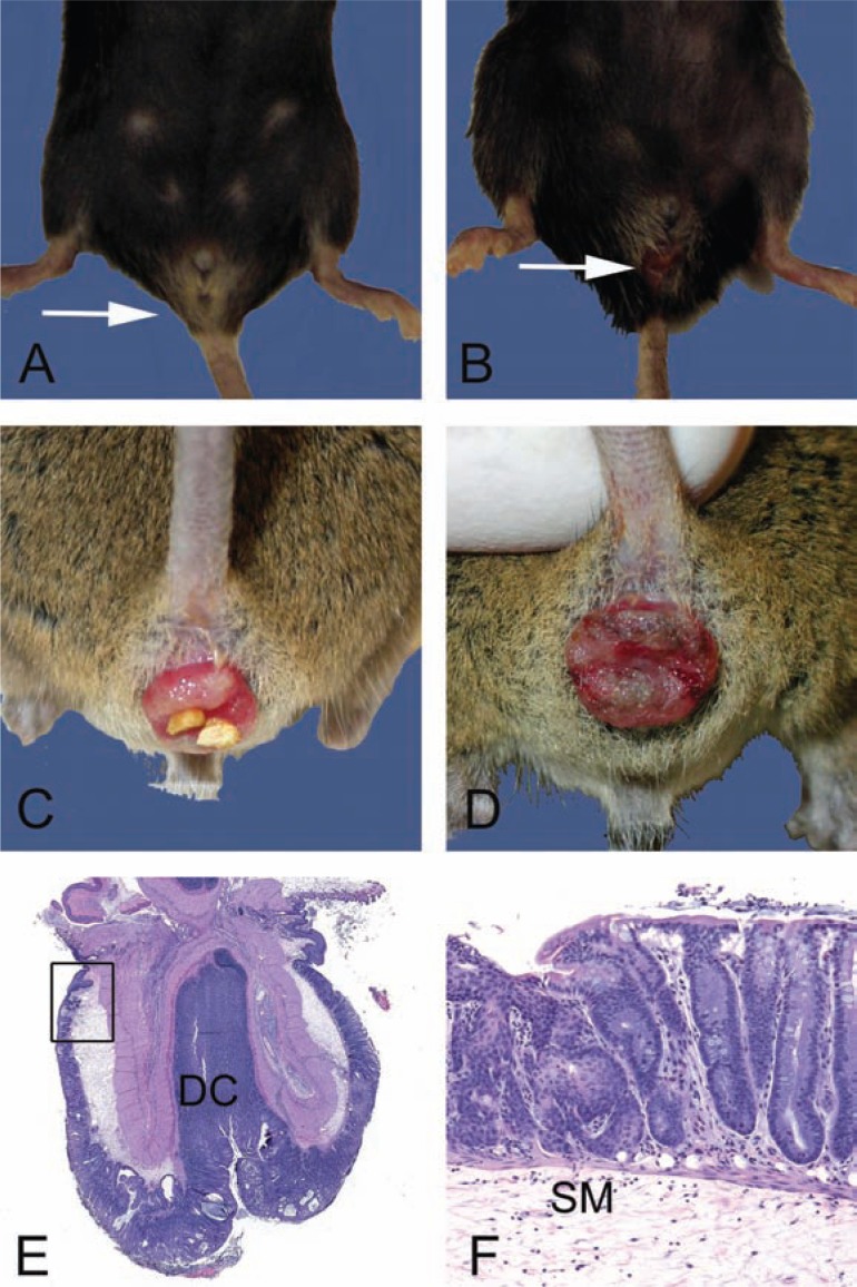
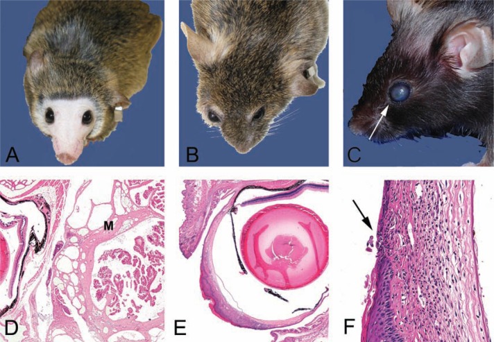
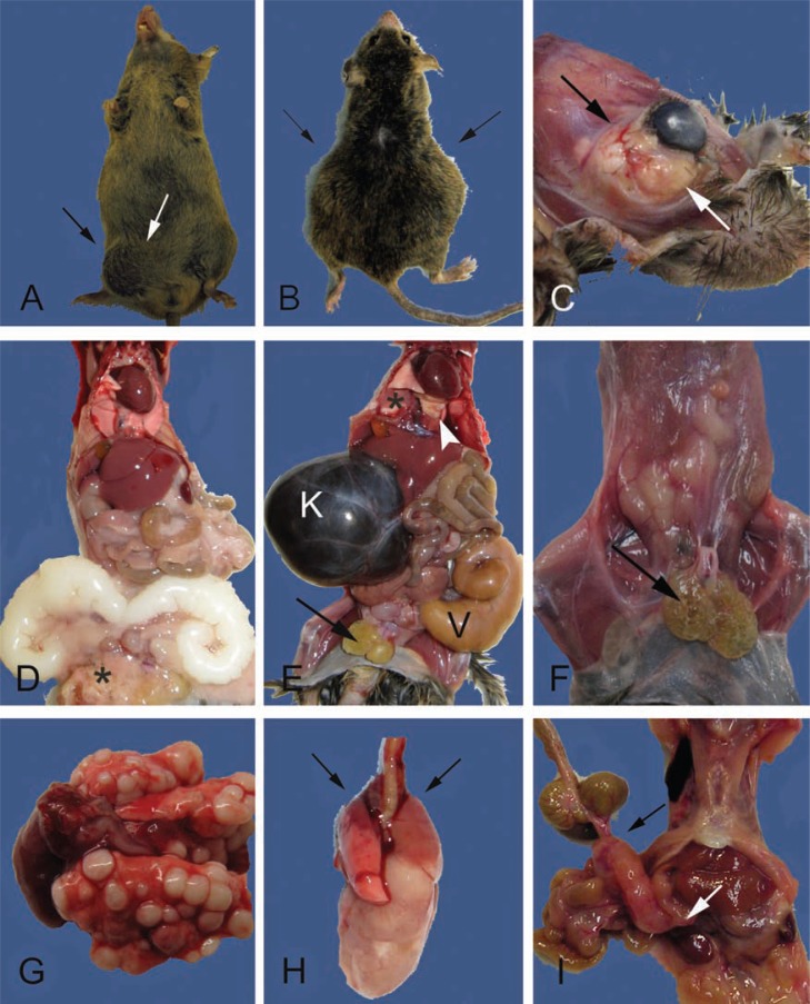
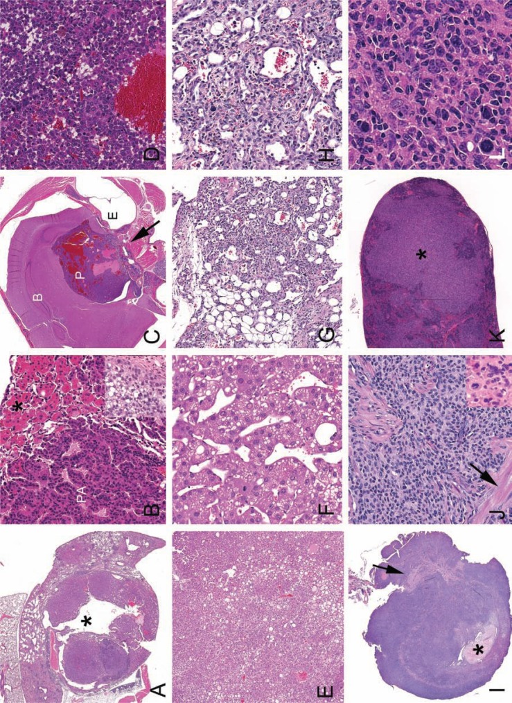
Old mice will have a subset of lesions as part of the progressive decline in organ function that defines aging. External and palpable lesions will be noted by the research, husbandry, or veterinary staff during testing, cage changing, or physical exams. While these readily observable lesions may cause alarm, not all cause undue distress or are life-threatening. In aging research, mice are maintained until near end of life that, depending on strain and genetic manipulation, can be upwards of 33 months. Aging research has unique welfare issues related to age-related decline, debilitation, fragility, and associated pain of chronic diseases. An effective aging research program includes the collaboration and education of the research, husbandry, and veterinary staff, and of the members of the institution animal care and use committee. This collaborative effort is critical to humanely maintaining older mice and preventing excessive censorship due to non-lethal diseases. Part of the educational process is becoming familiar with how old mice appear clinically, at necropsy and histopathologically. This baseline knowledge is important in making the determination of humane end points, defining health span, contributing causes of death and effects of interventions. The goal of this paper is to introduce investigators to age-associated diseases and lesion patterns in mice from clinical presentation to pathologic assessment. To do so, we present and illustrate the common clinical appearances, necropsy and histopathological lesions seen in subsets of the aging colonies maintained at the University of Washington.

Keywords: aging; animal models; cancer; mice; pathology; veterinary pathology.


Figures

Fig. 1
Fig. 1
Gross and microscopic examples of rectal prolapses. A. Normal female 28-month-old B6 mouse with the anus indicated (arrow). B. Female 28-month-old B6 mouse with a mild acute rectal prolapse. Note the mild protrusion of congested and moist rectal mucosa (arrow). In female mice, rectal prolapse must be differentiated from uterine prolapse. C. Moderate to severe subacute rectal prolapse in a 4 to 6-month-old male genetically modified mouse, 129-Smad3 tm/Par/J, infected with Helicobacter species. D. Chronic severe rectal prolapse in a 4 to 6-month-old male 129-Smad3 tm/Par/J infected with Helicobacter species. The mucosa is dry and thickened with adherent crust. E. Hematoxylin and eosin-stained section of prolapsed rectum. The prolapsed mucosa is covered by a serocellular crust. Distal colon (DC) and rectoanal junction (box) are indicated. F. Higher power of boxed region in E. Squamous metaplasia (arrow) and hyperplasia of the rectal and distal colonic mucosa is common in chronically prolapsed tissues exposed to the cage environment including bedding, skin, and fecal organisms. The submucosa (SM) is markedly expanded by edema.
Fig. 2
Fig. 2
Clinical and histological presentations of common skin lesions. A. A 28–month-old female B6 mouse in relatively good health deemed clinically normal. Note mild alopecia on the head, normal eyes, and well-groomed pelage. B. A 28-month-old female B6 mouse with ill-kept fur due to lack of adequate grooming. Note the hunched posture, opaque cornea, the elongated shape of the nose, and relatively boney appearance of this mouse in contrast to the mouse pictured in 2A. Internally, this mouse had a large pituitary adenocarcinoma (see Fig. 5C,D). C. Moderate patch alopecia in a B6 mouse (age unknown). This clinical presentation may be a result of dermatitis or alopecia due to other causes such as primary follicular dysplasia. Histology would aid in determining a definitive cause. D. Ulcerative and likely pruritic dermatitis in an agouti mouse (background and age unknown). This historical clinical presentation was often seen associated with fur mites, although numerous causes have been implicated in a similar clinical spectrum in C57BL/6 mice. See text for details. E. Hematoxylin and eosin-stained section of mild alopecia with minimal chronic inflammation within the dermis (arrow). Note thickness of the subcutis (SC). Follicles are sparse and devoid of hair shafts. F. Hematoxylin and eosin-stained section of severe proliferative dermatitis (pseudoepitheliomatous). The epidermis is markedly thickened with irregular rete peg formation in this somewhat tangentially oriented section. Dyskeratosis and mild orthokeratotic hyperkeratosis are also present with moderate chronic dermatitis that extends into the SC. Pseudoepitheliomatous hyperplasia is frequently seen histologically at the thickened edges of chronic ulcerative foci (see 2D).
Fig. 3
Fig. 3
Clinical and histological presentations of common eye lesions. A. Normal periocular shape, eye size, and color in a 24-month-old CB6F1 mouse included here for contrast to the mouse in 3B. Note the lack of pelage and vibrissae due to extensive barbering by cage mates. Barbering is another relatively common skin presentation. It may lead to dermatitis in the victim and impaction of hair fragments into gingival sulcus of the barber. B. A 24-month-old CB6F1 mouse has periocular and retrobulbar swelling (between arrows) with bulopthalmia. This presentation may be a result of periocular cellulitis, conjunctivitis, or retrobulbar mass such as an abscesses or neoplasia. In this mouse, the swelling was due to a large retrobulbar mass (4C), which was diagnosed histologically as a Harderian adenoma (3D) C. Corneal opacity in a 28–month-old B6 mouse. The small foci (arrow) at the rostal aspect of the cornea is an ulcer, which was confirmed clinically with fluorescein dye test clinically and histologically (3F) D. Hematoxylin and eosin-stained section of the Harderian gland adenoma from the CB6F1 mouse in panel 3B. The mutliobulated and expansible mass (M) is located in the retro-orbital space compresses the eye (E). E. Hematoxylin and eosin-stained section of the eye from the B6 mouse in 3C. The opacity noted clinically is due to marked proliferative and ulcerative keratitis (box). The normal corneal epithelium is non-keratinized and 4–7 cell layers thick. F. Higher magnification of boxed region of a punctate corneal ulcer in 3C and E. The defect in the overlying corneal epithelium (arrow) exposes the stroma. The hydrophilic stroma will uptake fluorescein dye when the hydrophobic epithelial layer is breeched. Note the dense inflammatory infiltrate and neovascularization in the stroma. These also contribute to the corneal opacity noted clinically (3C).
Fig. 4
Fig. 4
External and internal masses may be due to degenerative, inflammatory, or neoplastic lesions. A. Swelling in the lower right abdomen and the inguinal region (between arrows) are present in this 24-month-old male CB6F1. The inguinal swelling is due to an infected preputial gland. The abdominal swelling is due to enlarged, but otherwise normal, seminal vesicles (see also D). B. A 27-month-old male CB6F1 with bilateral abdominal swelling (arrows) from severe unilateral hydronephrosis and enlarged and infected seminal vesicles (see also E). C. A large multilobulated retro-orbital mass causing proptosis of the eye (between arrows). The cornea is cloudy due to keratitis (see also Fig. 3B,D). D. Internal examination of the mouse in A demonstrates bilaterally enlarged seminal vesicles, a common finding in older male mice. The seminal vesicular fluid is normally white and will discolor with infection or infarction (compare to E). Additional gross lesions are an enlarged heart with mild hepatomegaly, pneumonopathy, and the preputial abscessation (*) noted in A (between arrows). E. The internal examination of the mouse in B demonstrates severe unilateral hydronephrosis (K) and enlarged and infected seminal vesicle (V). The kidney is filled with dark brown colored fluid, a severe sequelae of obstructive uropathy (mouse urologic syndrome). Other lesions include absence of body fat, enlarged and cystic preputial glands (arrow), enlarged heart and pulmonary atelectasis (*), and light yellow firm foci (arrowhead) diagnosed histologically as AMP (see Fig. 6K,L). F. A 29-month-old male CB6F1 with bilateral cystic preputial glands (arrow), scant body fat (body condition score of 2/5), and tacky appearing tissues (mild to moderate dehydration). G. Multiple pulmonary neoplastic foci in a 30-month-old female CB6F1 mouse. This unusual presentation is more consistent with metastatic rather than primary disease. Histopathological diagnosis, however, confirmed mucinous pulmonary adenocarcinoma (see inset Fig. 5B). H. Typical presentation of a CB6F1 pulmonary adenocarcinoma from 28 to 30-month-old mice. Grossly, one large white firm mass extends from the right lung. Microscopically, there may be multiple foci of neoplasia. Cranioventral atelectasis and consolidation of the lungs is also present (arrows). I. A large mesenteric mass in the region of a cluster of lymph nodes (bracketed by arrows) from a 28-month-old B6 mouse. Differentials include lymphoma and histiocytic sarcoma. Hepatic involvement with the neoplastic process is suggested by the roughened appearance of the liver (compare to D and E) and was confirmed histologically.
Fig. 5
Fig. 5
Histological presentations of neoplasia. A. Pulmonary adenocarcinoma with gross presentation similar to the mouse in 4H. Note large cystic space (*). B. Higher magnification of a pulmonary adenoma (PA) with associated AMP (*). Inset: Mucinous pulmonary adenocarcinoma from lungs pictures in Fig. 4G. C. Decalcified cross-section of the head with a pituitary adenocarcinoma (P) of the pars distalis from mouse pictured in 2B. Pars distalis tumors typically are hemorrhagic and develop blood filled cystic spaces. Malignancy, in this case, was evident with invasion into the sphenoid bone (arrow). Brain (B), pituitary tumor (P) and ear (E) are indicated. D. Higher magnification of the pituitary adenocarcinoma in C. E. Liver with solid and trabecular hepatocellular carcinoma (HCC) from a 30-month-old female CB6F1. F. Higher magnification of trabecular HCC. G. Dermal hemangiosarcoma from a 30-month-old male CB6F1. H. Higher magnification of G. Irregular vascular channels are formed by atypical and anisokaryotic endothelial cells. I. Uterus from a 28-month-old B6 mouse is effaced with histiocytic sarcoma (HSA). Broad ligament (arrow) and formed lumen (*) are indicated. J. Higher magnification of HSA in I. Neoplastic histiocytes may have elongated oval to round nuclei or form multinucleated giant cells (inset). Myometrial smooth muscle (arrow) is indicated. K. Spleen with pleomorphic/follicular malignant lymphoma (*) from a 30-month-old CB6F1 female. L. Higher magnification of K. Pleomorphic lymphoma can resemble HSA. Anatomic distribution, morphology, and immunohistochemistry can aid in differentiation.
Fig. 6
Fig. 6
Histological presentations of subclinical chronic systemic inflammation-associated lesions seen in C57BL/6 mice aged 16–36 months. A. Hyperplastic mesenteric milky spot. B. Higher magnification of A. The reactive milky spot contains large and small lymphocytes, plasma cells, and Mott cells (arrows). C. Low magnification overview of chronic renal lesions. Interstitial lymphoid aggregates (*) and tubular ectasia (arrow) with degeneration, regeneration, necrosis and membranous glomerulonephropathy. D. Thickened mesangial matrix (*) and periglomerular fibrosis (arrow) characterize membranous glomerulonephropathy. Glomerular amyloidosis should also be considered. PAS and Congo Red histochemical stains can aid in differentiation. E. Chronic adenitis occurs in numerous glands including the exorbital lacrimal gland. F. Higher magnification of E. Lymphoplasmacytic inflammation disrupts the gland architecture. Note ectatic duct (*). G. Amyloid accumulation in the small intestinal villi. H. Higher magnification of G. An accumulation of light pink homogenous extracellular material (amyloid) in the lamina propria widens the villi. I. Cross-section of the base of the tongue with poly arteritis nodosa (box and *). Lingual minor salivary glands are indicated (arrow). J. Movat's pentachrome staining of lingual artery in I. The affected segment (*) lacks in elastic lamina (thin black line) and the media is expanded by smooth muscle cells. Mild to moderate perivascular chronic inflammatory cells (arrow) and lumena (L) are indicated. Similar necroproliferative arteritis near the inner and middle ear may result in neurological signs (see text for details). K. Low power view of lung severely affected by AMP. L. Higher magnification of AMP demonstrates the intrahistiocytic crystalline material (*) and dense perivascular and peribronchiolar lymphoplasmacytic inflammation, which is frequently present in severe cases. Hemosiderophage (arrow) and hyalinized respiratory epithelium (arrowheads) are indicated.
Fig. 7
Fig. 7
Subclinical degenerative lesions diagnosed histologically. A. Decal cross-section of the nose and incisor roots (arrows) with unilateral multiple immature intrapulpal denticles from a 28-month-old B6 mouse. B. Higher magnification of the intrapulpal denticles. Denticles with an unusual presentation of a central column of ameloblasts (*) encircled with clear space, predentin (arrow, pink band) and odontoblasts (arrowhead). Denticles may represent dysplastic tooth development. Normal position of ameloblasts (*) and odontoblasts (arrow) are also indicated. C. Osteoarthritis of the temporomandibular joint from the mouse in A with incisor dysplasia. Mandibular condyle is indicated (*). D. Higher magnification of boxed region in C. The mandibular condyle and maxillary fossal cartilages are degenerative and irregular. The joint space (*) contains free floating debris and cartilage (joint mice).

References

    1. Nadon NL. Aged Rodents for biogenrontology research. In: Conn PM, editor. Handbook of models for human aging. Amsterdam: Elsevier Academic Press; 2006. pp. 393–401.
    1. Boguski MS. Comparative genomics: the mouse that roared. Nature. 2002;420:515–16. - PubMed
    1. Ladiges W, Van Remmen H, Strong R, Ikeno Y, Treuting P, Rabinovitch P, et al. Lifespan extension in genetically modified mice. Aging Cell. 2009;8:346–52. - PubMed
    1. Treuting PM, Hopkins HC, Ware CA, Rabinovitch PR, Ladiges WC. Generation of genetically altered mouse models for aging studies. Exp Mol Pathol. 2002;72:49–55. - PubMed
    1. Kirkland JL, Peterson C. Healthspan, translation, and new outcomes for animal studies of aging. J Gerontol A Biol Sci Med Sci. 2009;64A:209–12. - PMC - PubMed

LinkOut - more resources