

Papillary endolymphatic sac tumor: a case report
- PMID: 22953101
- PMCID: PMC3420372
- DOI: 10.1155/2012/163851
Papillary endolymphatic sac tumor: a case report
Abstract
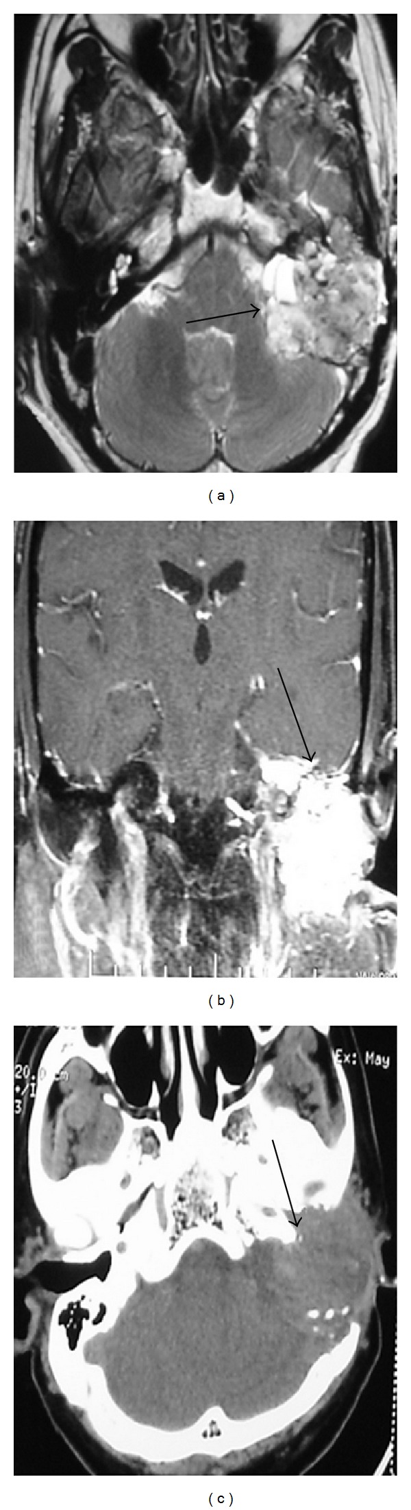
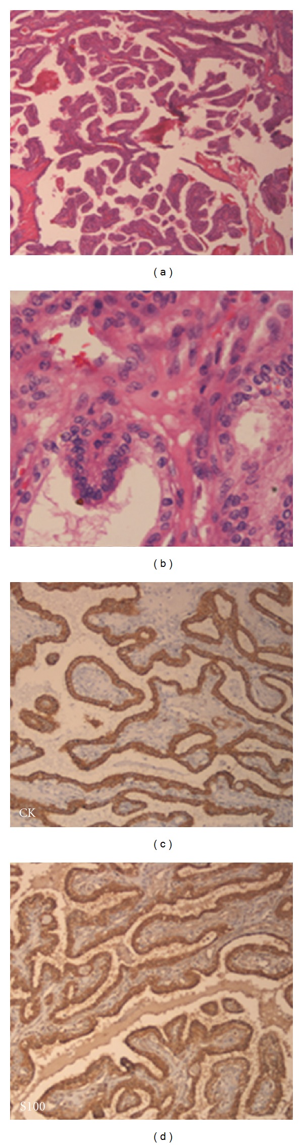
Glandular tumors involving the middle ear are rare and distinguishing between adenoma and adenocarcinoma remains difficult. A distinct subclass of these tumors demonstrates microscopic papillary architecture and has a propensity to erode the petrous bone and extend intracranially. The term "aggressive papillary middle ear tumor" has recently been proposed to describe this more invasive type of middle ear tumor. These tumors cause symptoms even when microscopic in size. Although histologically benign, they have been locally destructive with frequent intracranial extension and patients may die of uncontrolled local disease. These tumors do not metastasize but there is single case report of drop metastasis to the spine in the literature. Hence this tumor must be distinguished from other benign tumors of the middle ear. These rare neoplasms constitute a distinct pathological entity and deserve wider recognition.
Figures
References
-
- Tysome JR, Harcourt J, Patel MC, Sandison A, Michaels L. Aggressive papillary tumor of the middle ear: a true entity or an endolymphatic sac neoplasm? Ear, Nose and Throat Journal. 2008;87(7):378–393. - PubMed
-
- Bambakidis NC, Rodrigue T, Megerian CA, Ratcheson RA. Endolymphatic sac tumor metastatic to the spine. Journal of Neurosurgery. 2005;3(1):68–70. - PubMed
-
- Clark TD. Aggressive middle ear tumor. American Journal of Surgical Pathology. 1989;13(11):985–987. - PubMed
-
- Jagannathan J, Butman JA, Lonser RR, et al. Endolymphatic sac tumor demonstrated by intralabyrinthine hemorrhage. Journal of Neurosurgery. 2007;107(2):421–425. - PubMed
-
- Heffner DK. Low-grade adenocarcinoma of probable endolymphatic sac origin. A clinicopathologic study of 20 cases. Cancer. 1989;64(11):2292–2302. - PubMed
LinkOut - more resources
Full Text Sources
