

Case Reports
Epub 2010 Dec 1.
A giant occipital encephalocele
Affiliations
- PMID: 22953259
- PMCID: PMC3418000
Item in Clipboard
Case Reports
A giant occipital encephalocele
APSP J Case Rep.
2010 Jul.
Abstract
Giant occipital encephaloceles are rare lesions. Because of their enormous size they pose a surgical challenge. Herein we report a four months old female baby who presented with progressively increasing swelling over the occipital region. This swelling was present since birth. Surgery was planned to reduce the size of the swelling as well as its contents. The redundant sac was excised and reduced sufficiently enough to accommodate the healthy looking brain tissue. In contrast to the previous case reports where the neonates had poor prognosis, this infant did well postoperatively.
Keywords: Neurological development; Occipital encephalocele; Microcephaly.
Figures
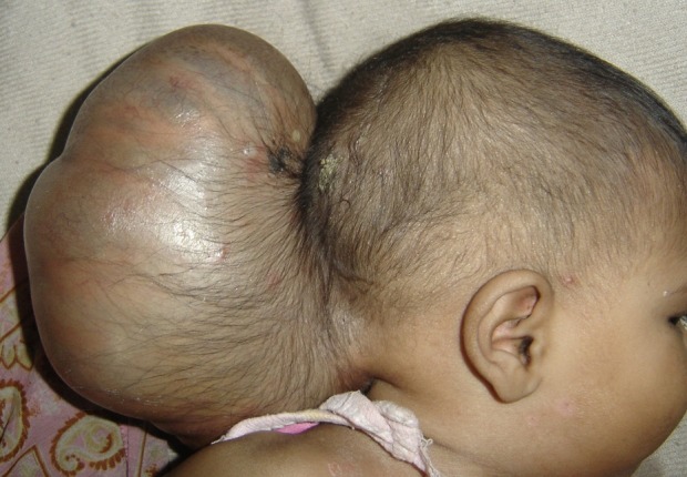
Figure 1: Clinical photograph showing giant occipital encephalocele.
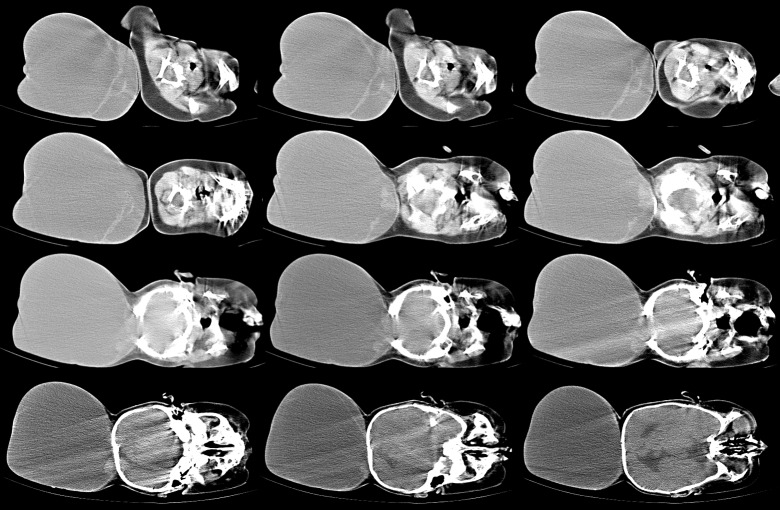
Figure 2: CT scan brain showing large encephalocele sac with protrusion of the contents.
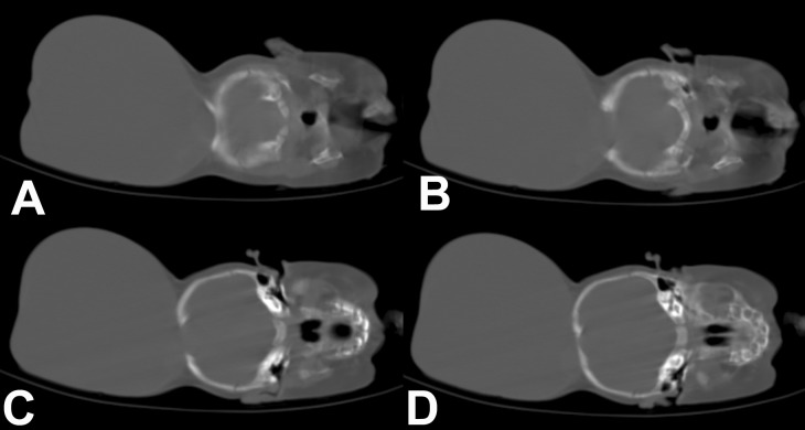
Figure 3: CT scan brain (bone window) showing large defect in the occipital bone with sclerosed margins.
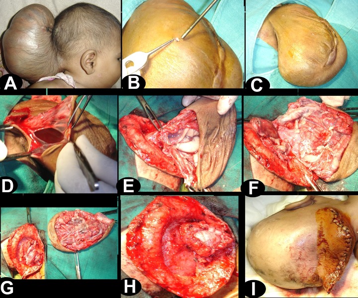
Figure 4: Intra-operative photographs showing operative steps (B) small amount of the CSF was let out through a small opening in the sac, (C) note the reduction in the size of swelling, (D) sac was opened, (E) base of sac was defined, note the presence of large vessels near to the opening in the skull, (F and G) redundant brain tissue was excised while preserving the large vessels, (H and I) dura and skin were closed.
References
-
- Shokunbi T, Adeloye A, Olumide A. Occipital encephalocoeles in 57 Nigerian children: a retrospective analysis. Childs Nerv Syst. 1990; 6: 99– 102. - PubMed
-
- Agrawal A, Lakhkar BB, Lakhkar B, Grover A. Giant occipital encephalocele associated with microcephaly and micrognathia. Pediatr Neurosurg. 2008; 44: 515– 6. - PubMed
-
- Lettau M, Halatsch ME, Hahnel S. [Occipital giant encephalocele] Rofo. 2007; 179: 971– 2. - PubMed
-
- Agrawal D, Mahapatra AK. Giant occipital encephalocele with microcephaly and micrognathia. Pediatr Neurosurg. 2004; 40: 205– 6. - PubMed
-
- Ozlu O, Sorar M, Sezer E, Bayraktar N. Anesthetic management in two infants with giant occipital encephalocele. Paediatr Anaesth. 2008; 18: 792– 3. - PubMed
Publication types
LinkOut - more resources
Full Text Sources