. 2013 Feb;19(3-4):437-47.
doi: 10.1089/ten.TEA.2012.0158. Epub 2012 Oct 19.

Pro-angiogenic and anti-inflammatory regulation by functional peptides loaded in polymeric implants for soft tissue regeneration

Affiliations

Pro-angiogenic and anti-inflammatory regulation by functional peptides loaded in polymeric implants for soft tissue regeneration

Angela L Zachman et al. Tissue Eng Part A. 2013 Feb.

Abstract

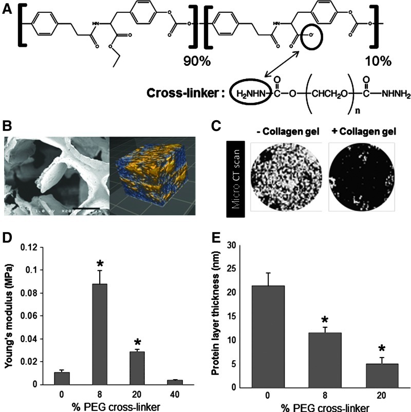
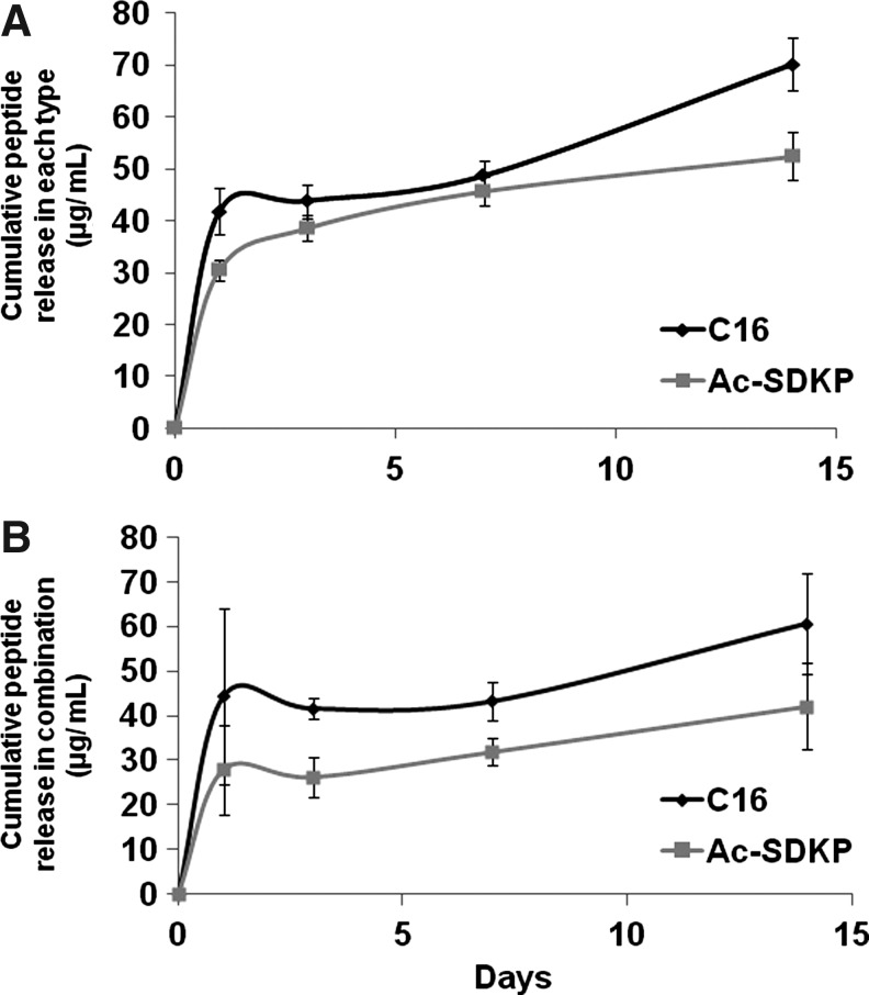
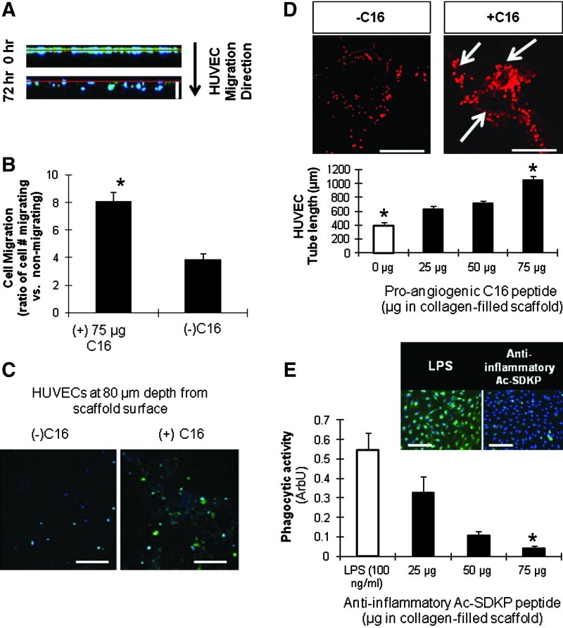
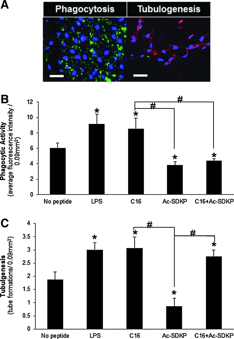
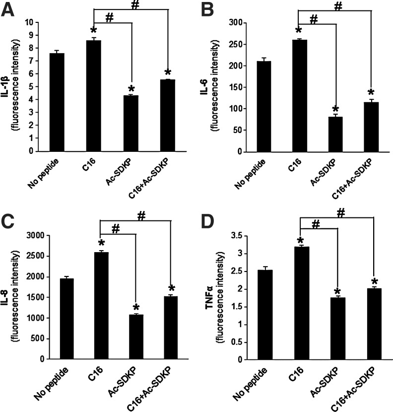
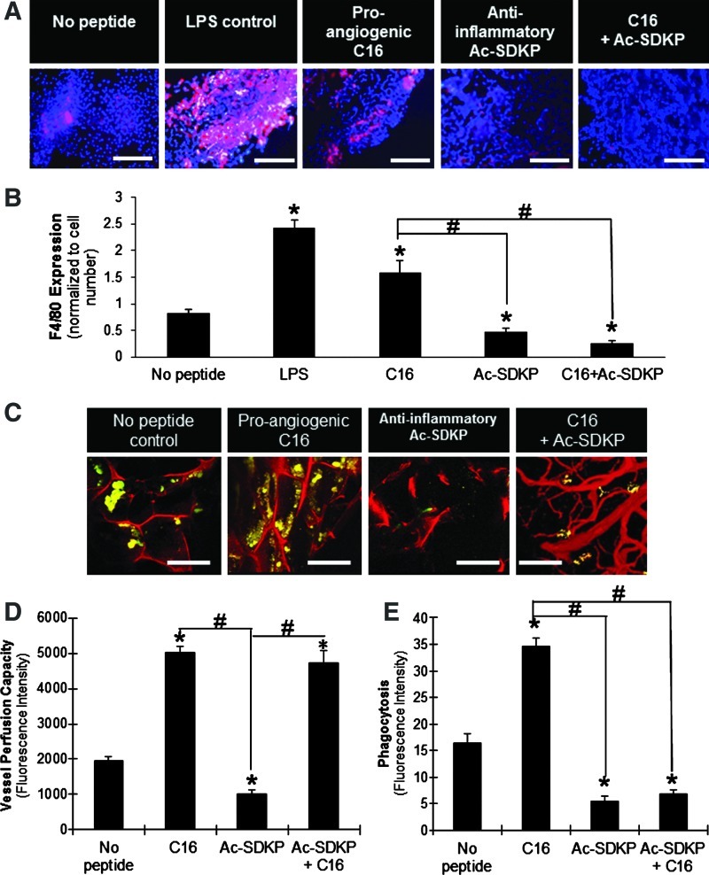
Inflammation and angiogenesis are inevitable in vivo responses to biomaterial implants. Continuous progress has been made in biomaterial design to improve tissue interactions with an implant by either reducing inflammation or promoting angiogenesis. However, it has become increasingly clear that the physiological processes of inflammation and angiogenesis are interconnected through various molecular mechanisms. Hence, there is an unmet need for engineering functional tissues by simultaneous activation of pro-angiogenic and anti-inflammatory responses to biomaterial implants. In this work, the modulus and fibrinogen adsorption of porous scaffolds were tuned to meet the requirements (i.e., ~100 kPa and ~10 nm, respectively), for soft tissue regeneration by employing tyrosine-derived combinatorial polymers with polyethylene glycol crosslinkers. Two types of functional peptides (i.e., pro-angiogenic laminin-derived C16 and anti-inflammatory thymosin β4-derived Ac-SDKP) were loaded in porous scaffolds through collagen gel embedding so that peptides were released in a controlled fashion, mimicking degradation of the extracellular matrix. The results from (1) in vitro coculture of human umbilical vein endothelial cells and human blood-derived macrophages and (2) in vivo subcutaneous implantation revealed the directly proportional relationship between angiogenic activities (i.e., tubulogenesis and perfusion capacity) and inflammatory activities (i.e., phagocytosis and F4/80 expression) upon treatment with either type of peptide. Interestingly, cotreatment with both types of peptides upregulated the angiogenic responses, while downregulating the inflammatory responses. Also, anti-inflammatory Ac-SDKP peptides reduced production of pro-inflammatory cytokines (i.e., interleukin [IL]-1β, IL-6, IL-8, and tumor necrosis factor alpha) even when treated in combination with pro-angiogenic C16 peptides. In addition to independent regulation of angiogenesis and inflammation, this study suggests a promising approach to improve soft tissue regeneration (e.g., blood vessel and heart muscle) when inflammatory diseases (e.g., ischemic tissue fibrosis and atherosclerosis) limit the regeneration process.


Figures

FIG. 1.
FIG. 1.
Scaffold characterization. (A) Chemical structure of backbone polymer and polyethylene glycol (PEG) dihydrazide crosslinkers. (B) Left: scanning electron microscopy image of pore architecture in a scaffold. Scale bar: 150 μm. Right: optical coherence tomography image of pore interconnectivity (2-×2-×2-mm scan), indicating a highly porous structure with a high degree of pore interconnectivity. Blue, pores; yellow, scaffold. (C) Microcomputed tomography scan images showing the entire surface of the scaffold (diameter=0.6 cm) without (left) and with (right) collagen gel at 7 days after gelation, proving the stability of collagen gel in the scaffold. White area, pores; black area, collagen gel or biomaterial scaffold. (D) Young's modulus obtained from compression testing of wet, collagen-filled scaffolds. (E) Protein adsorption on scaffolds, as measured by the thickness of adsorbed fibrinogen layer measured using quartz crystal microbalance with dissipation (QCM-D). (D, E) *p<0.05 versus 0% PEG crosslinker (n=3). Color images available online at www.liebertpub.com/tea
FIG. 2.
FIG. 2.
Cumulative release of peptides from scaffolds. Collagen-filled scaffolds were loaded with 75 μg of either Ac-SDKP or C16 peptides (A), or the combination of both peptides (75 μg each peptide) (B). After gelation, phosphate-buffered saline (PBS) was added on top of the gel-filled scaffolds and left to incubate at 37°C until collected at either 1, 3, 7, or 14 days after gelation. PBS releasate samples were then analyzed by high-performance liquid chromatography to quantify the amount of released peptide, as determined by fitting to a standard curve (n=4 per time point).
FIG. 3.
FIG. 3.
Peptide characterization. (A, B) Human umbilical vein endothelial cell (HUVEC) migration into scaffolds. Cell nuclei (blue) and proliferating cells with BrdU incorporation (green). (A) Z-sectional projection of HUVEC migration from the surface (red line). White scale bar: 120 μm. (B) Effect of C16 peptide (75 μg/scaffold) on HUVEC migration at 72 h. Ratio of migrated versus nonmigrated HUVECs was defined as the number of cells migrated a distance >0 μm into the scaffolds divided by the number of cells remaining at the surface. (C) Representative images of HUVECs that have migrated 80 μm into the scaffold after 72 h. Scale bar: 100 μm. (D) Tubulogenesis (as measured by total tube length) of HUVECs around 40 μm into the scaffold in response to varying doses of pro-angiogenic C16 peptide. Ethidium bromide stained HUVECs with (right) and without (right) C16 shown in top images. White arrows indicate points of tube formation. Scale bar: 100 μm. (E) MDM phagocytic activity. Macrophages (blue) and phagocytized Escherichia coli particles (green) shown in the top images. The phagocytic activity presented by the green fluorescence intensity normalized to cell number in the bottom graph. Scale bar: 100 μm. (B, D, E) *p<0.05 versus all the other conditions in same graph (n=5). MDM, monocyte-derived macrophage. Color images available online at www.liebertpub.com/tea
FIG. 4.
FIG. 4.
In vitro coculture of MDMs and HUVECs in scaffolds. (A) Representative images of phagocytic macrophages (green, left) and HUVECs stained for vascular cell adhesion molecule-1 (VCAM-1) (red, right) as an indicator of inflammatory-stimulated tubulogenesis in C16 containing scaffolds. Scale bar: 50 μm. (B) Macrophage phagocytic activity as measured by average green fluorescence intensity per image field. (C) Tubulogenesis of HUVECs as measured by the number of tube formations per image field. (B, C) *p<0.05 compared to no peptide treatment; #p<0.05 between groups connected by lines (n=8). Color images available online at www.liebertpub.com/tea
FIG. 5.
FIG. 5.
Pro-inflammatory cytokine secretion. (A) Interleukin (IL)-1β, (B) IL-6, (C) IL-8, (D) tumor necrosis factor alpha (TNF-α) cytokine release from a coculture of HUVECs and MDMs in peptide-loaded scaffolds as measured by BD Cytometric Bead Array. *p<0.05 compared to no peptide treatment; #p<0.05 between groups connected by lines (n=4).
FIG. 6.
FIG. 6.
In vivo implantation of scaffolds. (A) F4/80-positive macrophages (red) with cell nuclei (blue) infiltrated into implanted peptide-loaded scaffolds. Scale bar: 100 μm. (B) Quantification of F4/80 expression normalized to the corresponding cell number. (C) Blood vessel formation (red) visualized by fluorescent microangiography, and macrophages phagocytosing E. coli particles (yellow-green) in scaffold implants. Scale bar: 100 μm. (D) Vessel perfusion capacity as measured by red fluorescence intensity of perfused microspheres extracted from scaffolds. (E) Phagocytic activity (fluorescence intensity per image field). (B, D, E) *p<0.05 compared to no peptide treatment; #p<0.05 between groups connected by lines (n=4). Color images available online at www.liebertpub.com/tea

Similar articles

Cited by

References

    1. Bailey L.O. Washburn N.R. Simon C.G. Chan E.S. Wang F.W. Quantification of inflammatory cellular responses using real-time polymerase chain reaction. J Biomed Mater Res A. 2004;69A:305. - PubMed
    1. Hu W.J. Eaton J.W. Tang L.P. Molecular basis of biomaterial-mediated foreign body reactions. Blood. 2001;98:1231. - PubMed
    1. Ratner B.D. Gladhill K.W. Horbett T.A. Analysis of in vitro enzymatic and oxidative degradation of polyurethanes. J Biomed Mater Res. 1988;22:509. - PubMed
    1. Silva M.M. Cyster L.A. Barry J.J. Yang X.B. Oreffo R.O. Grant D.M., et al. The effect of anisotropic architecture on cell and tissue infiltration into tissue engineering scaffolds. Biomaterials. 2006;27:5909. - PubMed
    1. Robbins C.S. Swirski F.K. The multiple roles of monocyte subsets in steady state and inflammation. Cell Mol Life Sci. 2010;67:2685. - PMC - PubMed

Publication types

MeSH terms