Review
. 2012 Sep:1267:18-23.
doi: 10.1111/j.1749-6632.2012.06593.x.

The tricky path to recombining X and Y chromosomes in meiosis

Affiliations
Review

The tricky path to recombining X and Y chromosomes in meiosis

Liisa Kauppi et al. Ann N Y Acad Sci. 2012 Sep.

Abstract

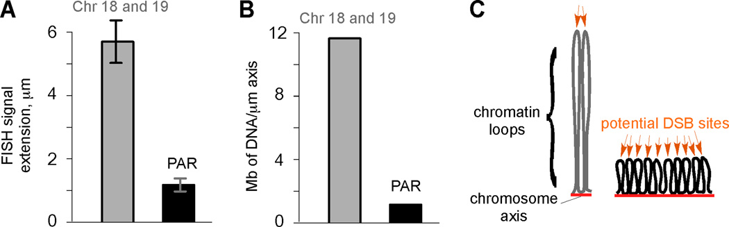
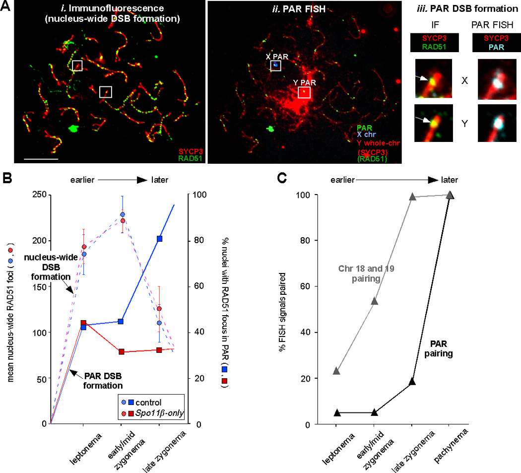
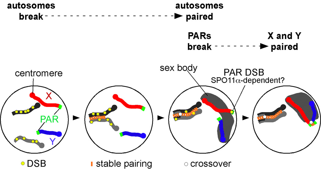
Sex chromosomes are the Achilles' heel of male meiosis in mammals. Mis-segregation of the X and Y chromosomes leads to sex chromosome aneuploidies, with clinical outcomes such as infertility and Klinefelter syndrome. Successful meiotic divisions require that all chromosomes find their homologous partner and achieve recombination and pairing. Sex chromosomes in males of many species have only a small region of homology (the pseudoautosomal region, PAR) that enables pairing. Until recently, little was known about the dynamics of recombination and pairing within mammalian X and Y PARs. Here, we review our recent findings on PAR behavior in mouse meiosis. We uncovered unexpected differences between autosomal chromosomes and the X-Y chromosome pair, namely that PAR recombination and pairing occurs later, and is under different genetic control. These findings imply that spermatocytes have evolved distinct strategies that ensure successful X-Y recombination and chromosome segregation.


Figures

Figure 1
Figure 1
Higher-order structure of the PAR compared to autosomes. A) Measurements of the maximal distance of FISH signals from chromosome axes (average for subtelomeric chromosome 18 and 19 probes, gray bar; PAR probe, black bar) in cells at pachynema (mean ± SD). B) DNA content (Mb) per µm of chromosome axis in the subtelomeric regions of chromosomes 18 and 19, and the PAR. C) Model of how organization of DNA into chromatin loops can influence DSB frequency (after ref. 18). Only one homolog (each consisting of a pair of sister chromatids) is shown, with chromatin loops tethered to chromosome axes (red lines). Each chromatin loop is envisioned to provide an opportunity for meiotic DSB formation (orange arrows). DNA organized into more and smaller loops per Mb (right) presents the DSB machinery with more opportunities for break formation than if the same length of DNA is organized into fewer and larger loops (left).
Figure 2
Figure 2
Many PAR DSBs form later than nucleus-wide DSBs, and are under different genetic control. A) Assay for nucleus-wide and PAR DSB formation. i-ii) Example of immunofluorescent (IF) and FISH staining of a spermatocyte in late zygonema. Nuclei were first stained with antibodies against RAD51 and SYCP3 and photographed (i), then FISH was performed using PAR, X and Y probes (as cartooned above the FISH image) and the same nuclei were photographed again (ii). Under the FISH conditions used in these experiments, some IF signal remains visible. The presence or absence of a RAD51 focus in the X and Y PAR was scored by comparing the IF image to an IF+FISH image overlay (iii). In this example, both the X and Y PAR (inside white squares in i-ii) display a RAD51 focus (white arrows in iii). Scale bar, 10 µm. B) Mean nucleus-wide RAD51 focus numbers (bars ± 95% confidence interval of the mean; left Y axis) and percentage of nuclei with a PAR RAD51 focus on the X and/or Y PAR (right Y axis) as prophase I progresses from leptonema to late zygonema, in mice of the indicated genotypes (see text). C) Percentage of nuclei with paired FISH signals for autosomal and PAR probes as prophase I progresses from leptonema to pachynema.
Figure 3
Figure 3
Model summarizing our findings on the distinct behavior of meiotic X and Y chromosomes. A pair of autosomal homologous chromosomes is shown as light and dark gray lines, the X and Y as red and blue lines, respectively. Most DSBs on autosomes form before PAR DSBs; as a consequence, autosomal chromosomes pair earlier than the X and Y. Only DSBs that can facilitate homolog pairing are depicted (yellow circles); not shown are the numerous DSBs that form on the non-PAR portion of the X chromosome (see ref. 4) but cannot mediate pairing. In many cells, PARs undergo DSB formation later, around the same time as the sex body (hatched area) begins to form. This chromatin domain brings the X and Y closer together and likely facilitates PAR pairing.

References

    1. Shi Q, et al. Single sperm typing demonstrates that reduced recombination is associated with the production of aneuploid 24, XY human sperm. Am. J. Med. Genet. 2001;99:34–38. - PubMed
    1. Shi Q, Martin RH. Aneuploidy in human spermatozoa: FISH analysis in men with constitutional chromosomal abnormalities, and in infertile men. Reproduction. 2001;121:655–666. - PubMed
    1. Raudsepp T, et al. The pseudoautosomal region and sex chromosome aneuploidies in domestic species. Sex. Dev. 2012;6:72–83. - PubMed
    1. Kauppi L, et al. Distinct properties of the XY pseudoautosomal region crucial for male meiosis. Science. 2011;331:916–920. - PMC - PubMed
    1. Zickler D, Kleckner N. Meiotic chromosomes: integrating structure and function. Annu. Rev. Genet. 1999;33:603–754. - PubMed

Publication types