. 2012 Sep 6;10(1):54.
doi: 10.1186/1477-5956-10-54.

Technical updates to basic proteins focalization using IPG strips

Affiliations

Technical updates to basic proteins focalization using IPG strips

Jordane Dépagne et al. Proteome Sci. .

Abstract

Background: Gel-based proteomic is a popular and versatile method of global protein separation and quantification. However, separation of basic protein still represents technical challenges with recurrent problems of resolution and reproducibility.

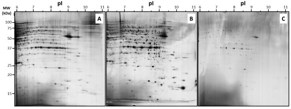
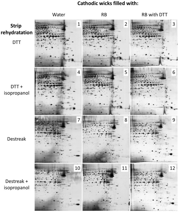
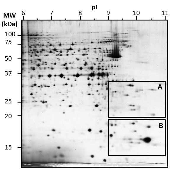
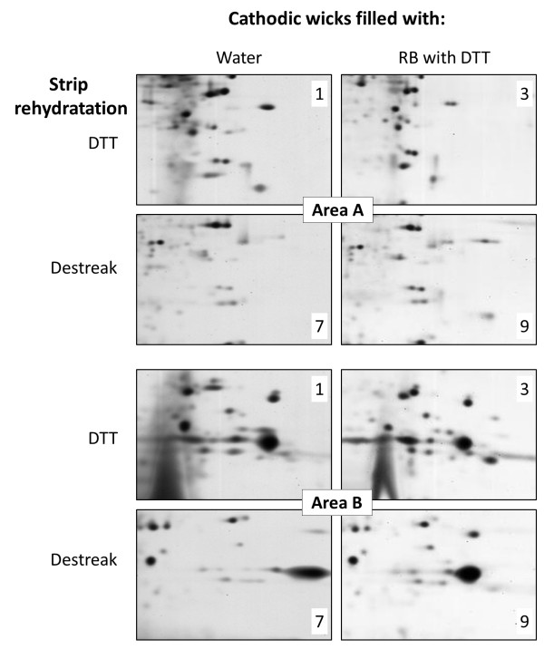
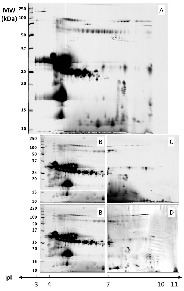
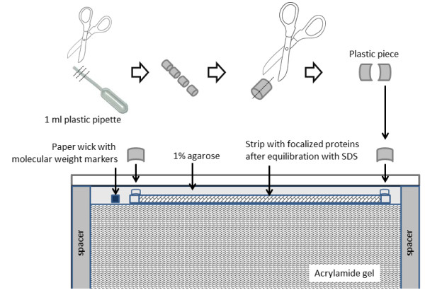
Results: Three different protocols of protein loading were compared using MCF7 cells proteins. In-gel rehydration, cup-loading and paper-bridge loading were first compared using 6-11 IPG strips, as attempted, in-gel rehydration gave large horizontal steaking; paper-bridge loading displayed an interesting spot resolution, but with a predominant loss of material; cup-loading was selected as the most relevant method, but still needing improvement. Twelve cup-loading protocols were compared with various strip rehydration, and cathodic wick solutions. Destreak appeared as better than DTT for strip rehydration; the use of isopropanol gave no improvement. The best 2DE separation was observed with cathodic wicks filled with rehydration solution complemented with DTT. Paper-bridge loading was finally analyzed using non-limited samples, such as bovine milk. In this case, new spots of basic milk proteins were observed, with or without paper wicks.

Conclusion: According to this technical study of basic protein focalization with IPG strips, the cup-loading protocol clearly displayed the best resolution and reproducibility: strips were first rehydrated with standard solution, then proteins were cup-loaded with destreak reagent, and focalisation was performed with cathodic wicks filled with rehydration solution and DTT. Paper-bridge loading could be as well used, but preferentially with non-limited samples.


Figures

Figure 1
Figure 1
2DE separation of MCF7 basic protein: iso-electric focalisation was performed with 50 μg proteins using 6–11 pH IPG strip and 12% acrylamide was used for the second dimension. Proteins were loaded by in-gel rehydration (A), anodic cup loading (B), and paper-bridge loading (C).
Figure 2
Figure 2
Comparison of MCF7 basic protein 2DE separation using twelve different anodic cup-loading protocols. Iso-electric focalisation was performed with 50 μg proteins using 6–11 pH IPG strip and 12% acrylamide was used for the second dimension. The number reported on each picture corresponded to the cup-loading condition used, as presented in detail in Table 1.
Figure 3
Figure 3
2DE map of MCF7 basic protein separation using anodic cup-loading protocol n°9, as presented in Figure 2 and Table 1. Specific areas A and B were further analysed.
Figure 4
Figure 4
Picture comparison of areas A and B from Figure 3 as a function of different anodic cup-loading protocols. The number of spots within each area was estimated by picture analysis and reported in Table 1.
Figure 5
Figure 5
2DE map comparison and alignment of bovine milk proteins using in-gel rehydration or paper-bridge loading. 600 μg of non-fat dry milk proteins were in-gel loaded using 3–10 IPG strips (A) or paper-bridge loaded using 6–11 IPG strips with (C) or without electrode wicks (D) during focalization. In a second time, paper-bridge previously used with 6–11 IPG strip was applied with 4–7 IPG strips (B) to focalize remaining proteins.
Figure 6
Figure 6
Schematic of IPG strip, electrodes and paper wicks positions as function of protein loading protocols. A: sample in-gel rehydration of IPG strip, with acrylamide face down. B: cup-loading of protein sample, with acrylamide face up, a plastic sample cup was placed at the anodic end, paper wicks connected strip with electrodes.
Figure 7
Figure 7
Schematic of IPG strip placement before the second dimension. A special precaution is presented to seal strips with 1% agarose and curved plastic pieces.

References

    1. Van den Bergh G, Arckens L. High Resolution Protein Display by Two-Dimensional Electrophoresis. Curr Anal Chem. 2009;5:106–115. doi: 10.2174/157341109787846199. - DOI
    1. Chevalier F. Highlights on the capacities of “Gel-based” proteomics. Proteome Science. 2010;8:23. doi: 10.1186/1477-5956-8-23. - DOI - PMC - PubMed
    1. Rabilloud T, Lelong C. Two-dimensional gel electrophoresis in proteomics: A tutorial. J Proteomics. 2011;74:1829–1841. doi: 10.1016/j.jprot.2011.05.040. - DOI - PubMed
    1. Luche S, Diemer H, Tastet C, Chevallet M, Van Dorsselaer A, Leize-Wagner E, Rabilloud T. About thiol derivatization and resolution of basic proteins in two-dimensional electrophoresis. Proteomics. 2004;4:551–561. doi: 10.1002/pmic.200300589. - DOI - PMC - PubMed
    1. Görg A, Drews O, Lück C, Weiland F, Weiss W. 2-DE with IPGs. Electrophoresis. 2009;30:S122–S132. doi: 10.1002/elps.200900051. - DOI - PubMed

LinkOut - more resources