. 2012 Sep 27;55(18):7998-8006.
doi: 10.1021/jm300804e. Epub 2012 Sep 18.

Small molecule inhibitors of Bacillus anthracis protective antigen proteolytic activation and oligomerization

Affiliations

Small molecule inhibitors of Bacillus anthracis protective antigen proteolytic activation and oligomerization

Alexander N Wein et al. J Med Chem. .

Abstract

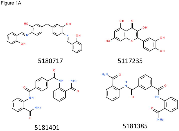
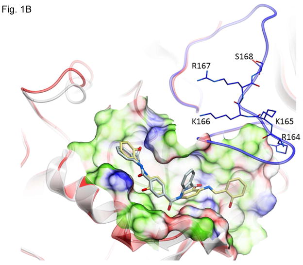
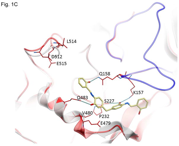
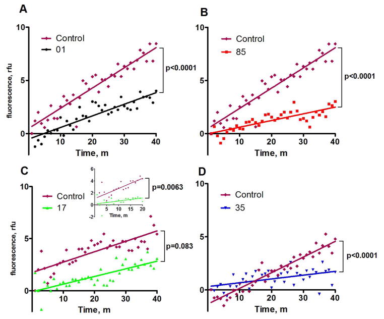
Protective antigen (PA), lethal factor, and edema factor, the protein toxins of Bacillus anthracis , are among its most important virulence factors and play a key role in infection. We performed a virtual ligand screen of a library of 10000 members to identify compounds predicted to bind to PA and prevent its oligomerization. Four of these compounds slowed PA association in a FRET-based oligomerization assay, and two of those protected cells from intoxication at concentrations of 1-10 μM. Exploration of the protective mechanism by Western blot showed decreased SDS-resistant PA oligomer on cells and, surprisingly, decreased amounts of activated PA. In vitro assays showed that one of the inhibitors blocked furin-mediated cleavage of PA, apparently through its binding to the PA substrate. Thus, we have identified inhibitors that can independently block both PA's cleavage by furin and its subsequent oligomerization. Lead optimization on these two backbones may yield compounds with high activity and specificity for the anthrax toxins.


Figures

Figure 1
Figure 1
Structures of inhibitors and modes of binding to PA. (A) The compounds characterized in this study. (B) PA crystal structure 1T6B (red ribbon) superimposed on the crystal structure 3TEW (grey ribbon) with the ordered furin loop in 3TEW highlighted in blue. The furin-type protease cleaves after the sequence 164RKKR which is shown in stick representation with carbon atoms colored blue. The predicted binding poses for inhibitors 17 and 01 are displayed in stick representation with carbon atoms colored yellow and grey, respectively. The binding pocket surface for 1T6B used for virtual screening is displayed (White = neutral surface, Green = hydrophobic surface, Red = hydrogen bonding acceptor potential, Blue = hydrogen bond donor potential). (C) and (D) Predicted interactions of 17 (yellow stick) and 01 (grey stick), respectively, with PA. Hydrogen bonds are displayed as small colored spheres and both ligands make common hydrogen bonds with Q158, Q483, and K157.
Figure 1
Figure 1
Structures of inhibitors and modes of binding to PA. (A) The compounds characterized in this study. (B) PA crystal structure 1T6B (red ribbon) superimposed on the crystal structure 3TEW (grey ribbon) with the ordered furin loop in 3TEW highlighted in blue. The furin-type protease cleaves after the sequence 164RKKR which is shown in stick representation with carbon atoms colored blue. The predicted binding poses for inhibitors 17 and 01 are displayed in stick representation with carbon atoms colored yellow and grey, respectively. The binding pocket surface for 1T6B used for virtual screening is displayed (White = neutral surface, Green = hydrophobic surface, Red = hydrogen bonding acceptor potential, Blue = hydrogen bond donor potential). (C) and (D) Predicted interactions of 17 (yellow stick) and 01 (grey stick), respectively, with PA. Hydrogen bonds are displayed as small colored spheres and both ligands make common hydrogen bonds with Q158, Q483, and K157.
Figure 2
Figure 2
FRET assays demonstrating inhibition of PA oligomerization. The dye-conjugated, nicked PA proteins nPA83 N645C*488 and nPA83 N645C*594 were added to a 96-well plate at 250 nM each. Inhibitors were added to a concentration of 100 μM to all wells except the control and oligomerization was driven by the addition of LF at a final concentration of 50 nM. Fluorescence at 610 nm was monitored in wells containing nPA83 and tested compounds in the presence of 50 nM LF. Plotted fluorescence is the difference in fluorescence of wells containing identical mixtures with and without inhibitors.
Figure 3
Figure 3
Protection of RAW264.7 and HT1080 cells by small molecule toxin inhibitors. RAW264.7 cells were exposed to 0.9 nM PA with 5.6 nM LF (A) or 0.06 nM PA with 1.88 nM FP59 (B) and HT1080 cells were exposed to 0.6 nM PA (C) or PA-L1 (D) with 1.88 nM FP59 for 5 h. The PA + LF cells were stained with MTT immediately (A) while the PA + FP59 and PA-L1 + FP59 cells (B-D) were treated with 10 mM ammonium chloride after 5 h to stop intoxication and MTT stained 48 h later. Data are plotted as mean +/- standard error of four independent experiments. Percent survival is compared to a non-intoxicated control.
Figure 4
Figure 4
Assessment of PA binding and oligomerization to cells, and in vitro furin activity. (A) CHO C4 cells were treated with PA or PA + LF for 1 h in the presence or absence of 100 μM inhibitor and the cells were lysed and analyzed by Western blotting for PA species. (B, C) Purified PA (B) or TGFα-PE38 (C) were mixed with furin at a 100:1 molar ratio and incubated for 1 h with the indicated concentrations of inhibitors. Wells labeled ND contained no drug, and wells labeled NT were not treated with toxins in (A) or not treated with furin in (B and C).

References

    1. Frankel AE, Kuo SR, Dostal D, Watson L, Duesbery NS, Cheng CP, Cheng HJ, Leppla SH. Pathophysiology of anthrax. Front Biosci. 2009;14:4516–4524. - PMC - PubMed
    1. Kintzer AF, Thoren KL, Sterling HJ, Dong KC, Feld GK, Tang II, Zhang TT, Williams ER, Berger JM, Krantz BA. The protective antigen component of anthrax toxin forms functional octameric complexes. J Mol Biol. 2009;392:614–629. - PMC - PubMed
    1. Moayeri M, Leppla SH. Cellular and systemic effects of anthrax lethal toxin and edema toxin. Mol Aspects Med. 2009;30:439–455. - PMC - PubMed
    1. Koehler TM. Bacillus anthracis physiology and genetics. Mol Aspects Med. 2009;30:386–396. - PMC - PubMed
    1. Liu S, Miller-Randolph S, Crown D, Moayeri M, Sastalla I, Okugawa S, Leppla SH. Anthrax toxin targeting of myeloid cells through the CMG2 receptor Is essential for establishment of Bacillus anthracis infections in mice. Cell Host Microbe. 2010;8:455–462. - PMC - PubMed

Publication types

MeSH terms

LinkOut - more resources