

Circulating miR-200c as a diagnostic and prognostic biomarker for gastric cancer
- PMID: 22954417
- PMCID: PMC3494541
- DOI: 10.1186/1479-5876-10-186
Circulating miR-200c as a diagnostic and prognostic biomarker for gastric cancer
Abstract
Background: MicroRNAs are aberrantly expressed and correlate with tumourigenesis and the progression of solid tumours. The miR-200 family determines the epithelial phenotype of cancer cells and regulates invasiveness and migration. Thus, we hypothesised that the quantitative detection of the miR-200 family as epithelial-specific microRNAs in the blood could be a useful clinical biomarker for gastric cancer (GC).
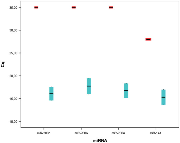
Methods: We initially validated the expression levels of miR-200a, 200b, 200c and 141 in GC cell lines (n = 2) and blood from healthy controls (n = 19) using real-time quantitative reverse transcription PCR (qRT-PCR). The microarray expression profiles of the miR-200 family in 160 paired samples of non-tumour gastric mucosae and GC were downloaded through ArrayExpress and analysed. MiR-200c was selected for clinical validation. The qRT-PCR prospective assessment of miR-200c was performed using 67 blood samples (52 stage I-IV GC patients and 15 controls); the area under the receiver operating characteristic curve (AUC-ROC) was estimated. The Kaplan-Meier and Breslow-Wilcoxon tests were used to assess the correlation of miR-200c with overall and progression-free survival (OS and PFS). Multivariate analyses were performed using the Cox model.
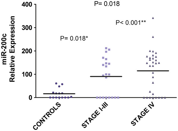
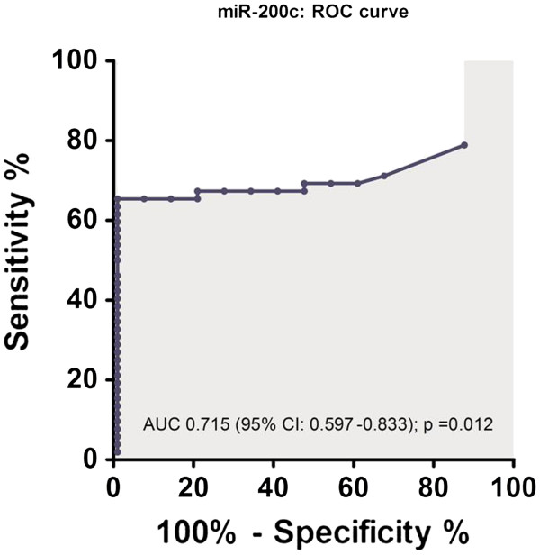
Results: The miR-200c blood expression levels in GC patients were significantly higher than in normal controls (p = 0.018). The AUC-ROC was 0.715 (p = 0.012). The sensitivity, specificity and accuracy rates of 65.4%, 100% and 73.1%, respectively, were observed. The levels of miR-200c in the blood above the cutoff defined by the ROC curve was found in 17.6% of stage I-II GC patients, 20.6% of stage III patients and 67.7% of stage IV patients (p < 0.001). The miR-200c expression levels were not associated with clinical or pathological characteristics or recent surgical procedures. There was a correlation (p = 0.016) with the number of lymph node metastases and the increased expression levels of miR-200c in blood were significantly associated with a poor OS (median OS, 9 vs 24 months; p = 0.016) and PFS (median PFS, 4 vs 11 months; p = 0.044). Multivariate analyses confirmed that the upregulation of miR-200c in the blood was associated with OS (HR = 2.24; p = 0.028) and PFS (HR = 2.27; p = 0.028), independent of clinical covariates.
Conclusions: These data suggest that increased miR-200c levels are detected in the blood of gastric cancer patients. MiR-200c has the potential to be a predictor of progression and survival.
Figures

Similar articles
-
Expression of serum miR-200a, miR-200b, and miR-200c as candidate biomarkers in epithelial ovarian cancer and their association with clinicopathological features.Clin Transl Oncol. 2015 Oct;17(10):779-87. doi: 10.1007/s12094-015-1303-1. Epub 2015 Jun 11. Clin Transl Oncol. 2015. PMID: 26063644
-
Association between miR-200c and the survival of patients with stage I epithelial ovarian cancer: a retrospective study of two independent tumour tissue collections.Lancet Oncol. 2011 Mar;12(3):273-85. doi: 10.1016/S1470-2045(11)70012-2. Epub 2011 Feb 21. Lancet Oncol. 2011. PMID: 21345725
-
Circulating microRNA-203 predicts metastases, early recurrence, and poor prognosis in human gastric cancer.Gastric Cancer. 2016 Jul;19(3):744-53. doi: 10.1007/s10120-015-0521-0. Epub 2015 Aug 2. Gastric Cancer. 2016. PMID: 26233325
-
Prognostic Value and Clinicopathology Significance of MicroRNA-200c Expression in Cancer: A Meta-Analysis.PLoS One. 2015 Jun 2;10(6):e0128642. doi: 10.1371/journal.pone.0128642. eCollection 2015. PLoS One. 2015. PMID: 26035744 Free PMC article.
-
Prognostic Role of MicroRNA-200c-141 Cluster in Various Human Solid Malignant Neoplasms.Dis Markers. 2015;2015:935626. doi: 10.1155/2015/935626. Epub 2015 Oct 18. Dis Markers. 2015. PMID: 26556949 Free PMC article. Review.
Cited by
-
Recent Trends and Advancements in the Diagnosis and Management of Gastric Cancer.Cancers (Basel). 2022 Nov 15;14(22):5615. doi: 10.3390/cancers14225615. Cancers (Basel). 2022. PMID: 36428707 Free PMC article. Review.
-
Potential Diagnostic, Prognostic and Therapeutic Targets of MicroRNAs in Human Gastric Cancer.Int J Mol Sci. 2016 Jun 16;17(6):945. doi: 10.3390/ijms17060945. Int J Mol Sci. 2016. PMID: 27322246 Free PMC article. Review.
-
Blood-Based Cancer Biomarkers in Liquid Biopsy: A Promising Non-Invasive Alternative to Tissue Biopsy.Int J Mol Sci. 2018 Sep 21;19(10):2877. doi: 10.3390/ijms19102877. Int J Mol Sci. 2018. PMID: 30248975 Free PMC article. Review.
-
The Diagnostic and Prognostic Value of miR-200c in Gastric Cancer: A Meta-Analysis.Dis Markers. 2019 Apr 4;2019:8949618. doi: 10.1155/2019/8949618. eCollection 2019. Dis Markers. 2019. PMID: 31089400 Free PMC article.
-
Recent advances in gastric cancer early diagnosis.World J Gastroenterol. 2019 May 7;25(17):2029-2044. doi: 10.3748/wjg.v25.i17.2029. World J Gastroenterol. 2019. PMID: 31114131 Free PMC article. Review.
References
-
- Sturgeon CM, Duffy MJ, Hofmann BR, Lamerz R, Fritsche HA, Gaarenstroom K, Bonfrer J, Ecke TH, Grossman HB, Hayes P, Hoffmann RF, Lerner SP, Löhe F, Louhimo J, Sawczuk I, Taketa K, Diamandis EP. National academy of clinical biochemistry laboratory medicine practice guidelines for use of tumor markers in liver, bladder, cervical, and gastric cancers. Clin Chem. 2010;56:e1–e48. doi: 10.1373/clinchem.2009.133124. - DOI - PubMed
Publication types
MeSH terms
Substances
LinkOut - more resources
Full Text Sources
Other Literature Sources
Medical
Miscellaneous
