Review
. 2012 Dec;13(14):1355-73.
doi: 10.4161/cbt.22020. Epub 2012 Sep 6.

The human proton-coupled folate transporter: Biology and therapeutic applications to cancer

Affiliations
Review

The human proton-coupled folate transporter: Biology and therapeutic applications to cancer

Sita Kugel Desmoulin et al. Cancer Biol Ther. 2012 Dec.

Abstract

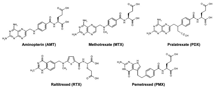
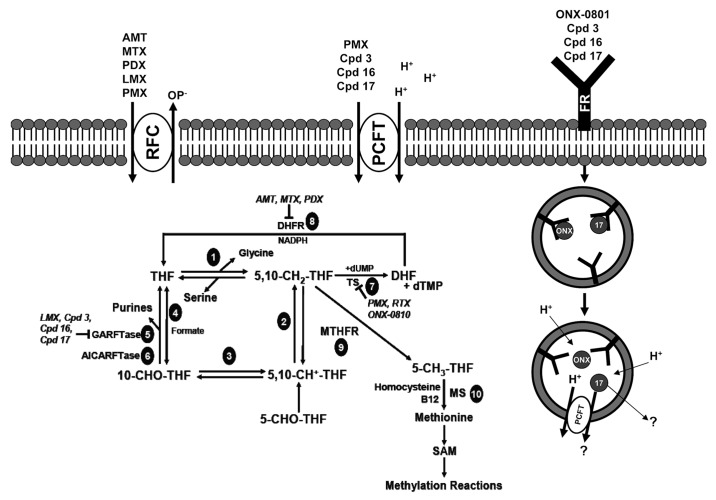
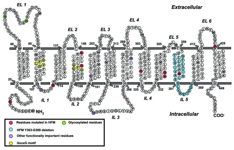
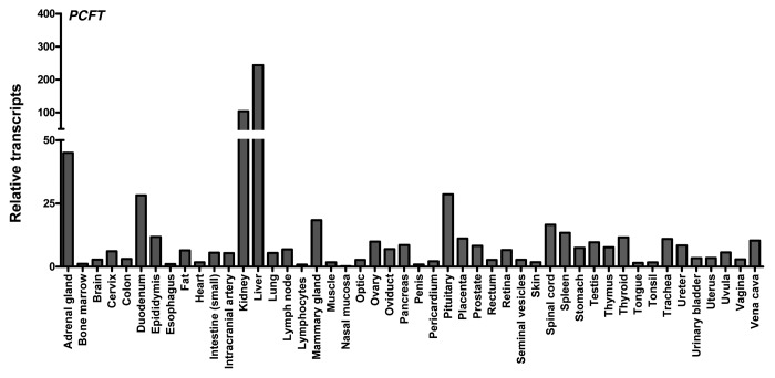
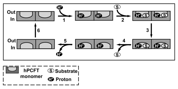
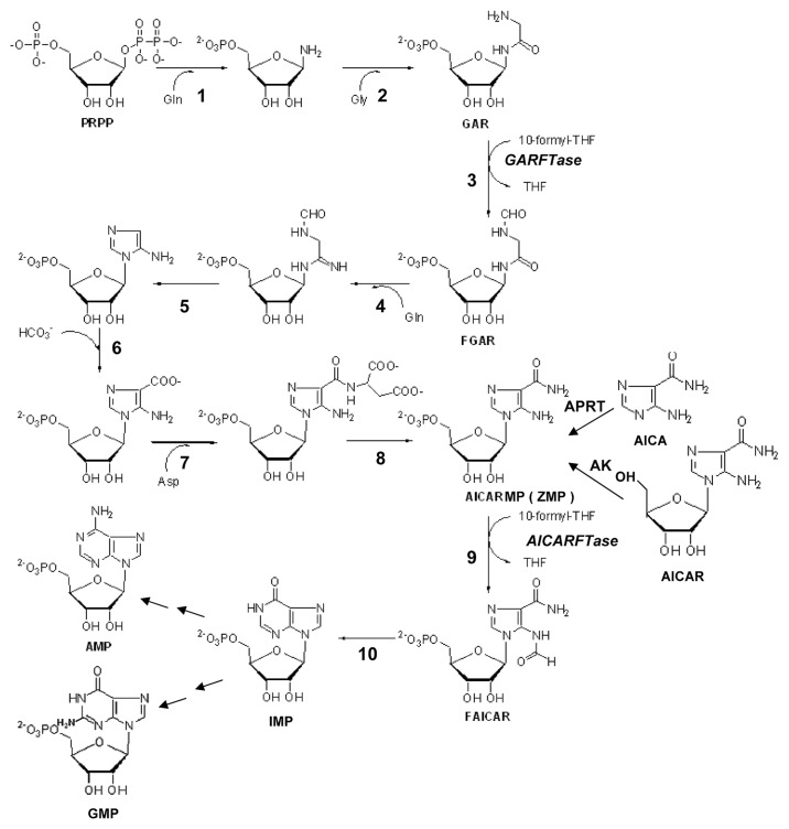
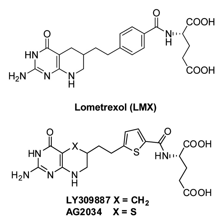
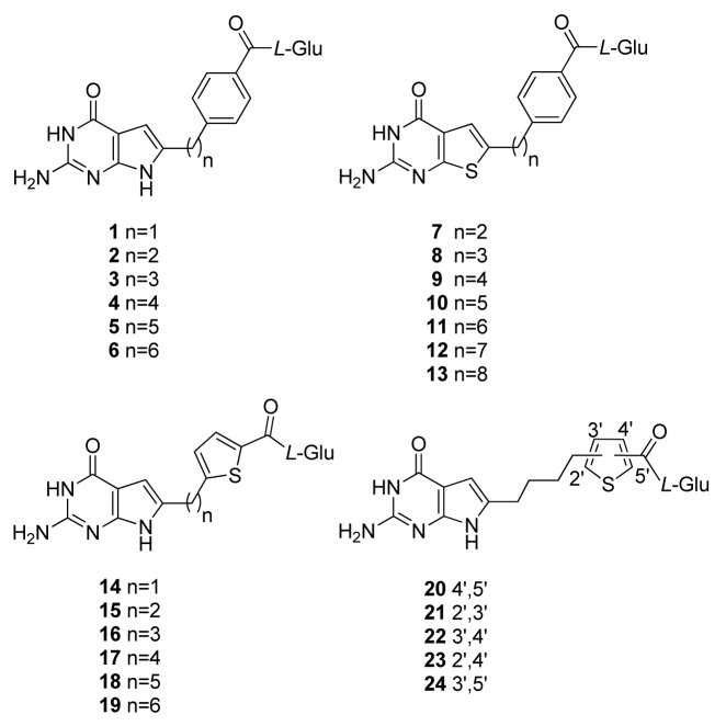
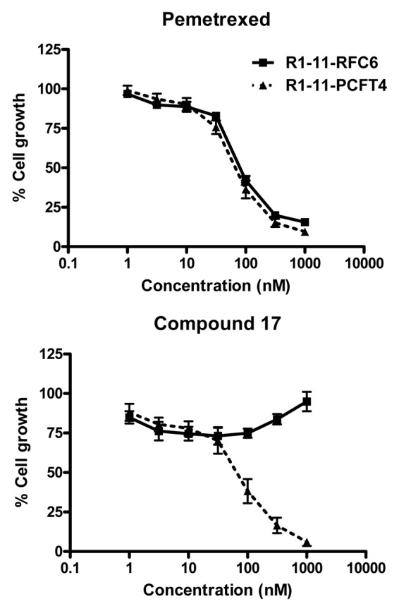
This review summarizes the biology of the proton-coupled folate transporter (PCFT). PCFT was identified in 2006 as the primary transporter for intestinal absorption of dietary folates, as mutations in PCFT are causal in hereditary folate malabsorption (HFM) syndrome. Since 2006, there have been major advances in understanding the mechanistic roles of critical amino acids and/or domains in the PCFT protein, many of which were identified as mutated in HFM patients, and in characterizing transcriptional control of the human PCFT gene. With the recognition that PCFT is abundantly expressed in human tumors and is active at pHs characterizing the tumor microenvironment, attention turned to exploiting PCFT for delivering novel cytotoxic antifolates for solid tumors. The finding that pemetrexed is an excellent PCFT substrate explains its demonstrated clinical efficacy for mesothelioma and non-small cell lung cancer, and prompted development of more PCFT-selective tumor-targeted 6-substituted pyrrolo[2,3-d]pyrimidine antifolates that derive their cytotoxic effects by targeting de novo purine nucleotide biosynthesis.


Figures

None
Figure 1. Structures of clinically relevant antifolates. Structures are shown for aminopterin (AMT), methotrexate (MTX), and pralatrexate (PDX), all dihydrofolate reductase (DHFR) inhibitors, and thymidylate synthase inhibitors raltitrexed (RTX) and pemetrexed (PMX).
None
Figure 2. Folate transporters, folate metabolic pathways and intracellular enzyme targets of antifolates. Folate and antifolate transport across biological membranes is mediated by the reduced folate carrier (RFC), the proton-coupled folate transporter (PCFT) and folate receptors (FR). While RFC and PCFT are facilitative transporters, FRs mediate transport by a non-classical endocytosis involving formation of endosomes that migrate to the cytoplasm. Exit of the folate ligand from the endosome involves diffusion or a mediated process. PCFT has been proposed to facilitate endosomal exit, however this does not appear to be obligatory. Intracellular folates including tetrahydrofolate (THF), dihydrofolate (DHF), 10-formyl tetrahydrofolate (10-CHO-THF), 5, 10-methylene tetrahydrofolate (5,10-CH2-THF), 5,10-methenyl tetrahydrofolate (5,10-CH+-THF), and 5-methyl tetrahydrofolate (5-CH3-THF) participate in folate interconverting and biosynthetic reactions. Reactions 1–4 are in both the cytoplasmic and mitochodrial compartments. Reactions 1 and 9 are also present in the nucleus. Reaction 1 designates serine hydroxymethyltransferase. In the mitochondria, reactions 2 and 3 are catalyzed by bifunctional 5,10-CH2-THF dehydrogenase (MTHFD) 2 or MTHFD2L and 4 is catalyzed by monofunctional MTHFD1L. In the cytoplasm, reactions 4, 3 and 2 are catalyzed by the 10-CHO-THF synthetase, 5,10-CH+-THF cyclohydrolase and MTFD activities, respectively, of the trifunctional C1-THF synthase (MTHFD1). Other steps shown are catalyzed by β-glycinamide ribonucleotide formyltransferase (GARFTase; reaction 5), 5-amino-4-imidazolecarboxamide ribonucleotide formyltransferase (AICARFTase; reaction 6), thymidylate synthase (TS; reaction 7), dihydrofolate reductase (DHFR; reaction 8), 5,10-methyleneTHF reductase (MTHFR; reaction 9), and methionine synthase (MS; reaction 10). Antifolates inhibit folate biosynthetic reactions as shown and include aminopterin (AMT), methotrexate (MTX), pralatrexate (PDX), raltitrexed (RTX), lometrexol (LMX), pemetrexed (PMX), ONX-0801 (ONX), and compounds 3, 16 and 17.
None
Figure 3. Schematic structure of PCFT topology. Structurally or functionally important amino acids, as determined from published mutagenesis studies, are shown as purple circles. Amino acids mutated in patients with hereditary folate malabsorption (HFM) are shown as red and blue circles. GxxxG putative oligomerization motifs are shown as yellow circles and glycosylated residues Asn58 and Asn68 are shown as green circles.
None
Figure 4. PCFT and RFC transcript expression in human normal tissues. PCFT transcripts were measured by real-time RT-PCR using an Origene cDNA array prepared from 48 pathologist-verified human normal tissues as previously described. Transcript levels were normalized to GAPDH transcripts.
None
Figure 5. Proposed reaction scheme for hPCFT-mediated cellular uptake involving cooperative interactions between hPCFT monomers. Based on the “alternate access model” for secondary transporters such as Lac Y, adapted from that of Unal et al. for monomeric PCFT, an analogous reaction scheme is depicted for hPCFT-mediated transport which incorporates the functional impact of hPCFT oligomerization. The model starts from the outward-facing unloaded dimer, followed by the ordered binding of the co-transported protons (step 1) and (anti)folate substrates (step 2), which triggers a conformational change resulting in simultaneous transition of the two hPCFT monomers to an inward-facing state (step 3). This is followed by an ordered release of substrates (step 4) and then protons (step 5) into the cytoplasm. The unloaded homo-oligomeric unit then returns to the outward-facing state (step 6) to complete the transport cycle. In this model, the two hPCFT monomers are suggested to function cooperatively in facilitating substrate and proton binding, conformational changes, and substrate and proton release. From Hou et al.
None
Figure 6. De novo purine nucleotide biosynthesis pathway. The de novo purine nucleotide biosynthetic pathway from phosphoribosyl pyrophosphate (PRPP) to IMP is shown. The numbered reactions are catalyzed by the following monofunctional enzymes: 1, glutamine phosphoribosylpyrophosphate amidotransferase (GPAT); 4, formylglycinamide ribonucleotide synthase (FGAM synthetase); and 8, adenylosuccinate lyase (ASL). Reactions 2, 3 and 5 are catalyzed by the trifunctional glycinamide ribonucleotide (GAR) formyltransferase (GARFTase) which contains GAR synthase (GARS; reaction 2), GAR formyltransferase (GARFTase; reaction 3) and 5-amino-4-imidazole ribonucleotide synthase (AIRS; reaction 5) activities. Reactions 6 and 7 are catalyzed by the bifunctional phosphoribosylaminoimidazole carboxylase/ phosphoribosylaminoimidazole succinocarboxamide synthetase (PAICS) enzyme, which contains carboxyaminoimidazole ribonucleotide synthase (CAIRS; reaction 6) and 5-aminoimidazole-4-(N-succinylocarboxamide) ribonucleotide synthase (SAICARS; reaction 7) activities. Reactions 9 and 10 are catalyzed by a bifunctional enzyme, 5-amino-4-imidazolecarboxamide ribonucleotide (AICAR) formyltransferase (AICARFTase)/IMP cyclohydrolase (ATIC) that sequentially catalyzes the last two steps in the pathway for de novo synthesis of IMP. Folate-dependent reactions (reactions 3 and 9) in which 10-CHO-THF serves as the one-carbon donor are catalyzed by GARFTase and AICARFTase. 5-Aminoimidazole-4-carboxamide (AICA) and AICAR can be metabolized to AICAR monophosphate (ZMP) by adenine phosphoribosyl transferase (APRT) and adenosine kinase (AK), respectively, thus circumventing the reaction catalyzed by GARFTase.
None
Figure 7. Structures of GARFTase inhibitors. Structures are shown for Lometrexol (LMX) and LY309887 and AG2034.
None
Figure 8. 6-Substituted pyrrolo- and thieno[2,3-d]pyrimidine antifolates with hPCFT and FR specificity over hRFC. Structures are shown for 6-substituted pyrrolo[2,3-d]pyrimidine benzoyl antifolates with carbon bridge length variations of 1- to 6-carbons (compounds 1–6, respectively), 6-substituted thieno[2,3-d]pyrimidine benzoyl antifolates with bridge length variations from 2–8 carbons (compounds 7–13, respectively), 6-substituted pyrrolo[2,3-d]pyrimidine antifolates with a thienoyl replacement for the benzoyl moiety and bridge length variations from 1–6 carbons (compounds 14–19, respectively), and 6-substituted pyrrolo[2,3-d]pyrimidine thienoyl regioisomers of compound 17 (compounds 20–24).
None
Figure 9. Characterization of compound 17 and PMX growth inhibition in R1–11 sublines differing in hPCFT and hRFC expression. Cell proliferation inhibition was measured for R1–11-PCFT4 (expresses hPCFT but not hRFC) and -RFC6 (expresses hRFC but not hPCFT) cells treated with PMX (A) or compound 17 (B) over a range of concentrations in complete folate-free RPMI1640 in the presence of 5-CHO-THF at 25 nM for 96 h. Cell densities were measured with CellTiter BlueTM fluorescence dye and a fluorescence plate reader. Results were normalized to cell densities in the absence of drug. From Kugel Desmoulin et al.

References

    1. Farber S, Cutler EC, Hawkins JW, Harrison JH, Peirce EC, 2nd, Lenz GG. The Action of Pteroylglutamic Conjugates on Man. Science. 1947;106:619–21. doi: 10.1126/science.106.2764.619. - DOI - PubMed
    1. Farber S, Diamond LK. Temporary remissions in acute leukemia in children produced by folic acid antagonist, 4-aminopteroyl-glutamic acid. N Engl J Med. 1948;238:787–93. doi: 10.1056/NEJM194806032382301. - DOI - PubMed
    1. Farber S. Some observations on the effect of folic acid antagonists on acute leukemia and other forms of incurable cancer. Blood. 1949;4:160–7. - PubMed
    1. Monahan BP, Allegra CJ. Antifolates. In: Chabner BA, Longo, D.L., ed. Cancer Chemotherapy and Biotherapy. Philadelphia, PA: Lippincott Williams and Wilkins, 2011:109-38.
    1. Zhao R, Goldman ID. Resistance to antifolates. Oncogene. 2003;22:7431–57. doi: 10.1038/sj.onc.1206946. - DOI - PubMed

Publication types

MeSH terms

LinkOut - more resources