. 2012 Nov 15;371(2):235-45.
doi: 10.1016/j.ydbio.2012.08.017. Epub 2012 Aug 30.

Signaling by FGF4 and FGF8 is required for axial elongation of the mouse embryo

Affiliations

Signaling by FGF4 and FGF8 is required for axial elongation of the mouse embryo

Anne M Boulet et al. Dev Biol. .

Abstract

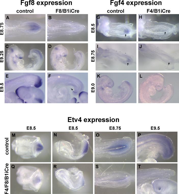
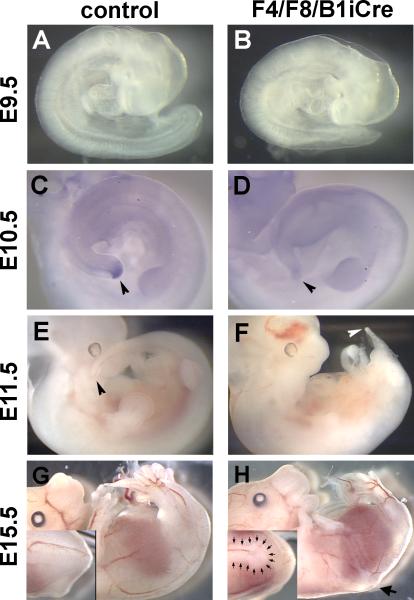
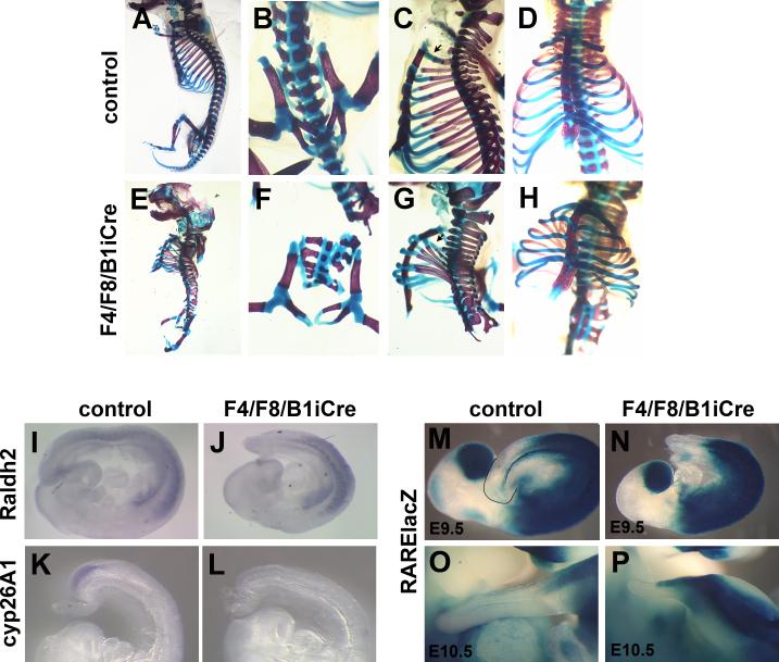
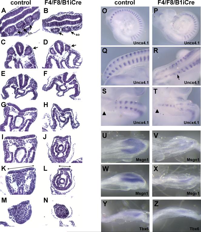
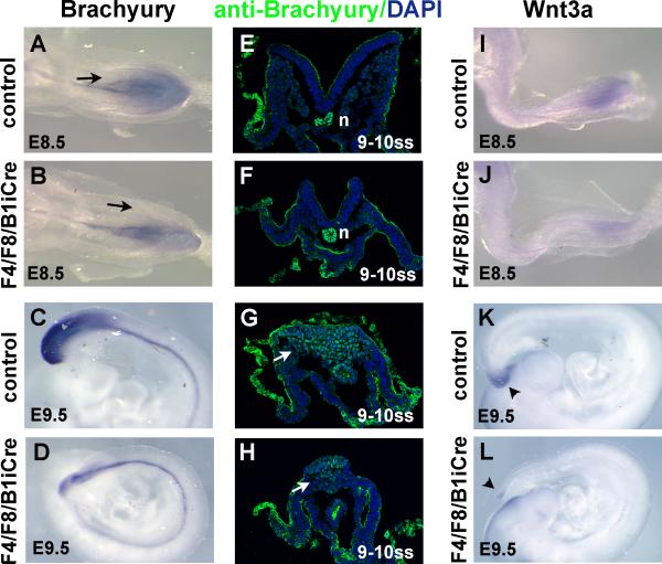
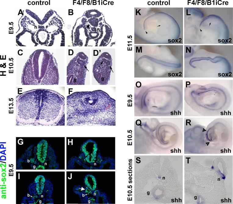
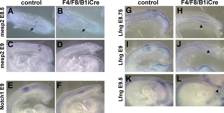
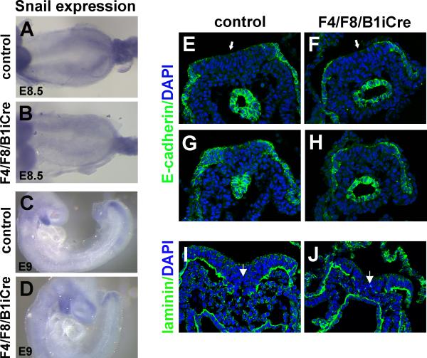
Fibroblast growth factor (FGF) signaling has been shown to play critical roles in vertebrate segmentation and elongation of the embryonic axis. Neither the exact roles of FGF signaling, nor the identity of the FGF ligands involved in these processes, has been conclusively determined. Fgf8 is required for cell migration away from the primitive streak when gastrulation initiates, but previous studies have shown that drastically reducing the level of FGF8 later in gastrulation has no apparent effect on somitogenesis or elongation of the embryo. In this study, we demonstrate that loss of both Fgf8 and Fgf4 expression during late gastrulation resulted in a dramatic skeletal phenotype. Thoracic vertebrae and ribs had abnormal morphology, lumbar and sacral vertebrae were malformed or completely absent, and no tail vertebrae were present. The expression of Wnt3a in the tail and the amount of nascent mesoderm expressing Brachyury were both severely reduced. Expression of genes in the NOTCH signaling pathway involved in segmentation was significantly affected, and somite formation ceased after the production of about 15-20 somites. Defects seen in the mutants appear to result from a failure to produce sufficient paraxial mesoderm, rather than a failure of mesoderm precursors to migrate away from the primitive streak. Although the epiblast prematurely decreases in size, we did not detect evidence of a change in the proliferation rate of cells in the tail region or excessive apoptosis of epiblast or mesoderm cells. We propose that FGF4 and FGF8 are required to maintain a population of progenitor cells in the epiblast that generates mesoderm and contributes to the stem cell population that is incorporated in the tailbud and required for axial elongation of the mouse embryo after gastrulation.


Figures

Fig. 1
Fig. 1. Loss of Fgf4 and Fgf8 expression after recombination mediated by B1iCre results in a reduction in FGF signaling in the posterior embryo
(A-F) Whole mount in situ hybridization with an Fgf8 probe to E8.75 control (A) and F8/B1i Cre mutant (B), E9.25 control (C) and F8/B1iCre mutant (D), and E9.5 control (E) and F8/B1iCre mutant (F) embryos. Arrow in (F) points to hindgut. (G-L) Fgf4 expression in E8.5 control (G) and F4/B1iCre mutant (H), E8.75 control (I) and F4/B1iCre mutant (J), and E9 control (K) and F4/B1iCre mutant (L) embryos. Arrows in (G), (H), (I) and (J) point to the site of Fgf4 expression in the primitive streak. (M-T) Etv4 expression, detected by whole mount in situ hybridization, in control embryos at E8.5 (M,N), E8.75 (O), and E9.5 (P), and F4/F8/B1iCre mutant embryos at E8.5 (Q,R), E8.75 (S), and E9.5 (T). (M) and (Q) are ventral views of the primitive streak region; (O) and (S) are dorsal views; and (N), (P), (R) and (T) are lateral views.
Fig. 2
Fig. 2. F4/F8/B1iCre embryos show obvious morphological defects and loss of Wnt5b expression in the tail
E9.5 control (A) and double mutant (B) embryos. E10.5 control (C) and F4/F8/B1iCre mutant (D) embryos hybridized with a Wnt5b probe. Control (E) and double mutant (F) embryos at E11.5. Arrowheads point to the tip of the tail in (C), (D), (E), and (F). E15.5 control (G) and double mutant (H) embryos. Insets in (G) and (H) are higher magnification dorsal views of the lumbar region showing truncation of the neural tube in the mutant (H inset). Large arrow in (H) marks the level of neural tube truncation. Small arrows in (H inset) outline the posterior end of the neural tube.
Fig. 3
Fig. 3. F4/F8/B1iCre mutants show severe skeletal defects and reduction of Cyp26A1 expression
Skeleton preparations of control (A-D) and F4/F8/B1iCre mutant newborns (E-H). (B,F) Higher magnification ventral views of lumbar/sacral region. Lateral views (C,G) and ventral views (D,H) of thoracic region. (I,J) Whole mount in situ hybridization with a Raldh2 probe to control (I) and F4/F8/B1iCre mutant (J) E9.5 embryos. (K,L) Expression of the Cyp26A1 gene in control (K) and F4/F8/B1iCre mutant (L) E9 embryos. (M-P) X-gal stained E9.5 control (M), E9.5 mutant (N), E10.5 control (O) and E10.5 mutant (P) embryos carrying the RARE-lacZ transgene for detection of endogenous RA activity.
Fig. 4
Fig. 4. Reduction in paraxial mesoderm and segmentation defects in F4/F8/B1iCre mutant embryos
H&E-stained longitudinal (A,B) or transverse (C-N) sections of control (A,C,E,G,I,K,M) and F4/F8/B1iCre mutant (B,D,F,H,J,L,N) embryos at E9 (13 somite stage). Arrows in (A-D) point to somites (so). Lines in (K) and (L) highlight difference in epiblast width between control and double mutant. (O,Q,S) Whole mount in situ hybridization of the Uncx4.1 probe to control E9.5 embryos. (P,R,T) Expression of Uncx4.1 in F4/F8/B1iCre mutant E9.5 embryos. (O,P) Lateral views and (Q-T) dorsal views of posterior embryo at higher magnification. Arrow in (R) points to region where Uncx4.1 stripes are asymmetric. Arrowheads in (S,T) mark the tip of the tail. (U-X) Msgn1 expression in E8.5 control (U,W) and F4/F8/B1iCre mutant (V,X) embryos. (U,V) Dorsal views and (W,X) dorsal-lateral views of primitive streak region. (Y,Z) Expression of Tbx6 in control (Y) and F4/F8/B1iCre mutant (Z) embryos at E8.5.
Fig. 5
Fig. 5. Expression of Brachyury and Wnt3a in F4/F8/B1iCre mutant embryos
(A-D) Whole mount in situ hybridization of E8.5 control (A), E8.5 F4/F8/B1iCre mutant (B), E9.5 control (C) and E9.5 mutant (D) embryos with a Brachyury probe. Black arrows in (A) and (B) point to nascent mesoderm emerging from the primitive streak. (E-H) Immunofluorescence of control (E,G) and mutant (F,H) cryosections of 9-10 somite stage embryos stained with an anti-Brachyury antibody (green). The anti-Brachyury antibody gives backgound staining in epithelial tissues which is readily distinguished from the nuclear Brachyury staining. Sections were counterstained with DAPI (blue). White arows point to nascent mesoderm beneath the primitive streak expressing Brachyury protein. n, notochord. (I-L) Expression of Wnt3a in E8.5 control (I), E8.5 F4/F8/B1iCre mutant (J), E9.5 control (K), and E9.5 mutant (L) embryos. Arrowheads in (K) and (L) point to expression of Wnt3a in the tail.
Fig. 6
Fig. 6. Neural tube and notochord abnormalities in F4/F8/B1iCre mutants
(A-F) H&E-stained paraffin sections of E9.5 control (A), E9.5 F4/F8/B1iCre mutant (B), E10.5 control (C), E10.5 mutant (D, D’), E13.5 control (E), and E13.5 mutant (F) embryos. Arrows in (B), (D) and (D’) mark structures resembling ectopic neural tubes. (G-J) Sox2 protein expression in cryosections of control (G,I) and F4/F8/B1iCre mutant (H,J) E10.5 embryos. Large white arrow in (J) points to structure resembling an ectopic neural tube. Small white arrows in (I,J) point to blood cells (b). n, notochord; g, hindgut. (K-N) Sox2 expression in E11.5 control (K,M) and E11.5 F4/F8/B1iCre mutant (L,N) embryos. (K,L) lateral views and (M,N) dorsal views of tail region. Small arrows in (K) and (L) point to posterior edge of hind limb bud and arrowheads point to tip of the tail. (O-R) Shh expression in E9.5 (O) and E10.5 (Q) control embryos and E9.5 (P) and E10.5 (R) F4/F8/B1iCre mutant embryos. Arrowheads in (R) delimit expanded region of Shh expression. (S,T) Sections from whole mount E10.5 control (S) and mutant (T) embryos hybridized with the Shh probe.
Fig. 7
Fig. 7. Expression of segmentation clock components is altered in F4/F8/B1iCre mutant embryos
(A-D) Whole mount in situ hybridization of E8.5 control (A) and F4/F8/B1iCre mutant (B), and E9 control (C) and mutant (D) embryos with the Mesp2 probe. Arrows in (A) and (B) point to stripes of Mesp2 expression. (E,F) Notch1 expression in E9 control (E) and E9 mutant (F) embryos. (G-L) Expression of Lunatic fringe (Lfng) in E8.75 control (G), E8.75 mutant (H), E9 control (I), E9 mutant (J), E9.5 control (K) and E9.5 mutant (L) embryos. Arrowheads in (H), (J), and (L) point to regions of weak Lfng expression in F4/F8/B1iCre mutants.
Fig. 8
Fig. 8. Expression of Snail mRNA and E-cadherin protein in F4/F8/B1iCre mutant embryos
(A-D) Snail expression in E8.5 control (A), E8.5 mutant (B), E9 control (C) and E9 mutant (D) embryos hybridized with a Snail probe. (E-H) Anti-E-cadherin antibody (green) staining of cryosections of control (E,G, rostral to caudal) and F4/F8/B1iCre mutant (F,H, rostral to caudal) embryos at the 7 somite stage with DAPI counterstain (blue). (I,J) Anti-laminin staining (green) of sections from control (I) and mutant (J) embryos at the 8 somite stage, counterstained with DAPI (blue). White arrows mark the primitive streak region.

References

    1. Abu-Abed S, Dolle P, Metzger D, Beckett B, Chambon P, Petkovich M. The retinoic acid-metabolizing enzyme, CYP26A1, is essential for normal hindbrain patterning, vertebral identity, and development of posterior structures. Genes Dev. 2001;15:226–40. - PMC - PubMed
    1. Arenkiel BR, Gaufo GO, Capecchi MR. Hoxb1 neural crest preferentially form glia of the PNS. Dev Dyn. 2003;227:379–86. - PubMed
    1. Aulehla A, Wehrle C, Brand-Saberi B, Kemler R, Gossler A, Kanzler B, Herrmann BG. Wnt3a plays a major role in the segmentation clock controlling somitogenesis. Dev Cell. 2003;4:395–406. - PubMed
    1. Beddington RS, Rashbass P, Wilson V. Brachyury--a gene affecting mouse gastrulation and early organogenesis. Dev Suppl. 1992:157–65. - PubMed
    1. Boulet AM, Capecchi MR. Targeted disruption of hoxc-4 causes esophageal defects and vertebral transformations. Dev Biol. 1996;177:232–49. - PubMed

Publication types

MeSH terms

LinkOut - more resources