. 2012 Nov 1;303(9):R929-40.
doi: 10.1152/ajpregu.00356.2012. Epub 2012 Sep 5.

Responses of neurons in the caudal medullary lateral tegmental field to visceral inputs and vestibular stimulation in vertical planes

Affiliations

Responses of neurons in the caudal medullary lateral tegmental field to visceral inputs and vestibular stimulation in vertical planes

Jennifer D Moy et al. Am J Physiol Regul Integr Comp Physiol. .

Abstract

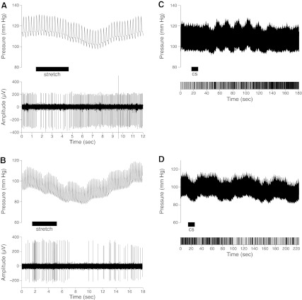
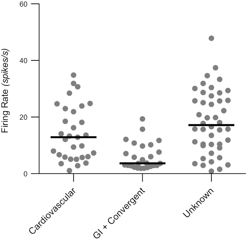
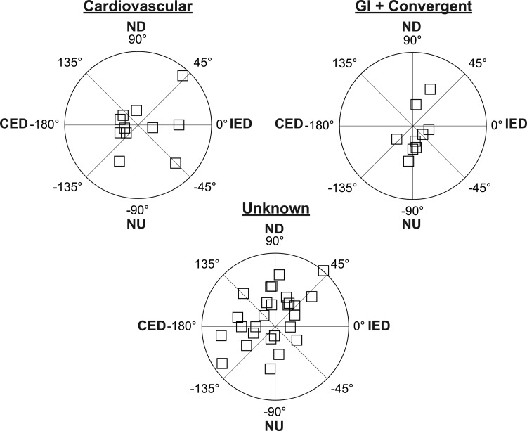
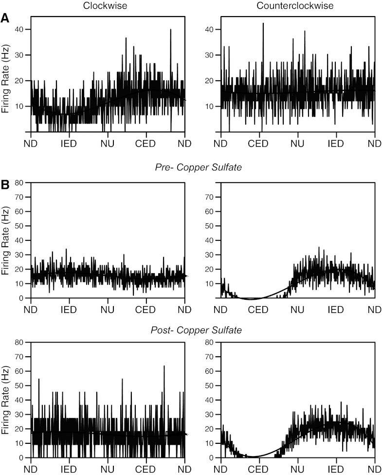
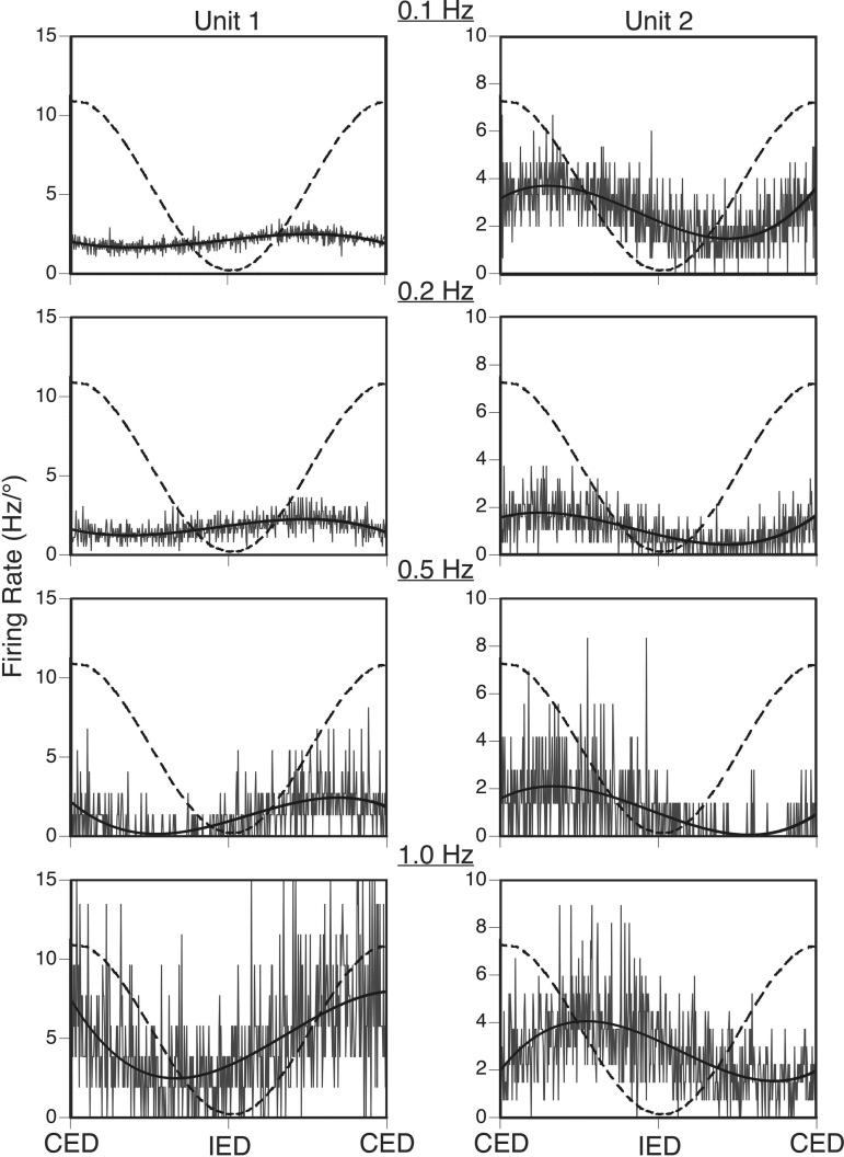
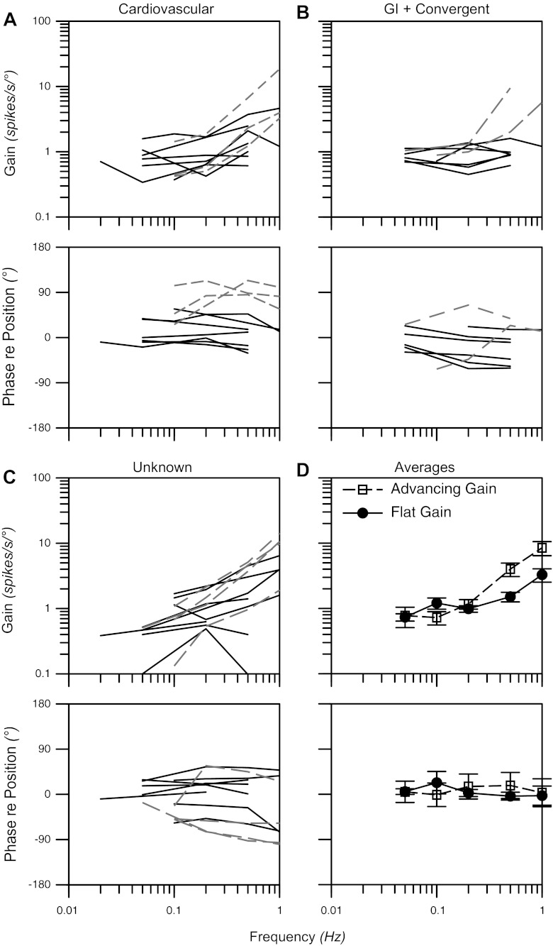
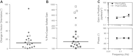
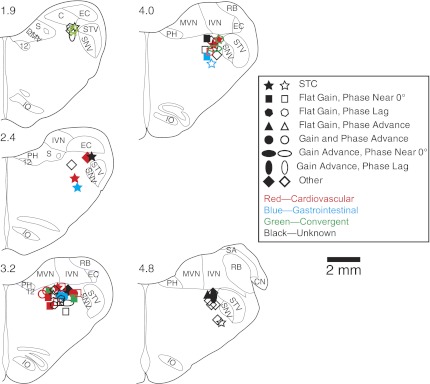
The dorsolateral reticular formation of the caudal medulla, or the lateral tegmental field (LTF), has been classified as the brain's "vomiting center", as well as an important region in regulating sympathetic outflow. We examined the responses of LTF neurons in cats to rotations of the body that activate vestibular receptors, as well as to stimulation of baroreceptors (through mechanical stretch of the carotid sinus) and gastrointestinal receptors (through the intragastric administration of the emetic compound copper sulfate). Approximately half of the LTF neurons exhibited graviceptive responses to vestibular stimulation, similar to primary afferents innervating otolith organs. The other half of the neurons had complex responses, including spatiotemporal convergence behavior, suggesting that they received convergent inputs from a variety of vestibular receptors. Neurons that received gastrointestinal and baroreceptor inputs had similar complex responses to vestibular stimulation; such responses are expected for neurons that contribute to the generation of motion sickness. LTF units with convergent baroreceptor and vestibular inputs may participate in producing the cardiovascular system components of motion sickness, such as the changes in skin blood flow that result in pallor. The administration of copper sulfate often modulated the gain of responses of LTF neurons to vestibular stimulation, particularly for units whose spontaneous firing rate was altered by infusion of drug (median of 459%). The present results raise the prospect that emetic signals from the gastrointestinal tract modify the processing of vestibular inputs by LTF neurons, thereby affecting the probability that vomiting will occur as a consequence of motion sickness.


Figures

Fig. 1.
Fig. 1.
Activity of lateral tegmental field (LTF) neurons during mechanical stimulation of baroreceptor afferents (A and B) and associated with the infusion of copper sulfate (cs) into the stomach (C and D). A and B: each panel contains a record of blood pressure (top, sampled at 100 Hz) and a recording of unit activity (bottom, sampled at 25,000 Hz), indicating the associated firing pattern of an LTF unit. When the carotid sinus was stretched (indicated by horizontal bar), the activity of the unit in A decreased, whereas that in B increased. A silencing of unit activity occurred in B following the carotid stretch, as blood pressure declined as a consequence of the baroreceptor reflex. Because spike size and shape did not change appreciably during the trial, it is likely that this reduction in unit activity resulted from the unloading of baroreceptors, and not a mechanical artifact. C and D: effect of copper sulfate infusion into the stomach (indicated by horizontal bar) on arterial blood pressure (top) and activity of an LTF unit (bottom raster plot). In C, a sustained increase in unit firing occurred after copper sulfate was present in the stomach for over a minute. A transient perturbation in blood pressure was also observed, which was not synchronized with the activity of the cell. In D, administration of copper sulfate resulted in a sustained reduction in the activity of an LTF unit, with a latency >1 min.
Fig. 2.
Fig. 2.
Spontaneous firing rates of different unit types tested for responses to vertical vestibular stimulation. Gray circles indicate data for each neuron, whereas black horizontal lines designate median firing rates. GI, gastrointestinal.
Fig. 3.
Fig. 3.
Polar plots showing response vector orientations and gains of cardiovascular units, units with gastrointestinal (GI) inputs (both GI and convergent neurons), and unknown units. Response vector orientations were determined using wobble stimuli, usually delivered at 0.2 Hz. The maximal radius of each plot designates a response gain of 3 spikes·s−1·deg−1. The response vector orientations were plotted using a head-centered coordinate system, with 0° corresponding to ipsilateral ear-down (IED) roll tilt, 90° corresponding to nose-down (ND) pitch, 180° corresponding to contralateral ear-down (CED) roll, and −90° corresponding to nose-up (NU) pitch.
Fig. 4.
Fig. 4.
Averaged responses of two STC neurons whose activity was modulated by only one direction of wobble stimulation. Responses illustrated were elicited by 0.2-Hz wobble stimulation delivered at 7.5°. Averaged unit activity is indicated by traces, whereas overlain black curves are sine waves fit to the responses. A: responses of one unit prior to the administration of copper sulfate. The unit responded to clockwise (CW) wobble stimulation, but not counterclockwise (CCW) rotations (the signal-to-noise ratio was 0.82 for the CW response, and 0.01 for the CCW response). B: responses of a different unit before (top) and after (bottom) the administration of copper sulfate. During both conditions, the unit responded to CCW wobble, but not CW rotations. The response to CCW stimuli was so powerful that the activity of the unit was completely abolished during one phase of the rotation (contralateral ear down). The response gain for CCW trials was similar before (2.3 spikes·s−1·deg−1) and after (2.2 spikes·s−1·deg−1) copper sulfate administration. See Fig. 3 for definitions of abbreviations. The number of sweeps averaged to generate each trace was A: 33 (CW), 30 (CCW); B, top: 53 (CW), 51 (CCW); B, bottom: 11 (CW), 44 (CCW).
Fig. 5.
Fig. 5.
Averaged responses of two neurons to sinusoidal tilts in the roll plane at frequencies of 0.1–1 Hz. Both units were excited by CED and inhibited by IED. Averaged unit activity is indicated by gray traces, overlain solid black curves are sine waves fit to the responses, and dashed curves indicate tilt table position. The following tilt amplitudes were used for each trial: Unit 1, 7.5° at 0.1 Hz, 5° at 0.2–0.5 Hz, and 2.5° at 1 Hz; Unit 2, 7.5° at 0.1–0.2 Hz, 5° at 0.5 Hz, and 2.5° at 1 Hz. Because smaller tilt amplitudes were used for higher-frequency rotations, firing rate is expressed in Hz/° to facilitate comparisons. The response gain for Unit 1 increased ∼10-fold as the stimulus frequency advanced from 0.1 to 1 Hz, and the response phase led stimulus position by ∼90° at all frequencies. In contrast, the response gain for Unit 2 was relatively flat across stimulus frequencies, whereas the response phase lagged stimulus position by 50–75° across the range of frequencies tested.
Fig. 6.
Fig. 6.
Bode plots illustrating the dynamic properties of responses of LTF neurons to rotations in a fixed plane at multiple frequencies. Response gain and phase are plotted with respect to stimulus position. Bode plots for cardiovascular neurons (A), neurons with gastrointestinal (GI) inputs (gastrointestinal and convergent unit types) (B), and unknown units (C) are provided separately. Solid lines designate data for flat-gain neurons, whereas dashed lines show responses for advancing gain neurons. D: average Bode plots for all flat and advancing gain neurons; error bars designate SE.
Fig. 7.
Fig. 7.
Effects of injection of copper sulfate into the stomach on the responses of LTF neurons to vestibular stimulation. A: changes in response vector orientation produced by administration of copper sulfate. Data are restricted to the subset of units for which a response vector orientation could be calculated before and after copper sulfate was provided. Open symbols designate values for neurons with gastrointestinal inputs, whereas filled symbols designate cardiovascular and unknown units. Horizontal lines show median values. B: Changes in response gain produced by delivery of copper sulfate. The absolute value of the percent change in gain following injection of the compound is indicated. Squares designate units whose response gain decreased after copper sulfate was infused, while circles indicate units whose response gain increased. C: bode plots comparing average response dynamics for neurons before and after copper sulfate administration. Error bars indicate means ± SE.
Fig. 8.
Fig. 8.
Locations of neurons that responded to vertical vestibular stimulation, either before or after the intragastric administration of copper sulfate. Unit locations, which were generated through reference to Berman's atlas (11), are plotted on standard transverse sections through the caudal medulla. Numbers above each section designate distance in millimeters rostral to the obex. Distinct symbols are used to designate the characteristics of responses to tilts at different frequencies: STC behavior (responds to only one direction of wobble rotations); flat gain (increasing <5-fold per stimulus decade) and phase near (within 45° of) stimulus position; flat gain and a phase lag (>45°) from stimulus position at higher stimulus frequencies; flat gain and a phase advance (>45°) from stimulus position at higher frequencies; both a gain advance (>5-fold per stimulus decade) and phase advance (>45° from stimulus position) at higher frequencies; a gain advance and a phase near (within 45° of) stimulus position at all frequencies; a gain advance and a phase lag (>45°) from stimulus position at higher frequencies; or others (including neurons that were lost before response dynamics could be determined). Different colors are used to designate cardiovascular, gastrointestinal, convergent, and unknown unit types. Solid symbols indicate neurons whose response characteristics changed after administration of copper sulfate (response gains changed over 30%, or which developed or lost STC behavior). 12, hypoglossal nucleus; C, cuneate nucleus; CN, cochlear nucleus; DMV, dorsal motor nucleus of the vagus; EC, external cuneate; IO, inferior olivary nucleus; PH, prepositus hypoglossi; IVN, inferior vestibular nucleus; MVN, medial vestibular nucleus; RB, restiform body; S, solitary nucleus; SA, stria acoustica; SNV, spinal trigeminal nucleus; STV, spinal trigeminal tract.

Similar articles

Cited by

References

    1. Anderson JH, Blanks RHI, Precht W. Response characteristics of semicircular canal and otolith systems in the cat. I Dynamic responses of primary vestibular fibers. Exp Brain Res 32: 491–507, 1978 - PubMed
    1. Baker J, Goldberg J, Hermann G, Peterson B. Spatial and temporal response properties of secondary neurons that receive convergent input in vestibular nuclei of alert cats. Brain Res 294: 138–143, 1984 - PubMed
    1. Balaban CD. Vestibular autonomic regulation (including motion sickness and the mechanism of vomiting). Curr Opin Neurol 12: 29–33, 1999 - PubMed
    1. Balaban CD. Vestibular nucleus projections to the parabrachial nucleus in rabbits: implications for vestibular influences on the autonomic nervous system. Exp Brain Res 108: 367–381, 1996 - PubMed
    1. Balaban CD, Jacob RG, Furman JM. Neurologic bases for comorbidity of balance disorders, anxiety disorders and migraine: neurotherapeutic implications. Exp Rev Neurother 11: 379–394, 2011 - PMC - PubMed

Publication types

LinkOut - more resources