. 2013 Feb;44(2):189-98.
doi: 10.1016/j.humpath.2012.05.004. Epub 2012 Sep 3.

The role of TGF-β and myofibroblasts in the arteritis of Kawasaki disease

Affiliations

The role of TGF-β and myofibroblasts in the arteritis of Kawasaki disease

Chisato Shimizu et al. Hum Pathol. 2013 Feb.

Abstract

Inflammation of medium-sized, muscular arteries and coronary artery aneurysms are hallmarks of Kawasaki disease (KD), an acute, self-limited vasculitis of children. We previously reported that genetic variation in transforming growth factor (TGF)-β pathway genes influences both susceptibility to KD and coronary artery aneurysm (CAA) formation. TGF-β signaling has been implicated in the generation of myofibroblasts that influence collagen lattice contraction, antigen presentation, and recruitment of inflammatory cells as well as the generation of regulatory T-cells (Tregs). These processes could be involved in aneurysm formation and recovery in KD. Coronary artery tissues from 8 KD patient autopsies were stained to detect proteins in the TGF-β pathway, to characterize myofibroblasts, and to detect Tregs. Expression of proteins in the TGF-β pathway was noted in infiltrating mononuclear cells and spindle-shaped cells in the thickened intima and adventitia. Coronary arteries from an infant who died on Illness Day 12 showed α-smooth muscle actin (SMA)-positive, smoothelin-negative myofibroblasts in the thickened intima that co-expressed IL-17 and IL-6. CD8+ T-cells expressing HLA-DR+ (marker of activation and proliferation) were detected in the aneurysmal arterial wall. Forkhead box P3 (FOXP3), whose expression is essential for Tregs, was also detected in the nucleus of infiltrating mononuclear cells, suggesting a role for Tregs in recovery from KD arteritis.TGF-β may contribute to aneurysm formation by promoting the generation of myofibroblasts that mediate damage to the arterial wall through recruitment of pro-inflammatory cells. This multi-functional growth factor may also be involved in the induction of Tregs in KD.


Figures

Figure 1
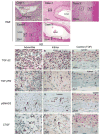
Figure 1. TGF-β2, TGF-βR2, pSMAD3 and CTGF expression in coronary arterial wall in KD and control autopsy tissues
Case 1, 2, 3, 6 (KD), and 9 (Tetrology of Fallot (TOF) negative control) (×100): H&E staining of coronary arteries. Boxed area indicates location of section used for IHC shown in A-L. TGF-β2 (A–C): Positive staining of mononuclear and spindle-shaped cells in KD arteries (A and B). Control shows rare positively stained spindle-shaped cell in adventitia (C). TGF-βR2 (D–F): Positive staining of spindle-shaped cells (arrow head) only in KD arteries (D and E) but not control tissues (F). pSMAD3 (G–I): Positive nuclear staining of spindle-shaped cells (arrow head) in adventitia and mononuclear cells (arrow) in intima from KD artery (G and H) but not control (I). CTGF (J–L): Positive staining of spindle-shaped cells (arrow head) in adventitia and intima of KD artery (J and K) and media of control artery (L). TGF-β2: transforming growth factor-β2, TGF-βR2: TGF-β receptor 2, pSMAD3: phosphorylated SMAD3, CTGF: connective tissue growth factor.
Figure 2
Figure 2
Expression of TGF-β2, TGF-βR2, pSMAD3 and CTGF in KD and control tissues.
Figure 3
Figure 3. α-SMA and smoothelin staining of coronary artery from Case 1 (Illness Day 12)
A–B, Coronary artery aneurysm from Case 1 (H&E, A: ×40, B: ×400), A: One side of the coronary arterial wall is well-preserved with normal architecture. Opposite wall shows disruption of arterial wall structures, thickened initma, and focal breaks in internal elastic lamina (iel) and infiltration of inflammatory cells (boxed area magnified in B). C–I: α-SMA(green) and smoothelin (red) staining of coronary artery(boxed area 1 magnified in D–F, boxed area 2 magnified in G–I), C, D and G: merged fluorescence α-SMA+/smoothelin-cells (myofibroblasts) in thickened intima(arrowhead in D). Double-staining smooth muscle cells (orange) in media(D and G). α-SMA: α-smooth muscle actin.
Figure 4
Figure 4
Fluorescent double staining for α-SMA with either IL-17 or IL-6 of coronary artery from Case 1 (Illness Day 12). A–C, α-SMA (green) with IL-17 (red). D–F, α-SMA (green) with IL-6 (red). α-SMA positive myofibroblasts expressing IL-17 (A) and IL-6 (D).
Figure 5
Figure 5. HLA-DR+/CD8+ mononuclear cells in arterial wall of a KD aneurysm
Thickened intima of coronary arterial wall from Case 1 was stained. A–C, double staining for CD8 (green) with HLA-DR (red). Many of the HLA-DR+ round, mononuclear cells are also CD8+ (white arrows). D–F, double staining for α-SMA (green) with HLA-DR (red). α-SMA+ myofibroblasts adjacent to HLA-DR+ mononuclear cells (white arrows).
Figure 6
Figure 6. Immunohistochemical staining of mononuclear cells with nuclear FOXP3 in KD arterial wall
A and B: Thickened intima of coronary arteries from Case 1 (A) and Case 4 (B) showing mononuclear cells expressing FOXP3 in the nucleus (arrows). C: Rabbit IgG negative antibody control. FOXP3: Forkhead box P3, L: lumen.
Figure 7
Figure 7. Proposed mechanism of aneurysm formation mediated by TGF-β
Local secretion of TGF-β generates myofibroblasts from endothelial cells, fibroblasts in adventitia, smooth muscle cells in media, and fibrocytes in the peripheral circulation. Myofibroblasts secrete cytokines (IL-17, IL-6) and chemokines that recruit inflammatory cells. Myofibroblasts also present antigen leading to activation and proliferation of T-cells. Cessation of inflammation is mediated by T-cell regulation (FOXP3 + cells). Genetic variation in TGF-β pathway genes may modulate this cascade either up (more damage) or down (less damage). ec: endothelial cell; e: internal elastic lamina; m: media; a: adventitia. EMT: endothelial/epithelial to mesenchymal transition, Treg: regulatory T-cell.

Similar articles

Cited by

References

    1. Burns JC, Glode MP. Kawasaki syndrome. Lancet. 2004;364:533–544. - PubMed
    1. Tremoulet AH, Best BM, Song S, et al. Resistance to intravenous immunoglobulin in children with Kawasaki disease. J Pediatr. 2008;153:117–121. - PMC - PubMed
    1. Newburger JW, Takahashi M, Beiser AS, et al. A single intravenous infusion of gamma globulin as compared with four infusions in the treatment of acute Kawasaki syndrome. N Engl J Med. 1991;324:1633–1639. - PubMed
    1. Shimizu C, Jain S, Davila S, et al. Transforming growth factor-beta signaling pathway in patients with Kawasaki disease. Circ Cardiovasc Genet. 2011;4:16–25. - PMC - PubMed
    1. Franco A, Shimizu C, Tremoulet AH, Burns JC. Memory T-cells and characterization of peripheral T-cell clones in acute Kawasaki disease. Autoimmunity. 2010;43:317–324. - PMC - PubMed

Publication types

MeSH terms