. 2012 Nov 2;287(45):38020-7.
doi: 10.1074/jbc.M112.348599. Epub 2012 Sep 6.

Interleukin-10 inhibits lipopolysaccharide-induced tumor necrosis factor-α translation through a SHIP1-dependent pathway

Affiliations

Interleukin-10 inhibits lipopolysaccharide-induced tumor necrosis factor-α translation through a SHIP1-dependent pathway

Catherine S Chan et al. J Biol Chem. .

Abstract

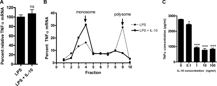
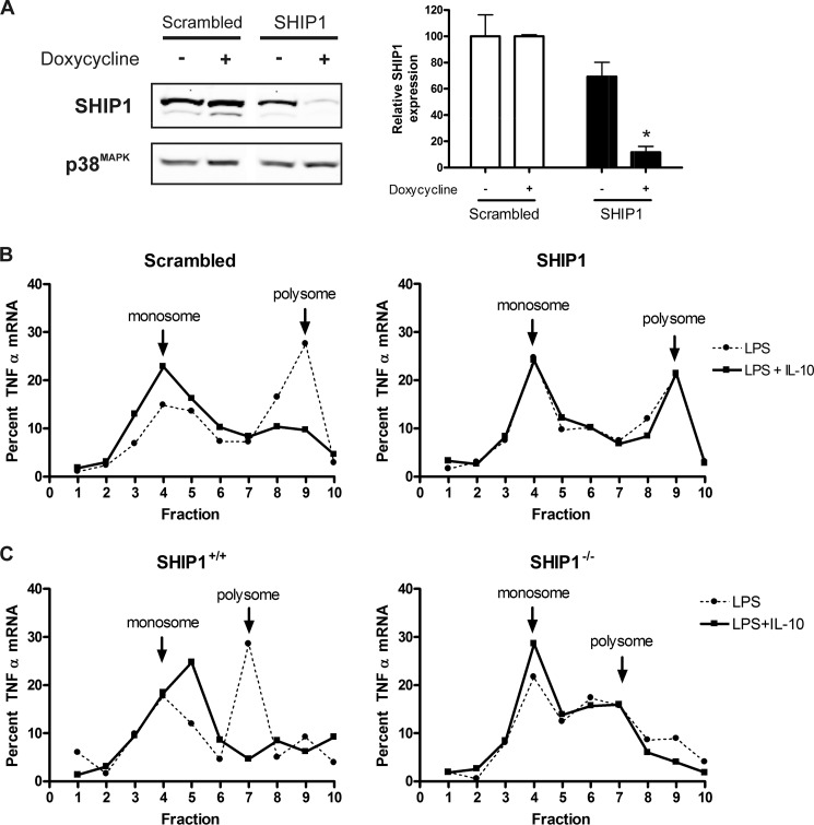
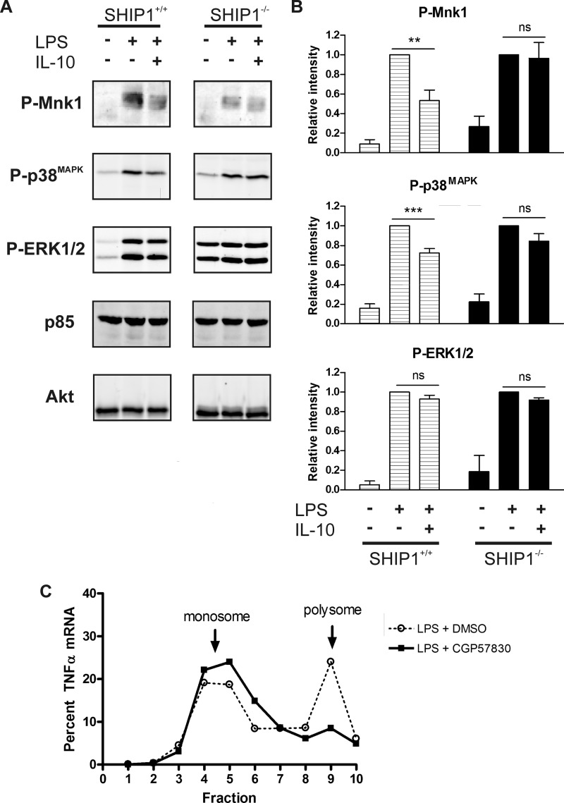
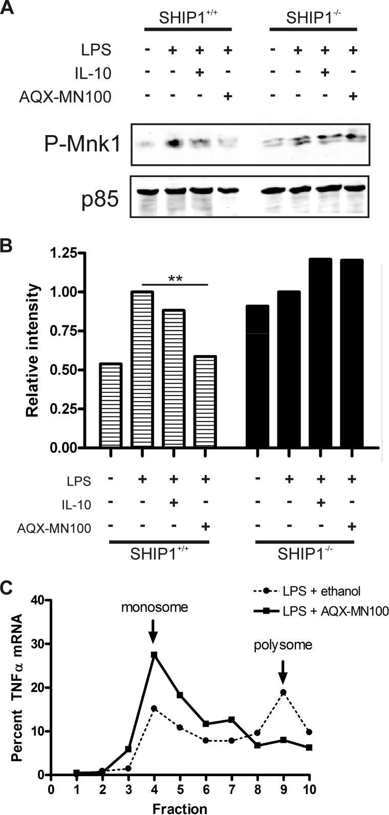
Production of the proinflammatory cytokine TNFα by activated macrophages is an important component of host defense. However, TNFα production must be tightly controlled to avoid pathological consequences. The anti-inflammatory cytokine IL-10 inhibits TNFα mRNA expression through activation of the STAT3 transcription factor pathway and subsequent expression of STAT3-dependent gene products. We hypothesized that IL-10 must also have more rapid mechanisms of action and show that IL-10 rapidly shifts existing TNFα mRNA from polyribosome-associated polysomes to monosomes. This translation suppression requires the presence of SHIP1 (SH2 domain-containing inositol 5'-phosphatase 1) and involves inhibition of Mnk1 (MAPK signal-integrating kinase 1). Furthermore, activating SHIP1 using a small-molecule agonist mimics the inhibitory effect of IL-10 on Mnk1 phosphorylation and TNFα translation. Our data support the existence of an alternative STAT3-independent pathway through SHIP1 for IL-10 to regulate TNFα translation during the anti-inflammatory response.


Figures

FIGURE 1.
FIGURE 1.
IL-10 inhibits TNFα synthesis through polysome disassembly in pre-activated cells. A, relative total TNFα mRNA from RAW 264.7 cells pre-activated with 1 ng/ml LPS for 45 min and then treated with 100 ng/ml IL-10 for an additional 15 min. Relative TNFα mRNA was normalized against mouse GAPDH mRNA and then expressed as a percentage against cells that were not treated. Statistical significance between treatments was calculated by Student's unpaired two-tailed t test with 95% confidence. ns, not significant (p = 0.3077). B, TNFα mRNA distribution in total mRNA from A fractionated over a sucrose density gradient. Fraction 10 indicates the bottom of the gradient. mRNA levels were measured by real-time quantitative PCR in each fraction and expressed as a percentage of the sum of the TNFα mRNA signal from all 10 fractions. Results are representative of two independent experiments. C, TNFα protein production in cell supernatants was measured by ELISA from pre-activated cells from A except that cells were treated with a range of IL-10 concentrations. Results are representative of three experiments. Statistical significance between treatments was calculated by one-way analysis of variance with 95% confidence. *, p < 0.05; ***, p < 0.001.
FIGURE 2.
FIGURE 2.
IL-10 regulates TNFα translation through SHIP1 in pre-activated cells. A, siRNA-inducible knockdown of SHIP1. Stably transduced cells carrying siRNA constructs of SHIP1 or a scrambled sequence were placed behind a doxycycline-inducible promoter, and the level of SHIP1 expression was assessed by immunoblotting and densitometric analysis after 48 h of induction. Protein levels were normalized against p38 MAPK, and relative SHIP1 levels were plotted as a percentage of SHIP1 from the scrambled cells. Statistical significance between treatments was calculated by Student's unpaired two-tailed t test with 95% confidence. *, p < 0.05. B, gradient fractionation of total mRNA from scrambled or SHIP1 siRNA knockdown pre-activated cells as described in the legend to Fig. 1. Results are representative of two experiments. C, gradient fractionation of total mRNA from wild-type (SHIP1+/+) or SHIP1 knock-out (SHIP1−/−) pre-activated peritoneal macrophages as described in the legend to Fig. 1. Results are representative of two experiments.
FIGURE 3.
FIGURE 3.
IL-10 inhibits TNFα translation through SHIP1 via Mnk1. A, immunoblot analysis of cell lysates from peritoneal macrophages isolated from wild-type (SHIP1+/+) or SHIP1 knock-out (SHIP1−/−) mice stimulated with 1 ng/ml LPS in the absence or presence of 100 ng/ml IL-10 for 1 h. Results are representative of three to five experiments. B, densitometric analyses were done by normalizing the intensity to Akt or PI3K p85 protein levels. The average relative intensities from four experiments were then plotted relative to the LPS-only fractions. Statistical significance between LPS ± IL-10 samples was calculated by Student's paired two-tailed t test with 95% confidence. **, p < 0.01; ***, p < 0.001; ns, not significant (p = 0.8280 (phospho-Mnk1), p = 0.1059 (phospho-p38), and p = 0.4069 (phospho-ERK1/2) for SHIP+/+ mice and p = 0.1722 (phospho-ERK1/2) for SHIP−/− mice). C, gradient fractionation of total mRNA from RAW 264.7 cells pretreated with vehicle (dimethyl sulfoxide (DMSO)) or 75 μm Mnk1 inhibitor (CGP57380) as described in the legend to Fig. 1, except without IL-10 treatment.
FIGURE 4.
FIGURE 4.
Small-molecule agonist-activated SHIP1 inhibits TNFα translation through Mnk1. A, immunoblot analysis of peritoneal macrophage cell lysates isolated from wild-type (SHIP1+/+) or SHIP1 knock-out (SHIP1−/−) mice stimulated with 10 ng/ml LPS in the absence or presence of 100 ng/ml IL-10 or 10 μm AQX-MN100 for 1 h. Results are representative of two experiments. B, densitometric analysis of phospho-Mnk1 levels in A normalized to the levels of PI3K p85 protein and plotted relative to the LPS-only samples. Statistical significance between treatments was calculated by one-way analysis of variance with 95% confidence. **, p < 0.01. C, gradient fractionation of total mRNA from pre-activated RAW 264.7 cells as described in the legend to Fig. 1, except with 10 μm AQX-MN100 treatment instead of IL-10 treatment.

Similar articles

Cited by

References

    1. Chow J. C., Young D. W., Golenbock D. T., Christ W. J., Gusovsky F. (1999) Toll-like receptor-4 mediates lipopolysaccharide-induced signal transduction. J. Biol. Chem. 274, 10689–10692 - PubMed
    1. Kruys V., Kemmer K., Shakhov A., Jongeneel V., Beutler B. (1992) Constitutive activity of the tumor necrosis factor promoter is canceled by the 3′-untranslated region in nonmacrophage cell lines; a trans-dominant factor overcomes this suppressive effect. Proc. Natl. Acad. Sci. U.S.A. 89, 673–677 - PMC - PubMed
    1. Lai W. S., Carballo E., Strum J. R., Kennington E. A., Phillips R. S., Blackshear P. J. (1999) Evidence that tristetraprolin binds to AU-rich elements and promotes the deadenylation and destabilization of tumor necrosis factor-α mRNA. Mol. Cell. Biol. 19, 4311–4323 - PMC - PubMed
    1. Piecyk M., Wax S., Beck A. R., Kedersha N., Gupta M., Maritim B., Chen S., Gueydan C., Kruys V., Streuli M., Anderson P. (2000) TIA-1 is a translational silencer that selectively regulates the expression of TNFα. EMBO J. 19, 4154–4163 - PMC - PubMed
    1. Buxadé M., Parra J. L., Rousseau S., Shpiro N., Marquez R., Morrice N., Bain J., Espel E., Proud C. G. (2005) The Mnks are novel components in the control of TNFα biosynthesis and phosphorylate and regulate hnRNP A1. Immunity 23, 177–189 - PubMed

Publication types

MeSH terms

Substances

LinkOut - more resources