. 2012 Nov 1;186(9):846-56.
doi: 10.1164/rccm.201206-1056OC. Epub 2012 Sep 6.

Assessment of F/HN-pseudotyped lentivirus as a clinically relevant vector for lung gene therapy

Affiliations

Assessment of F/HN-pseudotyped lentivirus as a clinically relevant vector for lung gene therapy

Uta Griesenbach et al. Am J Respir Crit Care Med. .

Abstract

Rationale: Ongoing efforts to improve pulmonary gene transfer thereby enabling gene therapy for the treatment of lung diseases, such as cystic fibrosis (CF), has led to the assessment of a lentiviral vector (simian immunodeficiency virus [SIV]) pseudotyped with the Sendai virus envelope proteins F and HN.

Objectives: To place this vector onto a translational pathway to the clinic by addressing some key milestones that have to be achieved.

Methods: F/HN-SIV transduction efficiency, duration of expression, and toxicity were assessed in mice. In addition, F/HN-SIV was assessed in differentiated human air-liquid interface cultures, primary human nasal epithelial cells, and human and sheep lung slices.

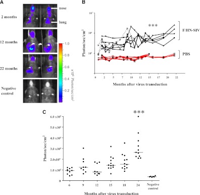
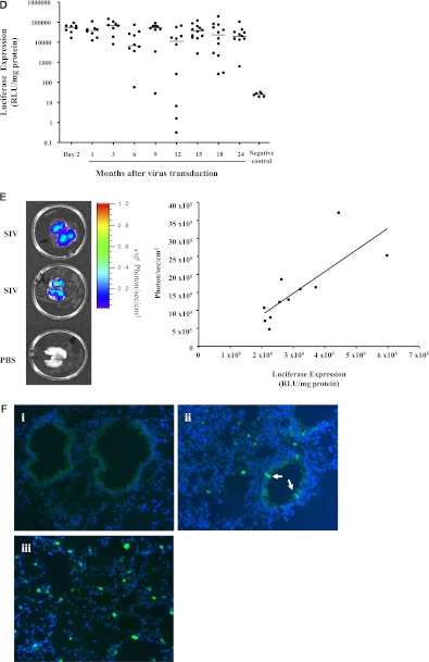
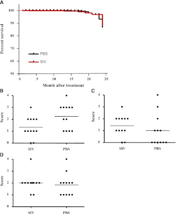
Measurements and main results: A single dose produces lung expression for the lifetime of the mouse (~2 yr). Only brief contact time is needed to achieve transduction. Repeated daily administration leads to a dose-related increase in gene expression. Repeated monthly administration to mouse lower airways is feasible without loss of gene expression. There is no evidence of chronic toxicity during a 2-year study period. F/HN-SIV leads to persistent gene expression in human differentiated airway cultures and human lung slices and transduces freshly obtained primary human airway epithelial cells.

Conclusions: The data support F/HN-pseudotyped SIV as a promising vector for pulmonary gene therapy for several diseases including CF. We are now undertaking the necessary refinements to progress this vector into clinical trials.


Figures

Figure 1.
Figure 1.
F/HN simian immunodeficiency virus (SIV) transduction leads to persistent gene expression in mouse lung. Mice were transduced with F/HN-SIV-luciferase (Lux) (5 × 10⁸ TU/mouse) by nasal sniffing (n = 8 in two independent experiments) or received phosphate-buffered saline (PBS) (negative controls, n = 6). Lux expression was quantified using bioluminescence imaging (BLI) or in tissue homogenate at regular intervals for up to 24 months. (A) BLI 2–22 months after transduction. Representative images of two mice reaching the 22 months time point are shown. (B) Quantification of in vivo BLI over time (black lines, n = 8) or PBS (red lines, n = 6). Each line represents photon emission over time in one animal. Solid and dotted lines represent independent experiments. ***P < 0.005 when compared with Month 2. BLI (C) and Lux expression in lung tissue homogenate (D) was repeated at regular intervals over 24 months in a third independent experiment (107 TU/mouse, = 8–12 per time-point). For technical reasons BLI could only be performed between 6 and 24 months after gene transfer. ***P < 0.005 when compared with Month 6. Quantification of Lux expression in lung tissue homogenate was performed at regular intervals over the 24-month study period. Each dot represents one animal. Horizontal bars represent the group median. (E) Correlation between Lux expression quantified in tissue homogenates and photons emitted from extracted lungs (representative images from 11 extracted lungs are shown). Each dot represents one animal. (F) Detection of green fluorescent protein (GFP) expression in lung after transduction with F/HN-SIV-GFP (108 TU/mouse). Transduced GFP-positive cells were identified using fluorescent microscopy (original magnification, ×20). (i) PBS-treated negative control. (ii and iii) Tissue sections from F/HN-SIV-GFP–treated mice showing GFP-positive cells in airways (arrows) and alveoli. Representative images from four mice per group are shown. RLU = relative light units.
Figure 1.
Figure 1.
F/HN simian immunodeficiency virus (SIV) transduction leads to persistent gene expression in mouse lung. Mice were transduced with F/HN-SIV-luciferase (Lux) (5 × 10⁸ TU/mouse) by nasal sniffing (n = 8 in two independent experiments) or received phosphate-buffered saline (PBS) (negative controls, n = 6). Lux expression was quantified using bioluminescence imaging (BLI) or in tissue homogenate at regular intervals for up to 24 months. (A) BLI 2–22 months after transduction. Representative images of two mice reaching the 22 months time point are shown. (B) Quantification of in vivo BLI over time (black lines, n = 8) or PBS (red lines, n = 6). Each line represents photon emission over time in one animal. Solid and dotted lines represent independent experiments. ***P < 0.005 when compared with Month 2. BLI (C) and Lux expression in lung tissue homogenate (D) was repeated at regular intervals over 24 months in a third independent experiment (107 TU/mouse, = 8–12 per time-point). For technical reasons BLI could only be performed between 6 and 24 months after gene transfer. ***P < 0.005 when compared with Month 6. Quantification of Lux expression in lung tissue homogenate was performed at regular intervals over the 24-month study period. Each dot represents one animal. Horizontal bars represent the group median. (E) Correlation between Lux expression quantified in tissue homogenates and photons emitted from extracted lungs (representative images from 11 extracted lungs are shown). Each dot represents one animal. (F) Detection of green fluorescent protein (GFP) expression in lung after transduction with F/HN-SIV-GFP (108 TU/mouse). Transduced GFP-positive cells were identified using fluorescent microscopy (original magnification, ×20). (i) PBS-treated negative control. (ii and iii) Tissue sections from F/HN-SIV-GFP–treated mice showing GFP-positive cells in airways (arrows) and alveoli. Representative images from four mice per group are shown. RLU = relative light units.
Figure 2.
Figure 2.
F/HN simian immunodeficiency virus (SIV) requires only short contact time to achieve efficient transduction. Luciferase (Lux) expression was compared in mice receiving F/HN-SIV-Lux (3–4 × 10⁸ TU/mouse in 100 μl) by slow perfusion (1.3 μl/min, n = 10) or as bolus administration by nasal sniffing (n = 8). (A) Bioluminescence in vivo signal in the mouse nose. Representative images of three mice are shown. (B) Quantification of in vivo bioluminescence in mouse nose after slow perfusion or nasal sniffing of the vector. Each dot represents one animal. Horizontal bars represent the group median. (C) Bioluminescence in vivo signal in mouse nose after intraperitoneal (IP) or intranasal (IN) administration of luciferin. Representative images of two of five mice are shown. Lux expression after slow perfusion of the F/HN-SIV-Lux has been published previously (11).
Figure 3.
Figure 3.
Daily and monthly repeat administration to the lung is feasible. (A) Daily readministration. Mice (n = 8 per group) were treated with either F/HN simian immunodeficiency virus (SIV) luciferase (Lux) for 10 days (106 TU/day) or with D phosphate buffered saline (PBS) for 5 days followed by 5 days of F/HN-SIV-Lux (106 TU/day) or with D-PBS for 9 days followed by 1 day of F/HN-SIV-Lux (106 TU). Lux expression was analyzed 28 days after F/HN-SIV-Lux administration. (B) Monthly readministration. Mice were either treated with one dose of F/HN-SIV-Lux (single-dose group) or two doses of F/HN-SIV-GFP (Day 0, Day 28), followed by F/HN-SIV-Lux on Day 56 (repeat-dose group). All mice receive 107 TU/dose (n = 20 per group) and gene expression was analyzed 28 days after F/HN-SIV-Lux administration. Each dot represents one animal. Horizontal bars represent the group median. Dotted line represents negative control values, ***P < 0.0001 when compared with 5 and 10 doses, **P < 0.01 when compared with 10 doses. RLU = relative light units.
Figure 4.
Figure 4.
No evidence of chronic toxicity during 2-year follow-up period. (A) Twenty-four months survival of mice treated with F/HN simian immunodeficiency virus (SIV) luciferase (Lux) (107 TU/mouse, n = 99 at start of the experiment) or phosphate-buffered saline (PBS) (n = 48 at start of the experiment) by nasal sniffing. Comparison of lymphocytes (B), edema (C), and inflammation (D) in mouse lung 24 months after F/HN-SIV-Lux or PBS treatment. Each dot represents one animal. Horizontal bars represent the group median.
Figure 5.
Figure 5.
Persistent gene expression in differentiated air–liquid interface (ALI) cultures. Differentiated human ALI cultures were transduced with F/HN simian immunodeficiency virus (SIV) luciferase (Lux) at multiplicity of infection (MOI) 25 or 250 (n = 5 per group), transfected with GL67A complexed to a eukaryotic expression plasmid expressing Lux (10 μg pCIKLux per ALI equivalent to 3 × 10¹² plasmids and an approximate MOI of 7 × 10⁶) (n = 3) or treated with phosphate-buffered saline (PBS) (negative control, n = 3). Lux expression was followed using bioluminescence imaging for 3 months. (A) Representative images are shown. (B) Quantification of Lux expression over 3 months.
Figure 6.
Figure 6.
F/HN simian immunodeficiency virus (SIV) transduces primary human pulmonary cells. (A) Microscopic section showing primary human airway epithelial cells obtained from nasal brushings (left, GC = goblet cell, CC = ciliated airway epithelial cell) or human lung slices (right, AW = airway, P = parenchyma). (B) Nasal brushings were transduced with F/HN-SIV-luciferase (Lux) at a multiplicity of infection (MOI) of 25 and 250 or treated with phosphate-buffered saline (PBS) (negative control) and Lux expression was quantified 24 hours after transduction. Each dot represents one sample. Horizontal bars represent the group median. ***P < 0.005 when compared with negative controls. (C) Human lung slices were transfected with F/HN-SIV-Lux (2 × 10⁷ TU/slice) or remained untransfected (negative control) and Lux expression quantified 2, 7, and 14 days after transduction. Two independent experiments were performed (open and closed symbols). (D) Sheep lung slices were transfected with F/HN-SIV-Lux (2 × 10⁷ TU/slice) or remained untransfected (negative control) and Lux expression quantified up to 26 days after transduction. Two independent experiments were performed (open and closed symbols). Each dot represents one animal. Horizontal bars represent the group median. *P < 0.05, **P < 0.01, ***P < 0.005 when compared with negative control. RLU = relative light units.

Similar articles

Cited by

References

    1. Griesenbach U, Alton EW. Progress in gene and cell therapy for cystic fibrosis lung disease. Curr Pharm Des 2012;18:642–662 - PubMed
    1. Yonemitsu Y, Kitson C, Ferrari S, Farley R, Griesenbach U, Judd D, Steel R, Scheid P, Zhu J, Jeffery PK, et al. Efficient gene transfer to airway epithelium using recombinant Sendai virus. Nat Biotechnol 2000;18:970–973 - PubMed
    1. Griesenbach U, Cassady RL, Ferrari S, Fukumura M, Muller C, Schmitt E, Zhu J, Jeffery PK, Nagai Y, Geddes DM, et al. The nasal epithelium as a factory for systemic protein delivery. Mol Ther 2002;5:98–103 - PubMed
    1. Harvey BG, Leopold PL, Hackett NR, Grasso TM, Williams PM, Tucker AL, Kaner RJ, Ferris B, Gonda I, Sweeney TD, et al. Airway epithelial CFTR mRNA expression in cystic fibrosis patients after repetitive administration of a recombinant adenovirus. J Clin Invest 1999;104:1245–1255 - PMC - PubMed
    1. Sumner-Jones SG, Davies LA, Varathalingam A, Gill DR, Hyde SC. Long-term persistence of gene expression from adeno-associated virus serotype 5 in the mouse airways. Gene Ther 2006;13:1703–1713 - PubMed

Publication types