Review
. 2013 Jun;70(11):1915-36.
doi: 10.1007/s00018-012-1146-7. Epub 2012 Sep 6.

Thyroid hormone actions in liver cancer

Affiliations
Review

Thyroid hormone actions in liver cancer

Sheng-Ming Wu et al. Cell Mol Life Sci. 2013 Jun.

Abstract

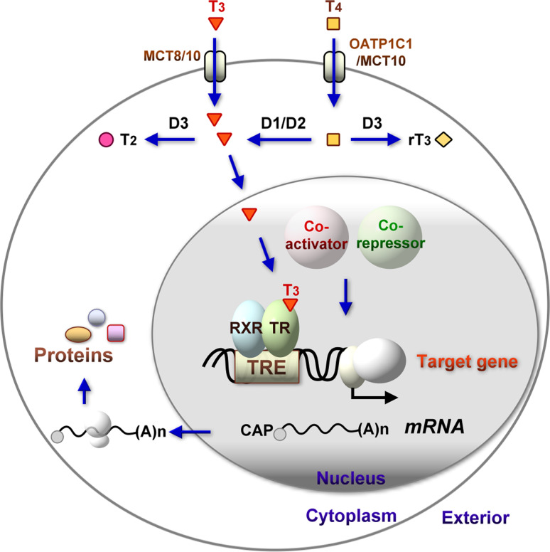
The thyroid hormone 3,3',5-triiodo-L-thyronine (T3) mediates several physiological processes, including embryonic development, cellular differentiation, metabolism, and the regulation of cell proliferation. Thyroid hormone receptors (TRs) generally act as heterodimers with the retinoid X receptor (RXR) to regulate target genes. In addition to their developmental and metabolic functions, TRs have been shown to play a tumor suppressor role, suggesting that their aberrant expression can lead to tumor transformation. Conversely, recent reports have shown an association between overexpression of wild-type TRs and tumor metastasis. Signaling crosstalk between T3/TR and other pathways or specific TR coregulators appear to affect tumor development. Since TR actions are complex as well as cell context-, tissue- and time-specific, aberrant expression of the various TR isoforms has different effects during diverse tumorigenesis. Therefore, elucidation of the T3/TR signaling mechanisms in cancers should facilitate the identification of novel therapeutic targets. This review provides a summary of recent studies focusing on the role of TRs in hepatocellular carcinomas (HCCs).


Conflict of interest statement

The authors have no conflicting financial interests.

Figures

Fig. 1
Fig. 1
Genomic actions of the thyroid hormone receptor. The ultimate effects of thyroid hormones (THs) on development, growth, and metabolism are attributed to signaling action at the cellular level. THs are transferred into target cells by specific transporter proteins. Monocarboxylate transporter 8 (MCT8), a membrane protein responsible for 3,3,5-triiodo-l-thyronine (T3) transport, is highly expressed in the liver and brain. Organic anion transporting polypeptide 1C1 (OATP1C1) is a member of the OATP family responsible for high-level transport of 3,3,5,5-tetraiodo-l-thyronine (T4; thyroxine) and low-level transport of reverse T3 (rT3). Other possible transporters include members of the l-type amino acid transporter family and MCT10. Although T4 is predominantly secreted from the thyroid gland, T3 displays ten times greater affinity and efficacy than T4 for the thyroid hormone receptor (TR). T4 requires deiodination and conversion to T3 to become biologically active. Deiodinating enzymes regulate the intracellular concentrations of T3. Type 1 or 2 deiodinase (D1 or D2) converts the precursor T4 to T3, whereas the major inactivator for the conversion of T4 and T3 to the inactive metabolites, rT3 and 3,3-diiodothyronine (T2), is type 3 deiodinase (D3). Several actions of TH are mediated by alterations in the regulation of gene transcription. In the absence or presence of the hormone, TRs (thyroid hormone receptors) bind to specific sequences, known as thyroid hormone response elements (TREs), in the regulatory regions of target genes, usually as heterodimers with RXR (retinoid X receptor). DNA-bound and unliganded TRs interact with corepressors that inhibit target gene transcription. Bioactive hormone T3 binding induces a conformational change in TR that leads to dissociation of the corepressor, recruitment of coactivators, and transcriptional activation
Fig. 2
Fig. 2
Schematic representation of TRα and TRβ isoforms. a Rat THRA and THRB encode the major functional receptor isoforms, TRα1, TRβ1, TRβ2, and TRβ3, as well as several dominant-negative antagonists (TRα2, TRα3, TR∆α1, TR∆α2, and TR∆β3). TRα2 and TRα3 isoforms generated via alternative splicing do not bind T3 and exhibit weak dominant-negative activity. TR∆α1 and TR∆α2 are truncated variants generated by alternative splicing and internal promoter usage. Truncated TRα variants do not bind T3, and inhibit transcriptional activation mediated by T3-liganded TRs. The TRβ2 isoform is generated by separate promoter usage and alternative splicing. Rat TRβ3 and TR∆β3 isoforms are generated from alternative splicing. The TR∆β3 variant lacks the DNA-binding domain and exhibits dominant negative antagonist activity. Similar to other nuclear receptors, these TR isoforms contain several functional domains, including an amino-terminal region (A/B), a conserved DNA-binding domain (DBD) or region C composed of two zinc fingers responsible for DNA binding, a hinge region D that links DBD with the ligand-binding domain (LBD), and an E region containing the LBD and residues responsible for receptor dimerization. A functional domain with similar amino acid sequences is presented in the same color. b Human THRA and THRB genes are located on chromosomes 17 and 3, respectively. Alternative splicing of each gene generates multiple isoforms, with TRα1 and TRα2 from TRα as well as TRβ1 and TRβ2 from TRβ identified in humans. TRα1, TRβ1 and TRβ2 are the main ligand-binding receptors. However, the TRα2 isoform lacking the ligand-binding domain does not bind hormone and suppress the expression of genes containing TREs. The first 246 amino acids of TRβ4 are identical to those of the functional receptor, TRβ1, while the carboxy (C)-terminal 215 residues are replaced by an entirely distinct sequence of 13 residues. The TRβ4 protein is unable to bind thyroid hormone (T3), and therefore does not mediate T3-dependent gene regulation. c TRs regulate target gene expression directly through thyroid hormone response elements (TREs). The consensus nucleotide sequence (half-site) of thyroid TRE is arranged as a direct repeat separated by four nucleotides (DR4; malic enzyme gene), palindrome (Pal; growth hormone gene), and inverted palindrome separated by six nucleotides (IP6; the chicken lysozyme gene). Two half-sites with a specific orientation or spacing are required for efficient binding and function
Fig. 3
Fig. 3
Non-genomic actions of the thyroid hormone. Nongenomic actions of the thyroid hormone initiated at the plasma membrane receptor on integrin αVβ3 or in the cytoplasm. a The thyroid hormone interacts with the integrin receptor, and stimulates activation and trafficking of extracellular signal-regulated kinase 1/2 (ERK1/2) through phospholipase C (PLC) and protein kinase Cα (PKCα). T4-activated ERK1/2 also modulates intracellular protein trafficking of estrogen receptor α (ERα) and TRβ1 from the cytoplasm to nucleus and acts locally at the plasma membrane to activate the sodium proton exchanger (NHE). Complex cellular events induced from the cell surface receptor include angiogenesis and tumor cell proliferation. T3 may also act via the integrin receptor, but the affinity of the receptor for T3 is lower than that for T4. b T3, but not T4, interacts with a T3-specific binding domain of integrin to activate the phosphatidylinositol 3-kinase (PI3K) signal pathway via Src kinase activation. The T3-specific site activates PI3K and leads to trafficking of TRα1 from the cytoplasm to nucleus and increased hypoxia-inducible factor-1α (HIF-1α) gene expression, but not cell proliferation. c In the cytoplasm, T3 rapidly activates the PI3K pathway and initiates the downstream transcription of specific genes. The T3-liganded TRβ1 interacts with the PI3K regulatory subunit, p85α, and induces phosphorylation of Akt (also known as protein kinase B, PKB). Activated Akt translocates to the nucleus and subsequently phosphorylates nuclear mammalian target of rapamycin (mTOR). Consequently, activation of mTOR triggers the expression of ZAKI-4α (also termed DSCR1L, Down syndrome critical region gene 1-like) and HIF-1α. Ultimately, the pathway may lead to alterations in numbers of pumps inserted in the membrane and increased activity of the sodium pump (Na,K-ATPase) in the plasma membrane. In addition, TRα1 interacts with the p85α subunit of PI3K in a T3-dependent manner, resulting into the activation of Akt and endothelial nitric oxide synthase (eNOS). d A truncated form of TRα1 (TR∆α1) in the cytoplasm mediates the actions of T4 and rT3 on polymerization of the actin cytoskeleton. Rapid thyroid hormone signaling induces enzyme inactivation by modulating actin-mediated internalization
Fig. 4
Fig. 4
Proposed model for the anti-oncogenic actions of thyroid hormone receptors. a Wild-type thyroid hormone receptors (TRs) regulate the expression of genes involved in tumor progression. In the absence of hormone, the unliganded receptor recruits corepressors (e.g., NCoR or SMRT) to positively regulated genes and histone deacetylases, which maintain chromatin in a compact and repressed state. Binding of T3 to TR induces conformational changes and corepressor dissociation, allowing ligand-bound TR to recruit coactivator and other associated proteins to modify chromatin structures, thus facilitating transcriptional activation. T3 binds to the TR and promotes transcription by releasing corepressors and recruiting coactivators (SRC-1), histone acetylases (CBP, p300, and pCAF), and other mediators (TRAP and DRIP), in turn, facilitating access of general transcriptional factors (GTFs including TBP, TFII, and TAFs) and RNA polymerase II (RNA Pol II) to the promoter. b Mutant receptors associated with somatic mutations in human tumors. i Expression of TR with mutations in the hormone-binding domain blocking T3 interactions but displaying intact DNA binding maintains chromatin in the repressed state. ii DNA-binding domain mutations leading to loss of DNA interactions in TR may eradicate crosstalk with other transcription factors or several cytosolic proteins, resulting in irregular function. TR mutants that induce tumors display strong dominant-negative activity and act by antagonizing the actions of native TRs. These mutant receptors lose the ability to regulate ligand-dependent transcription, leading to carcinogenesis. CBP CREB-binding protein, TRAP thyroid hormone receptor-associated protein, DRIP vitamin D receptor-interacting protein, HDAC histone deacetylase, NCoR nuclear receptor corepressor, pCAF mammalian homolog of the yeast transcriptional activator GCN5, GTFs general transcription factors, TBP TATA-binding protein, TAFs TBP-associated factors, TFII transcription factor II
Fig. 5
Fig. 5
T3/TR mediate growth inhibition but promote tumor metastasis in a subset of hepatoma cells. The schematic diagram summarizes some of our findings, including TR function in liver cancer cells. a T3 inhibits proliferation and stimulates transforming growth factor β (TGFβ) overexpression in HepG2-TR cells. T3 signaling mediated by TGFβ inhibits the proliferation of hepatoma cells expressing high levels of TR proteins. T3 suppresses hepatoma cell growth by lengthening the G1 phase of the cell cycle, concomitantly decreasing cyclin-dependent kinase 2 (cdk2) and cyclin E expression. The cdk2-cyclinE complex is important for progression from the G1 to S phase. b TRs play a suppressor role by reducing pituitary tumor-transforming gene 1 (PTTG1) and specificity protein 1 (Sp1) expression. Oncogene PTTG1 mRNA expression is mediated by the transcription factor Sp1 and indirectly downregulated by T3. Sp1 binds to the promoter of PTTG1 and stimulates transcriptional activity. Moreover, Sp1 is downregulated in HCC cells and thyroidectomized rats after T3 treatment. Knockdown of PTTG1 or Sp1 inhibits HepG2-TRα cell growth and increases the percentage of cells at the G0–G1 phases. a, b Show that T3 signaling blocks the cell cycle from the G1 to S phase, and leads to hepatoma cell growth inhibition. c T3/TR enhances Dickkopf 4 (DKK4) mRNA and protein expression. DKK4, a member of the DKK family, is a secreted protein that antagonizes the Wnt signal pathway. Overexpression of DKK4 in J7 or HepG2 cells decreases cell invasion in vitro. Conversely, knockdown of DKK4 restores cell invasiveness. DKK4-expressing J7 cells show increased degradation of β-catenin, and downregulation of CD44, cyclin D1, and c-Jun. Nude and severe-combined immune deficient (SCID) mice inoculated with J7-DKK4 and J7-TRα1 display growth arrest and lower metastatic ability than control mice, supporting an inhibitory role of DKK4 in tumor progression. T3/TR signaling mediates DKK4 expression and inhibits the Wnt signaling cascade leading to suppression of proliferation and migration of hepatoma cells during the metastasis process, thus supporting a tumor suppressor role of TR. d T3 regulates furin gene expression in cooperation with TGFβ to enhance tumor metastasis in vitro and in vivo. Furin is a member of the pro-protein convertase (PC) family, which plays a vital role in tumor malignancy. T3-activated TR directly transactivates furin expression in cooperation with TGFβ signaling, since T3 induction is increased following Smad3/4 addition. This regulation additionally involves crosstalk with the MEK (mitogen-activated protein kinase/ERK kinase)/ERK (extracellular signal-regulated kinase) signaling pathway. Furthermore, the invasiveness of HepG2-TR cells is significantly increased upon T3 treatment, possibly due to furin processing of matrix metalloproteinases-2 and -9 (MMP-2 and MMP-9). Furin is upregulated, either via stable overexpression or T3 and/or TGFβ induction, in SCID mice inoculated with HepG2-TRα1 cells. HepG2-furin mice display higher metastatic ability than HepG2-control mice. Notably, increased liver and lung tumor number or size in hyper-thyroid SCID as well as TGFβ mice are attributed specifically to furin overexpression in HepG2-TRα1 cells. e T3-mediated cathepsin H (CTSH) overexpression leads to MMP or ERK activation and increased cell migration. The cysteine protease, CTSH, is regulated directly by T3 in human TR-overexpressing hepatoma cell lines, and enhances the metastatic potential of hepatoma cells. T3 additionally stimulates MMP-2 and MMP-3 activation in parallel with CTSH-mediated migration via the increase in MMP activity. ERK has been implicated in the migration of numerous cell types. T3-mediated ERK activation is evident in HepG2-TRα1 cells in parallel with ERK phosphorylation in J7 CTSH-overexpressing cells. Overexpression of CTSH also leads to increased hepatoma cell (J7-CTSH) metastasis in vivo. f Tumor necrosis factor (TNF)-related apoptosis-inducing ligand (TRAIL) is a ligand protein that induces apoptosis. Recently, we reported that T3 upregulates TRAIL expression in TR-overexpressing hepatoma cells. However, TR-overexpressing hepatoma cells treated with T3 were apoptosis-resistant, even upon upregulation of TRAIL. This apoptosis resistance may be attributable to the simultaneous upregulation of B cell lymphoma-extra large (Bcl-xL) by T3. Bcl-xL further protects hepatoma cells against TRAIL-induced apoptotic death, consequently leading to metastasis. Moreover, T3-enhanced metastasis (J7-TRα1) in vivo was repressed by treatment with a TRAIL-blocking antibody. T3-mediated MMP activation and enhanced tumor metastasis are shown in (df)
Fig. 6
Fig. 6
Schematic representation depicting regulation of tumor cell growth and metastasis by T3 and/or other signaling pathways. The gene regulatory activity of TRs is affected by other coregulators and T3 signaling crosstalk with other pathways in tumorigenesis. a T3 suppresses oncogenic function through TR-mediated growth inhibition of hepatoma cells. T3/TR signaling enhances DKK4 expression and inhibits the Wnt pathway that reduces β-catenin degradation and targets gene expression, leading to inhibition of hepatoma cell proliferation and migration. Moreover, crosstalk of TRs with the TGFβ signaling pathway mediates a tumor suppressor function. T3/TR upregulates the TGFβ protein. TGFβ signaling, in turn, leads to expansion of the G1 phase of the cell cycle. Reduction of cdk2, cyclin E, PTTG1 and Sp1 expression via T3 signaling has the same effect. b Several T3-mediated proteases, including furin, cathepsin, plasminogen activator inhibitor-1 (PAI-1), and brain-specific serine protease 4 (BSSP4), that are highly expressed in hepatoma cells lead to MMP activation and tumor metastasis. The urokinase plasminogen activator (uPA) system includes PAI-1 and BSSP4 that are involved in degradation of the extracellular matrix, leading to tumor cell metastasis. Consequently, these secreted proteases in the tumor microenvironment degrade cell–cell junction and extracellular matrix proteins that facilitate tumor cell migratory activity and angiogenesis. Moreover, rapid and nongenomic signaling of THs has been shown to contribute to angiogenesis. Interestingly, our studies showed that this TGFβ signaling not only inhibits cell growth but also leads to furin induction and tumor metastasis, and concurrently crosstalks with T3 signaling. However, the mechanism underlying the T3 signaling-mediated switch from tumor suppression to promotion is unknown. c T3 signaling induces TRAIL overexpression, but does not ultimately cause apoptosis in hepatoma cells. Intriguingly, T3 signaling influences cell fate (i.e., survival or apoptosis), depending on the cell type. The anti-apoptotic protein, Bcl-xL, has been implicated in cancer cell survival. T3 leads to the induction of Bcl-xL, which may protect cells against death induced by simultaneous expression of TRAIL, thus explaining the lack of apoptosis. The nonapoptotic pathway, mediated by TRAIL and Bcl-xL, contributes to cancer cell migration and invasion following T3 signaling. T3/TR signaling crosstalk with other pathways may lead to the switch from tumor suppressor to promoter activity in different cell contexts, tissues and stages

References

    1. Huang YH, Tsai MM, Lin KH. Thyroid hormone-dependent regulation of target genes and their physiological significance. Chang Gung Med J. 2008;31(4):325–334. - PubMed
    1. Pilo A, Iervasi G, Vitek F, Ferdeghini M, Cazzuola F, Bianchi R. Thyroidal and peripheral production of 3,5,3′-triiodothyronine in humans by multicompartmental analysis. Am J Physiol. 1990;258(4 Pt 1):E715–E726. - PubMed
    1. Malik R, Hodgson H. The relationship between the thyroid gland and the liver. QJM. 2002;95(9):559–569. - PubMed
    1. Dayan CM, Panicker V. Novel insights into thyroid hormones from the study of common genetic variation. Nat Rev Endocrinol. 2009;5(4):211–218. - PubMed
    1. Oppenheimer JH, Schwartz HL, Surks MI. Nuclear binding capacity appears to limit the hepatic response to L-triiodothyronine (T3) Endocr Res Commun. 1975;2(4–5):309–325. - PubMed

Publication types

MeSH terms

Substances