

New strategy for automatic tumor segmentation by adaptive thresholding on PET/CT images
- PMID: 22955656
- PMCID: PMC5718227
- DOI: 10.1120/jacmp.v13i5.3875
New strategy for automatic tumor segmentation by adaptive thresholding on PET/CT images
Abstract
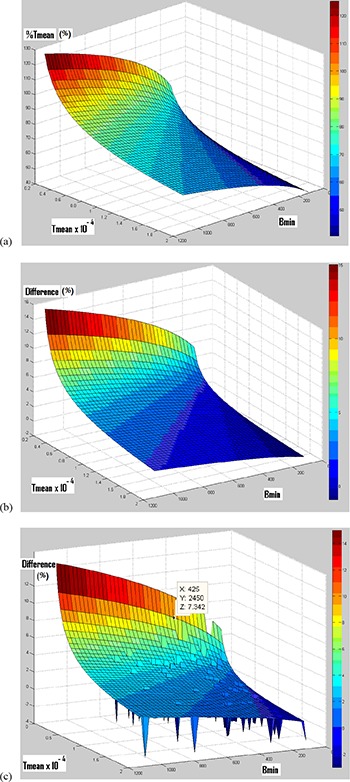
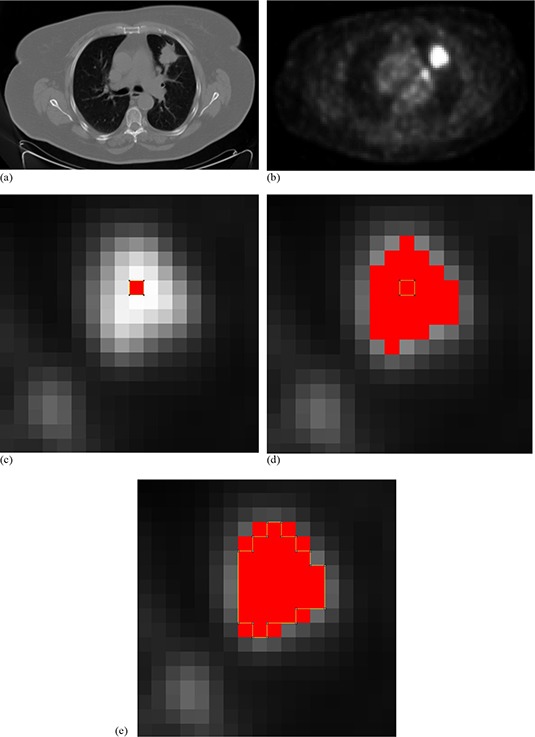
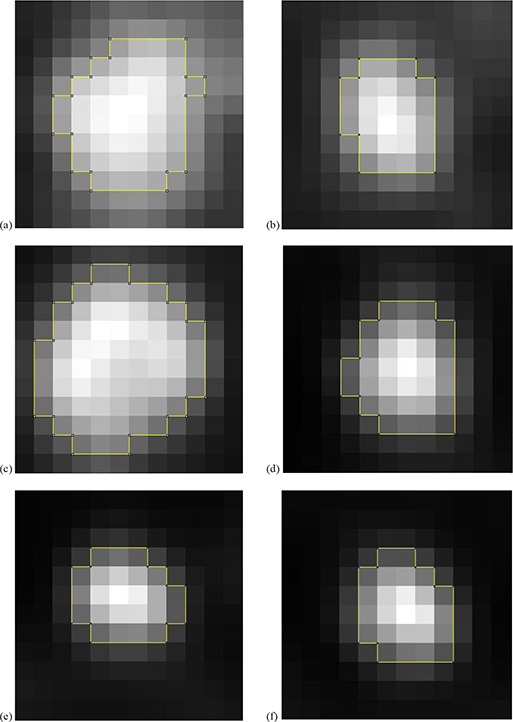
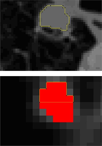
Tumor delineation is a critical aspect in radiotherapy treatment planning and is usually performed with the anatomical images of a computed tomography (CT) scan. For non-small cell lung cancer, it has been recommended to use functional positron emission tomography (PET) images to take into account the biological target characteristics. However, today, there is no satisfactory segmentation technique for PET images in clinical applications. In the present study, a solution to this problem is proposed. The development of the segmentation technique is based on the threshold's adjustment directly from patients, rather than from phantoms. To this end, two references were chosen: measurements performed on CT images of the selected lesions, and histological measurements of surgically removed tumors. The inclusion and exclusion criteria were chosen to produce references that are assumed to have measured tumor sizes equal to the true in vivo tumor sizes. In total, for the two references, 65 lung lesions of 54 patients referred for FDG-PET/CT exams were selected. For validation, measurements of segmented lesions on PET images using this technique were also compared to CT and histological measurements. For lesions greater than 20 mm, our segmentation technique showed a good estimation of histological measurements (mean difference between measured and calculated data equal to -0.8 ± 9.0%) and an acceptable estimation of CT measurements. For lesions smaller than or equal to 20 mm, the method showed disagreement with the measurements derived from histological or CT data. This novel segmentation technique shows high accuracy for the lesions with largest axes between 2 and 4.5 cm. However, it does not correctly evaluate smaller lesions, likely due to the partial volume effect and/or respiratory motions.
Figures

References
-
- Geets X, Daisne J, Arcangeli S, et al. Inter‐observer variability in the delineation of pharyngo‐laryngeal tumor, parotid glands and cervical spinal cord: comparison between CT‐scan and MRI. Radiother Oncol. 2005;77(1):25–31. - PubMed
-
- Strauss MA and Conti PS. The application of PET in clinical oncology. J Nucl Med. 1991;32(4):623–48. - PubMed
-
- Lecchi M, Fossati P, Elisei F, Orecchia R, Lucignani G. Current concepts on imaging in radiotherapy. Eur J Nucl Med Mol Imaging. 2008;35(4):821–37. - PubMed
-
- Gould MK, Maclean CC, Kuschner WG, Rydzak CE, Owens DK. Accuracy of positron emission tomography for diagnosis of pulmonary nodules and mass lesions: a meta analysis. JAMA. 2001;285(7):914–24. - PubMed
-
- Gambhir SS, Czernin J, Schwinner J, Silverman DH, Coleman RE, Phelps ME. A tabulated summary of the FDG PET literature. J Nucl Med. 2001;42(5 Suppl):1S–98S. - PubMed
Publication types
MeSH terms
Substances
LinkOut - more resources
Full Text Sources
Medical
