. 2012 Sep 25;109(39):E2635-44.
doi: 10.1073/pnas.1202526109. Epub 2012 Sep 5.

Mechanism for Hypocretin-mediated sleep-to-wake transitions

Affiliations

Mechanism for Hypocretin-mediated sleep-to-wake transitions

Matthew E Carter et al. Proc Natl Acad Sci U S A. .

Abstract

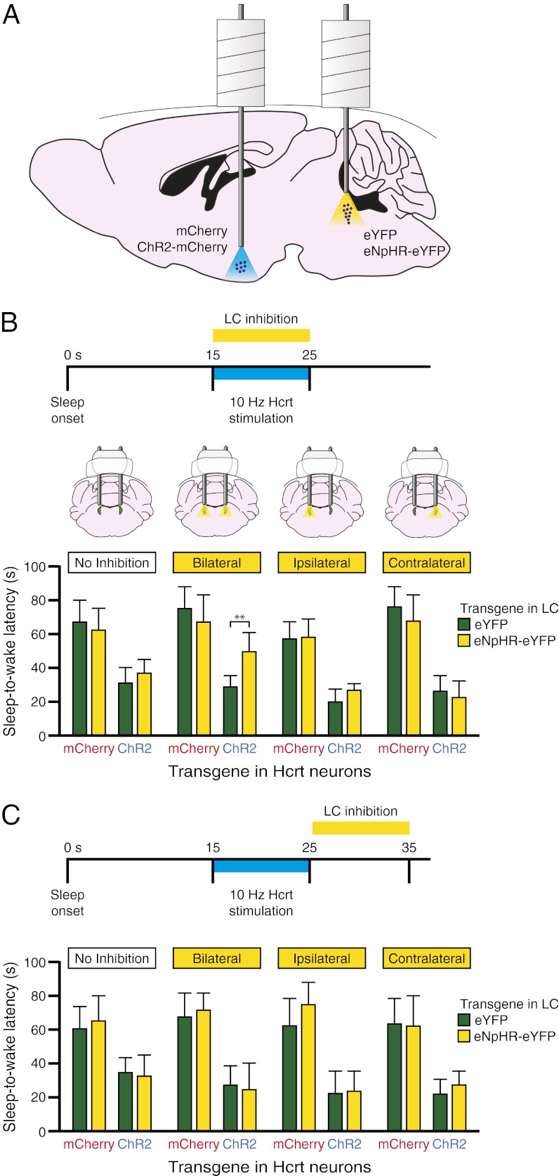
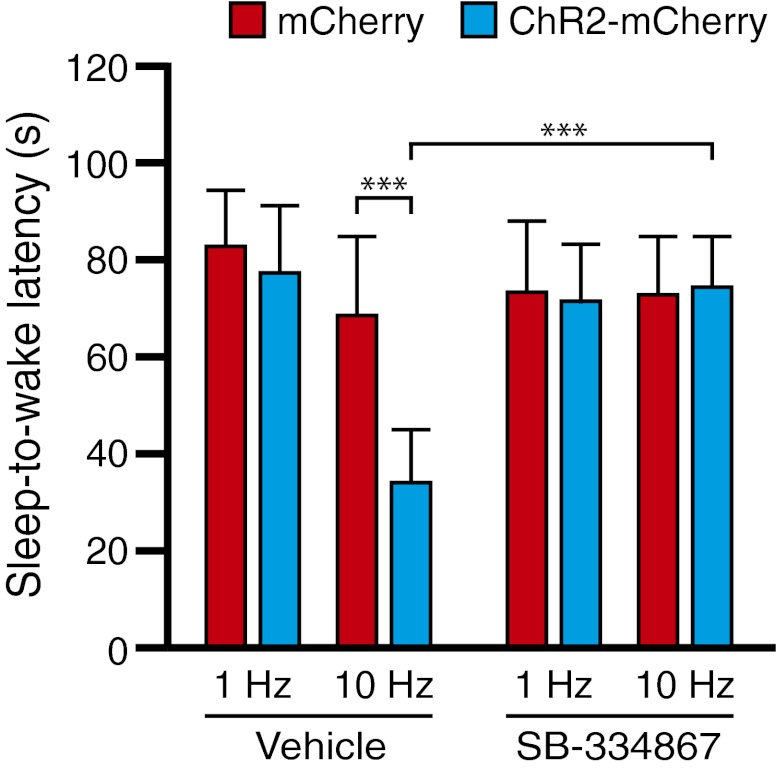
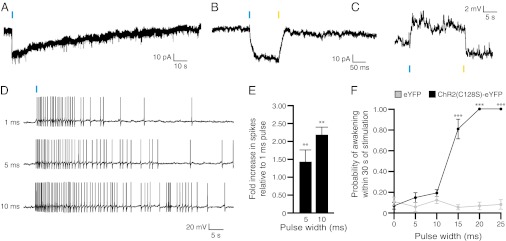
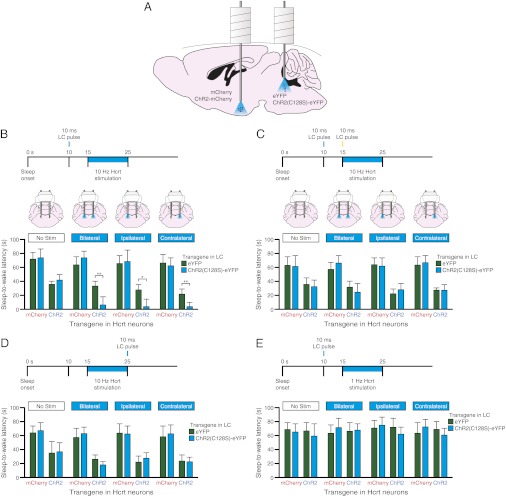
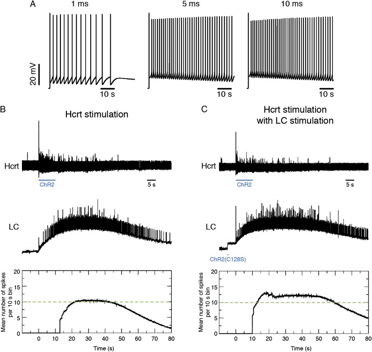
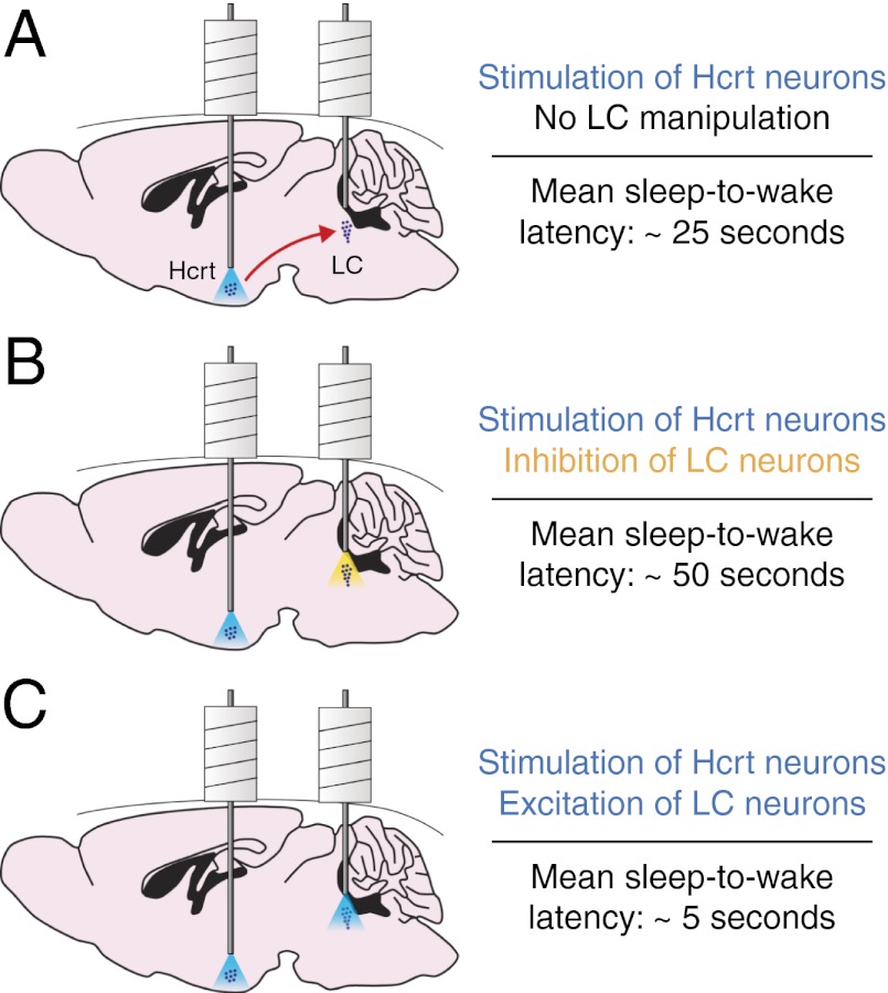
Current models of sleep/wake regulation posit that Hypocretin (Hcrt)-expressing neurons in the lateral hypothalamus promote and stabilize wakefulness by projecting to subcortical arousal centers. However, the critical downstream effectors of Hcrt neurons are unknown. Here we use optogenetic, pharmacological, and computational tools to investigate the functional connectivity between Hcrt neurons and downstream noradrenergic neurons in the locus coeruleus (LC) during nonrapid eye movement (NREM) sleep. We found that photoinhibiting LC neurons during Hcrt stimulation blocked Hcrt-mediated sleep-to-wake transitions. In contrast, when LC neurons were optically stimulated to increase membrane excitability, concomitant photostimulation of Hcrt neurons significantly increased the probability of sleep-to-wake transitions compared with Hcrt stimulation alone. We also built a conductance-based computational model of Hcrt-LC circuitry that recapitulates our behavioral results using LC neurons as the main effectors of Hcrt signaling. These results establish the Hcrt-LC connection as a critical integrator-effector circuit that regulates NREM sleep/wake behavior during the inactive period. This coupling of distinct neuronal systems can be generalized to other hypothalamic integrator nuclei with downstream effector/output populations in the brain.


Conflict of interest statement

The authors declare no conflict of interest.

Figures

Fig. 1.
Fig. 1.
Hcrt and LC neuroanatomy and functional connectivity. (A) Diagram depicting the sagittal mouse brain with relative locations of Hcrt and LC neurons. Hcrt neurons send projections throughout the brain but project most strongly to LC neurons in the brainstem. (B and C) Spatial distribution of tyrosine hydroxylase-immunoreactive soma in the brainstem (B) closely parallels the density of Hcrt-immunoreactive fibers in an adjacent section (C). (Scale bar, 50 μm.) (D) In vivo unilateral photostimulation of Hcrt neurons increases c-Fos immunoreactivity in the LC. Shown are representative images of the ipsilateral and contralateral LC costained for TH (red) and c-Fos (white). (Scale bar, 25 μm.) (E) Quantification of the percentage of neurons showing TH immunoreactivity that also express c-Fos. Data represent the mean ± SEM from animals transduced with ChR2-mCherry (n = 4) and mCherry (n = 4). **P < 0.001, two-way ANOVA followed by Tukey’s posthoc test.
Fig. 2.
Fig. 2.
Simultaneous bilateral photoinhibition of LC neurons blocks Hcrt-mediated sleep-to-wake transitions. (A) Diagram depicting experimental conditions. Hcrt neurons were transduced with either ChR2-mCherry or mCherry and LC neurons were transduced with either eNpHR-eYFP or eYFP. (B) Latencies to wake during NREM sleep following photostimulation in Hcrt neurons with simultaneous photoinhibition in LC neurons. Data represent the mean ± SEM sleep-to-wake latencies (15 trials per mouse per condition, six mice per condition). **P < 0.001, Student’s t test between eNpHR-eYFP and eYFP conditions. (C) Latencies to wake during NREM sleep following photostimulation in Hcrt neurons and subsequent photoinhibition in LC neurons. Data represent the mean ± SEM sleep-to-wake latencies (15 trials per mouse per condition, six mice per condition).
Fig. 3.
Fig. 3.
Pharmacological antagonism of Hcrt signaling in the LC region blocks Hcrt-mediated sleep-to-wake transitions. Photostimulation of Hcrt neurons at 10 Hz causes a significant decrease in sleep-to-wake latency between animals expressing ChR2-mCherry and control animals expressing mCherry; these effects are blocked by local injection of the Hcrt antagonist SB-334867 directly into the LC field. ***P < 0.0001, ANOVA between genotype and stimulation frequency, followed by Tukey’s posthoc test.
Fig. 4.
Fig. 4.
Functional expression of ChR2(C128S)-eYFP in LC neurons. (A) Voltage-clamp recording of an LC neuron expressing ChR2(C128S)-eYFP in an acute brainstem slice showing inward current in response to a single 10-ms pulse of blue light. (B) Voltage-clamp recording of a neuron showing inward current in response to a single 10-ms pulse of blue light and a return to baseline in response to a single 10-ms pulse of yellow light. (C) Current-clamp recording of a neuron showing a depolarization in response to a single 10-ms pulse of blue light and a return to baseline in response to a single 10-ms pulse of yellow light. (D) Action potential trains generated in current clamp in response to single 1-, 5-, and 10-ms pulses of blue light. (E) Quantification of the fold-increase number of spikes relative to 1-ms pulses of light. Data represent mean ± SD fold increase from n = 5 neurons. **P < 0.001, Student’s t test between conditions. (F) Probability of a sleep-to-wake transition within 30 s of stimulation with single pulses of light of different durations between ChR2(C128S)-eYFP and eYFP control animals. Data represent the mean ± SEM probability values for n = 6 mice, 10 trials per mouse. ***P < 0.0001, Student’s t test between transduced mice.
Fig. 5.
Fig. 5.
Stimulation of LC neurons with ChR2(C128S)-eYFP enhances Hcrt-mediated sleep-to-wake transitions. (A) Diagram depicting experimental conditions. Hcrt neurons were transduced with either ChR2-mCherry or mCherry and LC neurons were transduced with either ChR2(C128S)-eYFP or eYFP. (BE) (Upper) Experimental stimulation paradigms. (Lower) Latencies to wakefulness during NREM sleep following photostimulation in Hcrt neurons and simultaneous photostimulation in LC neurons. Data represent the mean ± SEM sleep-to-wake latencies (15 trials per mouse per condition, six mice per condition). In B, LC stimulation precedes Hcrt stimulation by 5 s. In C, LC stimulation precedes Hcrt stimulation but is followed with a 10-ms pulse of yellow light before Hcrt stimulation. In D, LC stimulation occurs at the offset of Hcrt stimulation. In E, LC stimulation precedes 1-Hz Hcrt stimulation by 5 s. *P < 0.05, **P < 0.001, Student’s t test between ChR2(C128S)-eYFP and eYFP conditions.
Fig. 6.
Fig. 6.
Conductance-based model of Hcrt-LC circuitry. (A) Model of the effect of a single pulse of blue light (1, 5, or 10 ms) in ChR2(C128S)-eYFP-transduced LC neurons. (B) Model of the effect of Hcrt stimulation on LC neurons. We executed our model with a paradigm of 10 s prestimulation baseline activity followed with 10 s ChR2-mediated stimulation of Hcrt neurons at 10 Hz. We then let the model run an additional 70 s to monitor poststimulation effects. (Top) Spike trains in Hcrt neurons throughout a run of the model. (Middle) Corresponding spike trains in LC neurons. (Bottom) Average number of spikes in LC neurons per 10-s bins during the run of the model. Dashed green line corresponds to the threshold of LC activity previously shown (24) to be sufficient to cause a sleep-to-wake transition. See also Movie S2. (C) Same as B but with an additional 10-ms blue-light pulse in ChR2(C128S)-eYFP–transduced LC neurons occurring 5 s before the onset of Hcrt stimulation.
Fig. P1.
Fig. P1.
Hcrt and LC neurons both promote wakefulness. Hcrt neurons send dense, excitatory projections to the LC (depicted on a sagittal mouse brain), raising the possibility that the Hcrt-mediated promotion of wakefulness depends on the LC. To determine the necessity and sufficiency of LC activity for Hcrt-mediated sleep-to-wake transitions, we used optogenetic probes to selectively inhibit or stimulate LC neurons during Hcrt stimulation. (A) When Hcrt neurons were stimulated during sleep, the mean sleep-to-wake latency was about 25 s. (B) Inhibiting the LC during Hcrt stimulation significantly increased the sleep-to-wake latency to about 50 s. (C) In contrast, increasing the membrane excitability of the LC during Hcrt stimulation decreased the sleep-to-wake latency to about 5 s. Our results demonstrate that the LC serves as a critical effector for Hcrt-mediated sleep-to-wake transitions.

References

    1. Saper CB, Scammell TE, Lu J. Hypothalamic regulation of sleep and circadian rhythms. Nature. 2005;437:1257–1263. - PubMed
    1. Pace-Schott EF, Hobson JA. The neurobiology of sleep: Genetics, cellular physiology and subcortical networks. Nat Rev Neurosci. 2002;3:591–605. - PubMed
    1. de Lecea L, et al. The hypocretins: Hypothalamus-specific peptides with neuroexcitatory activity. Proc Natl Acad Sci USA. 1998;95:322–327. - PMC - PubMed
    1. Sakurai T, et al. Orexins and orexin receptors: A family of hypothalamic neuropeptides and G protein-coupled receptors that regulate feeding behavior. Cell. 1998;92:573–585. - PubMed
    1. Sutcliffe JG, de Lecea L. The hypocretins: Setting the arousal threshold. Nat Rev Neurosci. 2002;3:339–349. - PubMed

Publication types