. 2012 Dec;19(12):1896-907.
doi: 10.1038/cdd.2012.108. Epub 2012 Sep 7.

A novel cellular stress response characterised by a rapid reorganisation of membranes of the endoplasmic reticulum

Affiliations

A novel cellular stress response characterised by a rapid reorganisation of membranes of the endoplasmic reticulum

S Varadarajan et al. Cell Death Differ. 2012 Dec.

Abstract

Canonical endoplasmic reticulum (ER) stress, which occurs in many physiological and disease processes, results in activation of the unfolded protein response (UPR). We now describe a new, evolutionarily conserved cellular stress response characterised by a striking, but reversible, reorganisation of ER membranes that occurs independently of the UPR, resulting in impaired ER transport and function. This reorganisation is characterised by a dramatic redistribution and clustering of ER membrane proteins. ER membrane aggregation is regulated, in part, by anti-apoptotic BCL-2 family members, particularly MCL-1. Using connectivity mapping, we report the widespread occurrence of this stress response by identifying several structurally diverse chemicals from different pharmacological classes, including antihistamines, antimalarials and antipsychotics, which induce ER membrane reorganisation. Furthermore, we demonstrate the potential of ER membrane aggregation to result in pathological consequences, such as the long-QT syndrome, a cardiac arrhythmic abnormality, arising because of a novel trafficking defect of the human ether-a-go-go-related channel protein from the ER to the plasma membrane. Thus, ER membrane reorganisation is a feature of a new cellular stress pathway, clearly distinct from the UPR, with important consequences affecting the normal functioning of the ER.


Figures
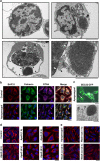
Figure 1
Figure 1
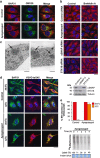
Apogossypol induces a reversible aggregation of ER membranes. (a) CLL cells, exposed for 16 h to apogossypol (10 μM), formed membrane aggregates (arrow, top right panel) in contrast to untreated CLL cells (top left panel). Severe swelling of the nuclear envelope was evident in Jurkat cells, exposed for 16 h to apogossypol (10 μM) (lower left panel) and the continuity of this envelope with an ER membrane aggregate is shown at higher magnification (lower right panel) (scale bars, 2 μm). The inner and outer nuclear membranes are indicated by white and black arrowheads, respectively. (b) HeLa cells exposed for 4 h to apogossypol (10 μM) and subsequently immunostained for ER membrane proteins (BAP31, calnexin and RTN4) exhibited dramatic clustering of the ER membrane proteins (scale bar, 20 μm). (c) Fluorescence microscopy can be used to detect ER membrane aggregates. HeLa cells transfected with CFP-tagged SEC22 and subsequently exposed for 4 h to apogossypol (10 μM) exhibited numerous fluorescent foci. A CFP-labelled cluster (white arrow upper panel, scale bar, 20 μm) was subsequently examined by electron microscopy, establishing that these foci were in fact ER membrane aggregates (lower panel, scale bar 2 μm). (d) BAP31-containing ER membrane aggregates undergo time-dependent formation and coalescence in HeLa cells exposed to apogossypol (ApoG, 10 μM) (scale bar, 20 μm). The ER membrane aggregates were visible within 1 h. (e) The BAP31-containing ER membrane aggregates, formed in HeLa cells following exposure for 4 h to apogossypol (10 μM), dispersed rapidly on wash out (scale bar 20 μm)
Figure 2
Figure 2
Apogossypol disrupts ER transport and function. (a) HeLa cells, exposed for 4 h to apogossypol (10 μM), were immunostained for membrane proteins in the ER (BAP31) and Golgi (GM130). Cells with BAP31 aggregates exhibited a near complete dispersal of Golgi (scale bar, 20 μm). (b) Disruption of trafficking, by downregulation of αSNAP and STX18, resulted in the formation of BAP31 membrane aggregates similar to those following exposure to apogossypol (scale bar, 20 μm). Exposure to brefeldin A (20 μM) for 0.5 h completely dispersed BAP31-containing aggregates in the STX18 but not in αSNAP downregulated or apogossypol-treated cells, consistent with defects in anterograde rather than retrograde trafficking (scale bar, 20 μm). The loss of the respective proteins was confirmed by western blotting (lower panel). (c) Downregulation of αSNAP (left panel) and STX18 (right panel) by RNAi resulted in the formation of ER membrane aggregates (black arrows) (scale bar, 1 μm) (d) ER membrane aggregates disrupt ER to Golgi trafficking. HeLa cells transfected with VSVG-ts045-Cherry construct and maintained at the restrictive (39.5 °C) temperature were exposed to apogossypol (10 μM) for 2 h and shifted to permissive (32 °C) temperature for another 2 h. The cells were then fixed and immunostained for BAP31, and assessed by confocal microscopy (scale bar, 20 μm). (e) The graph shows the ER retention of VSVG protein following a shift from the restrictive (39.5 °C) to permissive (32 °C) temperature in control and treated cells, from an average of a minimum of 200 cells from three independent experiments; error bars show the S.D. (f) Apogossypol induced a global attenuation of translation, as depicted in the autoradiogram. Autoradiography of [35S] methionine-labelled proteins in lysates of HeLa cells exposed to apogossypol (10 μM) for 1–4 h were resolved by NuPage. Data were expressed as a percentage of [35S]-labelling of HeLa cells not exposed to ApoG following Phosphorimager analysis. Lower panel is a photomicrograph of an instant blue-stained gel, showing equal loading
Figure 3
Figure 3
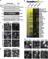
Apogossypol induces a novel form of ER stress distinct from the classical UPR. (a) Most biochemical changes associated with canonical ER stress occur much later than the induction of ER membrane aggregates at 1 h. Whole-cell lysates or total RNA of HeLa cells exposed to apogossypol (10 μM) or tunicamycin (20 μM) for the indicated times were either immunoblotted with the indicated antibodies, or analysed by RT-PCR using a primer set that amplified human XBP1 mRNA. Unspliced and IRE1-spliced XBP1 (*) transcripts yield 289  and 263-bp products, respectively. (b) Apogossypol (1 h) induced very few gene changes associated with canonical ER stress despite extensive ER membrane aggregation. Heat map analysis comparing gene changes following 6 h of tunicamycin (20 μM) or brefeldin A (20 μM) to 1 or 6 h of apogossypol (10 μM) treatment in MCF-7 cells, where yellow and blue indicate up- or downregulation, respectively. Tunicamycin and brefeldin A markedly altered many characteristic ER stress genes, whereas apogossypol changed a few of these genes and to a much lesser degree. Analysis from three separate experiments (n) was used to generate the heat map. (c) Apogossypol induces ER membrane aggregation in the absence of transcription or translation, as evidenced by a 4-h pre-treatment of actinomycin D (10 μM) or cycloheximide (20 μM) followed by another 4 h to apogossypol (10 μM) (scale bar, 20 μm). (d) Two classical inducers of canonical ER stress, tunicamycin (20 μM) and brefeldin A (20 μM), failed to induce ER membrane aggregation (scale bar, 20 μm). (e) Knockout of genes critical for canonical ER stress did not affect the ability of apogossypol to induce ER membrane aggregation. After 4 h, apogossypol (20 μM) induced ER membrane aggregation in PERK−/−, eIF2α S51A, IRE1α−/−, XBP1−/−, CHOP−/− and ATF6-deficient murine embryonic fibroblasts (scale bar, 10 μm)
Figure 4
Figure 4
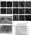
MCL-1 is a key anti-apoptotic BCL-2 family member that regulates ER membrane reorganisation. (a) Pan-BCL-2 family inhibitors induce ER membrane reorganisation more potently than BCL-2 and BCL-XL-specific inhibitors. Apogossypol (10 μM) and TW37 (20 μM) induced extensive membrane reorganisation, assessed by BAP31 staining in HeLa cells, within 4 h of exposure, whereas ABT-737 (20 μM) induced modest reorganisation after 8 h and the inactive enantiomer ABT-737E (20 μM) failed to induce reorganisation (scale bar, 20 μm). (b) MCL-1 is the most important anti-apoptotic BCL-2 family member involved in the regulation of ER membrane reorganisation. HeLa cells following 72 h silencing of BCL-2, BCL-XL or BCL-W did not exhibit detectable ER modification, whereas ER membrane reorganisation was induced by three independent siRNAs of MCL-1 and this was potentiated by ABT-737 (scale bar, 20 μm). (c) The nature of the ER membrane reorganisation resulting from MCL-1 knockdown in MCF7 cells was confirmed by electron microscopy. A detail of a membrane aggregate in the upper panel is shown in the lower panel (scale bars, 1 μm). (d) Western blots confirmed the efficiency of the siRNA oligoduplexes
Figure 5
Figure 5
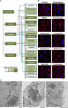
Chemical connectivity mapping identifies compounds from diverse chemical classes that induce ER membrane reorganisation. (a) The connectivity map identified 24 chemicals of which 20 were positive when tested for their ability to induce ER membrane reorganisation. Scheme representing the connectivity map of apogossypol (grey lines), NDGA (brown lines), THG (blue lines), astemizole (red lines) and ivermectin (green lines) with several compounds contained within the connectivity reference database. All boxes are colour coded in increasing shades of green to demonstrate the extent of ER membrane reorganisation, in HeLa cells after 4 h exposure to the indicated chemical, from no (−) to extensive aggregation (+++). (b) Exposure of HeLa cells for 4 h to apogossypol (10 μM), NDGA (50 μM), THG (10 μM), astemizole (5 μM), ivermectin (20 μM), troglitazone (20 μM in serum-free media), mefloquine (20 μM), suloctidil (20 μM) and terfenadine (10 μM) exhibited varying levels of ER membrane reorganisation assessed by BAP31 staining (scale bar, 20 μm). (c) Ultrastructural analysis confirmed that these BAP31 clusters induced by NDGA (left panel, scale bar, 1 μm), THG (middle panel, scale bar, 1 μm) and rottlerin (right panel, scale bar, 2 μm) were bona fide ER membrane aggregates
Figure 6
Figure 6
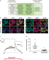
ER membrane reorganisation leads to hERG trafficking and functional defects. (a) Most agents that induce ER membrane reorganisation are associated with cardiotoxicity as illustrated by the degree of overlap in the scheme. The drugs implicated in causing LQTS are marked with an asterisk (*). Some of the ER membrane reorganisation agents in the left-hand panel have not been tested for their ability to induce LQTS. (b) hERG colocalised with ER membrane aggregates in apogossypol- and terfenadine- but not fexofenadine-treated cells. HEK293 cells, stably expressing hERG, were exposed for 4 h to either apogossypol (10 μM), terfenadine (10 μM) or fexofenadine (10 μM), and immunostained for BAP31 and hERG (scale bar, 20 μm). (c) Voltage clamp measurements reveal that hERG currents are greatly reduced in apogossypol-treated cells compared with untreated cells. Currents were elicited with 5-s voltage steps to +20 mV and peak tail currents measured upon repolarisation to −50 mV. (d) The decreases in hERG channel function are owing both to a decrease in cell size (as indicated by reductions in membrane surface area and capacitance) and also owing to reduced channel numbers. Tail current densities (peak tail current amplitude divided by capacitance) for individual cells are substantially attenuated in apogossypol-treated cells. Each data point in the scatter plots represents a measurement from a single cell. The mean current densities for each group are illustrated by the horizontal lines
Figure 7
Figure 7
Scheme representing the formation, regulation and consequences of ER membrane reorganisation. Under normal conditions, proteins, such as hERG, are synthesised in the ER lumen, and trafficked via the Golgi to the plasma membrane (PM) to express a functional channel (left-hand panel). Exposure to stress conditions, such as inhibition of the BCL-2 family (apogossypol and TW37), induced ER membrane reorganisation (right-hand panel). This ER membrane reorganisation is reversible and occurs independent of canonical ER stress, as tunicamycin (Tunic) and brefeldin A (BFA), two inducers of canonical ER stress, do not result in ER membrane reorganisation. However, at later times, the extensive ER membrane reorganisation may eventually lead to canonical ER stress and the UPR, thus placing ER membrane reorganisation upstream and/or independent of the UPR. Furthermore, ER membrane reorganisation results in the functional perturbation of the ER, characterised by a near complete dispersion of the Golgi and an accompanying trafficking defect. This novel cellular stress pathway could lead to important pathological consequences, including LQTS, mediated by the entrapment of the newly synthesised hERG in the aggregates resulting in a trafficking defect that is marked by the loss of functional hERG channels at the cell surface

References

    1. Hu J, Prinz WA, Rapoport TA. Weaving the web of ER tubules. Cell. 2011;147:1226–1231. - PMC - PubMed
    1. Voeltz GK, Prinz WA, Shibata Y, Rist JM, Rapoport TA. A class of membrane proteins shaping the tubular endoplasmic reticulum. Cell. 2006;124:573–586. - PubMed
    1. Baumann O, Walz B. Endoplasmic reticulum of animal cells and its organization into structural and functional domains. Int Rev Cytol. 2001;205:149–214. - PubMed
    1. Lynes EM, Simmen T. Urban planning of the endoplasmic reticulum (ER): how diverse mechanisms segregate the many functions of the ER. Biochim Biophys Acta. 2011;1813:1893–1905. - PMC - PubMed
    1. Ron D, Walter P. Signal integration in the endoplasmic reticulum unfolded protein response. Nat Rev Mol Cell Biol. 2007;8:519–529. - PubMed

Publication types

MeSH terms