. 2012 Oct 1;53(11):7159-66.
doi: 10.1167/iovs.12-10222.

Selective activation of ATF6 and PERK endoplasmic reticulum stress signaling pathways prevent mutant rhodopsin accumulation

Affiliations

Selective activation of ATF6 and PERK endoplasmic reticulum stress signaling pathways prevent mutant rhodopsin accumulation

Wei-Chieh Chiang et al. Invest Ophthalmol Vis Sci. .

Abstract

Purpose: Many rhodopsin mutations that cause retinitis pigmentosa produce misfolded rhodopsin proteins that are retained within the endoplasmic reticulum (ER) and cause photoreceptor cell death. Activating transcription factor 6 (ATF6) and protein kinase RNA-like endoplasmic reticulum kinase (PERK) control intracellular signaling pathways that maintain ER homeostasis. The aim of this study was to investigate how ATF6 and PERK signaling affected misfolded rhodopsin in cells, which could identify new molecular therapies to treat retinal diseases associated with ER protein misfolding.

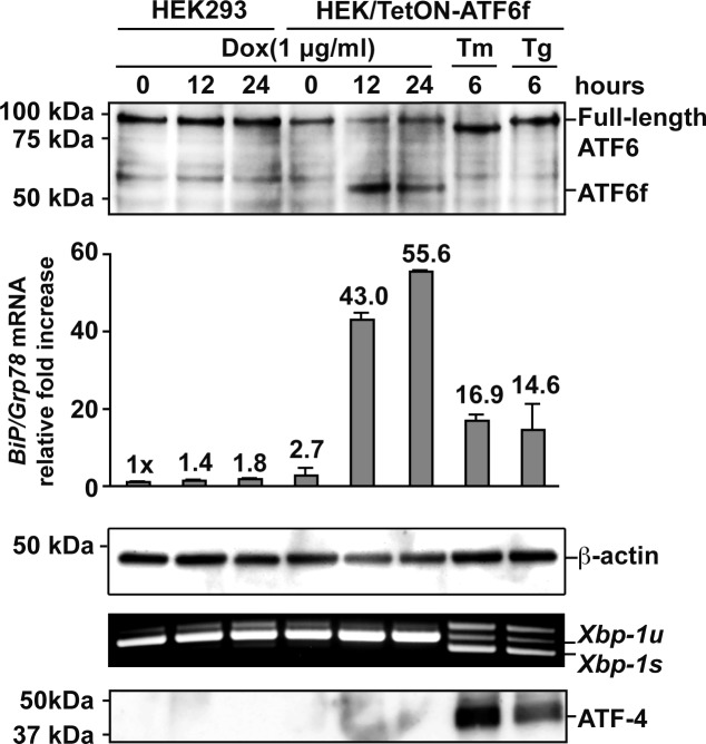
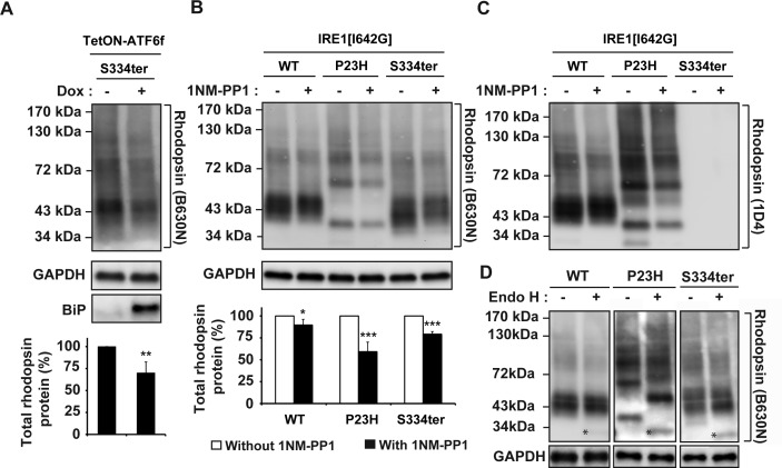
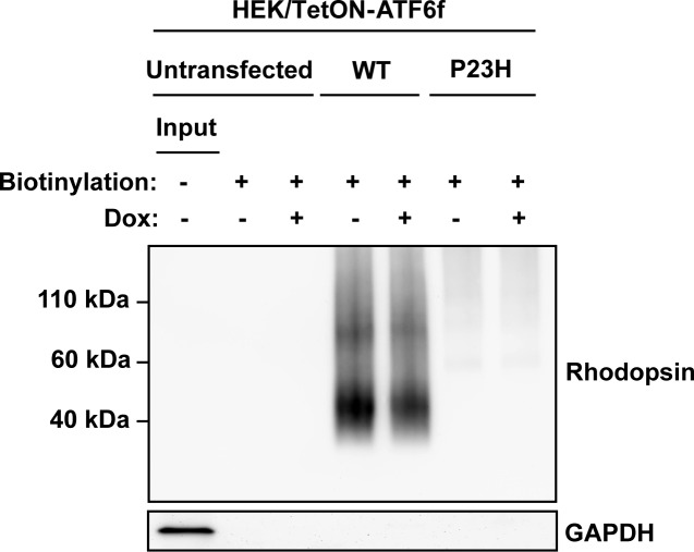
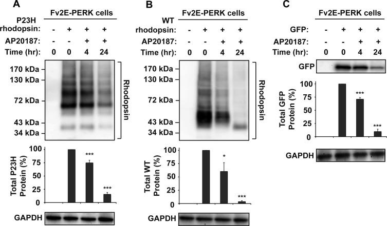
Methods: To examine the effect of ATF6 on rhodopsin, wild-type (WT) or mutant rhodopsins were expressed in cells expressing inducible human ATF6f, the transcriptional activator domain of ATF6. Induction of ATF6f synthesis rapidly activated downstream genes. To examine PERK's effect on rhodopsin, WT or mutant rhodopsins were expressed in cells expressing a genetically altered PERK protein, Fv2E-PERK. Addition of the dimerizing molecule (AP20187) rapidly activated Fv2E-PERK and downstream genes. By use of these strategies, it was examined how selective ATF6 or PERK signaling affected the fate of WT and mutant rhodopsins.

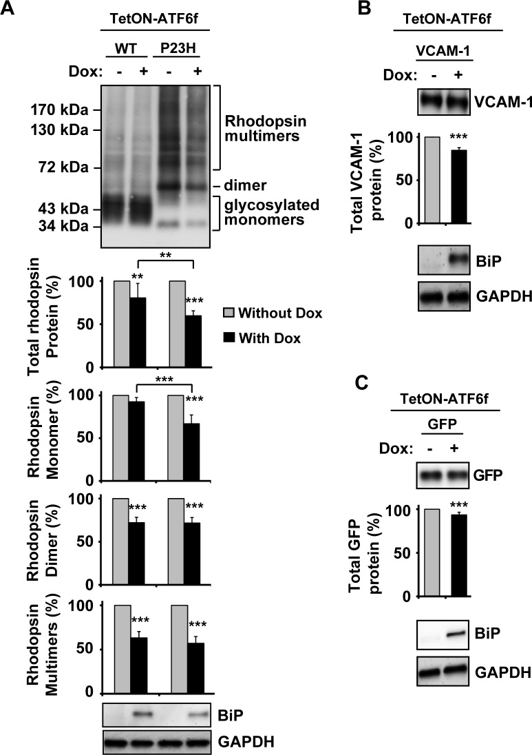
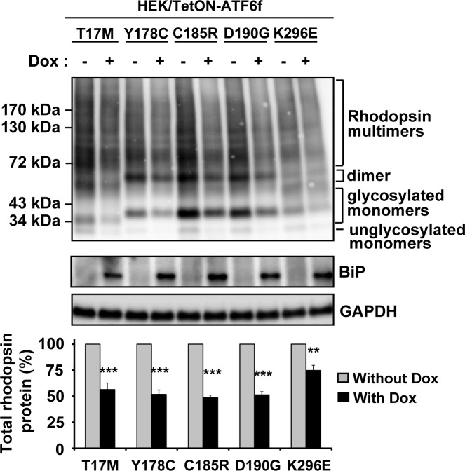
Results: ATF6 significantly reduced T17M, P23H, Y178C, C185R, D190G, K296E, and S334ter rhodopsin protein levels in the cells with minimal effects on monomeric WT rhodopsin protein levels. By contrast, the PERK pathway reduced both levels of WT, mutant rhodopsins, and many other proteins in the cell.

Conclusions: This study indicates that selectively activating ATF6 or PERK prevents mutant rhodopsin from accumulating in cells. ATF6 signaling may be especially useful in treating retinal degenerative diseases arising from rhodopsin misfolding by preferentially clearing mutant rhodopsin and abnormal rhodopsin aggregates.


Conflict of interest statement

Disclosure: W.-C. Chiang, None; N. Hiramatsu, None; C. Messah, None; H. Kroeger, None; J.H. Lin, None

Figures

Figure 1.
Figure 1.
Chemical–genetic activation of ATF6. WT and isogenic HEK293 cells stably expressing a tetracycline-inducible 373 amino acid cytosolic transcriptional activator domain of human ATF6 (ATF6f) were treated with doxycycline (Dox) (1 μg/mL), tunicamycin (Tm) (5 μg/mL), or thapsigargin (Tg) (1 μM) for the indicated durations. Endogenous full-length and induced ATF6 transcriptional activator domain, ATF6f, were detected by immunoblotting. β-Actin protein levels were assessed as a loading control. Levels of BiP/Grp78 mRNA, a gene robustly induced by ATF6, were assessed by real-time quantitative PCR. Xbp-1 mRNA splicing, a specific marker of IRE1 activation, was assessed by RT-PCR. Levels of ATF-4, a protein robustly induced by PERK signaling, were assessed by immunoblotting.
Figure 2.
Figure 2.
Chemical–genetic activation of ATF6 reduced misfolded P23H rhodopsin protein levels. (A) WT or P23H rhodopsin, (B) WT VCAM-1, or (C) GFP was transfected into cells expressing a tetracycline-inducible ATF6f (TetON-ATF6f), and Dox (1 μg/mL) was applied as indicated for 24 hours. Rhodopsin, VCAM-1, or GFP protein levels were detected by immunoblotting using 1D4 anti-rhodopsin, anti-VCAM-1, and anti-GFP antibody respectively and quantified by a commercial image acquisition and analysis software program (VisionWorks LS Software). Rhodopsin monomer, dimer, and multimers protein levels were determined by measuring the area density within the indicated line or bracket. Protein levels of BiP/Grp78, a downstream transcriptional target induced by ATF6 were assessed by immunoblotting. GAPDH levels were assessed as a loading control. Statistical significance was determined by one-way ANOVA with Bonferroni post hoc test (A, n = 7) or Student's t-test (B, C; n = 3), and is denoted by asterisks: **P < 0.01 and ***P < 0.001 compared with the cells expressing the transfected protein without the treatment of doxycycline or as indicated.
Figure 3.
Figure 3.
Chemical–genetic activation of ATF6 reduced five additional class II mutant rhodopsin protein levels. T17M, Y178C, C185R, D190G, or K296E class II mutant rhodopsins were transfected into cells expressing TetON-ATF6f, and Dox (1 μg/mL) was applied as indicated for 24 hours. Rhodopsin protein levels were detected by immunoblotting using 1D4 anti-rhodopsin antibody and quantified by a commercial image acquisition and analysis software program (VisionWorks LS Software). Protein levels of BiP/Grp78, a downstream transcriptional target induced by ATF6, were assessed by immunoblotting. GAPDH levels were assessed as a loading control. Immunoblots are representative of three independent experiments. Statistical significance (mean ± SD; n = 3) was determined by Student's t-test, and is denoted by asterisks: **P < 0.01 and ***P < 0.001 compared with the cells expressing transfected mutant rhodopsin without the treatment of doxycycline.
Figure 4.
Figure 4.
Chemical–genetic activation of ATF6 and IRE1 reduced S334ter rhodopsin protein levels. (A) S334ter rhodopsin was transfected into cells expressing TetON-ATF6f, and Dox (1 μg/mL) was applied as indicated for 24 hours. Rhodopsin protein levels were detected by immunoblotting using B630N anti-rhodopsin antibody and quantified by a commercial image acquisition and analysis software program (VisionWorks LS Software). Protein levels of BiP/Grp78, a downstream transcriptional target induced by ATF6, were assessed by immunoblotting. (B) WT or mutant rhodopsin was transfected into cells bearing IRE1[I642G], and 1NM-PP1 (5 μM) was applied as indicated for 24 hours. Rhodopsin protein levels were detected by immunoblotting using B630N anti-rhodopsin antibody and quantified by a commercial software program (VisionWorks LS Software). (A, B) GAPDH levels were assessed as a loading control. Immunoblots are representative of three independent experiments. Statistical significance (mean ± SD; n = 3) was determined by Student's t-test, and is denoted by asterisks: *P < 0.05, **P < 0.01, and ***P < 0.001 compared with the cells expressing WT or mutant rhodopsin without the treatment of doxycycline or 1NM-PP1. (C) For comparison, the same immunoblot from (B) was stripped and reprobed with 1D4 anti-rhodopsin antibody. (D) To measure the amount of Endo H–sensitive rhodopsin, 10 μg of total cell lysates expressing wild-type, P23H, or S334ter rhodopsin were treated with Endo H and the deglycosylated Endo H–sensitive species were resolved by SDS-PAGE and immunoblotting analysis. An asterisk (*) indicates the deglycosylated rhodopsin monomer isolated after Endo H treatment. GAPDH protein levels were assessed as a protein loading control.
Figure 5.
Figure 5.
Chemical–genetic activation of ATF6 does not promote the delivery of rhodopsin to the cell surface. WT or P23H rhodopsin was transfected into cells expressing Tet-On-ATF6f, with or without application of Dox (1 μg/mL) for 24 hours. Surface membrane proteins were biotinylated, and rhodopsin protein levels in the biotinylated fraction were assessed by immunoblotting using 1D4 anti-rhodopsin antibody. GAPDH protein levels in the biotinylated protein fractions were assessed as a control for the specificity of surface protein biotinylation. Input: total cell lysate of the untransfected cells.
Figure 6.
Figure 6.
Chemical–genetic activation of PERK reduced WT and P23H rhodopsin protein levels. (A) P23H rhodopsin was transfected into cells expressing drug-sensitized Fv2E-PERK, and AP20187 (2 nM) was applied as indicated. (B) WT rhodopsin was transfected into cells expressing Fv2E-PERK, and AP20187 (2 nM) was applied as indicated. (C) GFP was transfected into HEK293 cells expressing Fv2E-PERK, and AP20187 (2 nM) was applied as indicated. (AC) Rhodopsin or GFP protein levels were detected by immunoblotting and quantified by a commercial image acquisition and analysis software program (VisionWorks LS Software). GAPDH levels were assessed as a protein loading control. Immunoblots are representative of three independent experiments. Statistical significance (mean ± SD; n = 3) was determined by Student's t-test, and is denoted by asterisks: *P < 0.05 and ***P < 0.001 compared with the cells expressing WT or P23H rhodopsin without the treatment of AP20187.

Comment in

  • Unpicking the UPR.
    Cheetham ME. Cheetham ME. Invest Ophthalmol Vis Sci. 2012 Oct 1;53(11):7167. doi: 10.1167/iovs.12-11004. Invest Ophthalmol Vis Sci. 2012. PMID: 23065632 No abstract available.

References

    1. Krauss HR, Heckenlively JR. Visual field changes in cone-rod degenerations. Arch Ophthalmol. 1982;100:1784–1790 - PubMed
    1. Berson EL. Retinitis pigmentosa. The Friedenwald Lecture. Invest Ophthalmol Vis Sci. 1993;34:1659–1676 - PubMed
    1. Park PS, Lodowski DT, Palczewski K. Activation of G protein-coupled receptors: beyond two-state models and tertiary conformational changes. Annu Rev Pharmacol Toxicol. 2008;48:107–141 - PMC - PubMed
    1. Sung CH, Schneider BG, Agarwal N, Papermaster DS, Nathans J. Functional heterogeneity of mutant rhodopsins responsible for autosomal dominant retinitis pigmentosa. Proc Natl Acad Sci U S A. 1991;88:8840–8844 - PMC - PubMed
    1. Kaushal S, Khorana HG. Structure and function in rhodopsin. 7. Point mutations associated with autosomal dominant retinitis pigmentosa. Biochemistry. 1994;33:6121–6128 - PubMed

Publication types

MeSH terms