. 2012 Oct 9;53(11):7043-51.
doi: 10.1167/iovs.12-9664.

Existence of the canonical Wnt signaling pathway in the human trabecular meshwork

Affiliations

Existence of the canonical Wnt signaling pathway in the human trabecular meshwork

Weiming Mao et al. Invest Ophthalmol Vis Sci. .

Abstract

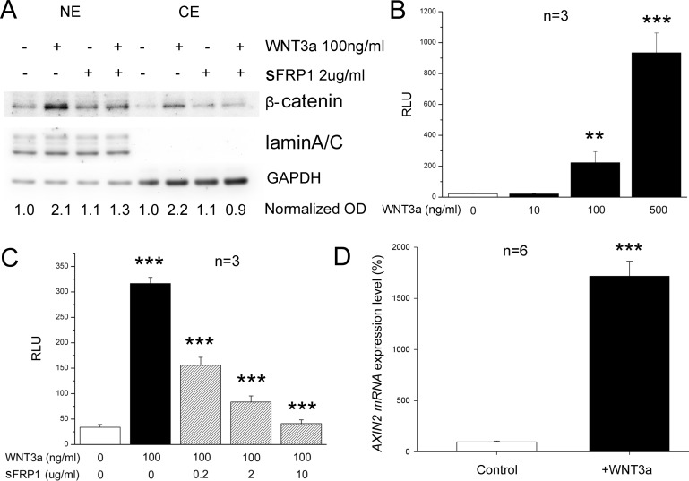
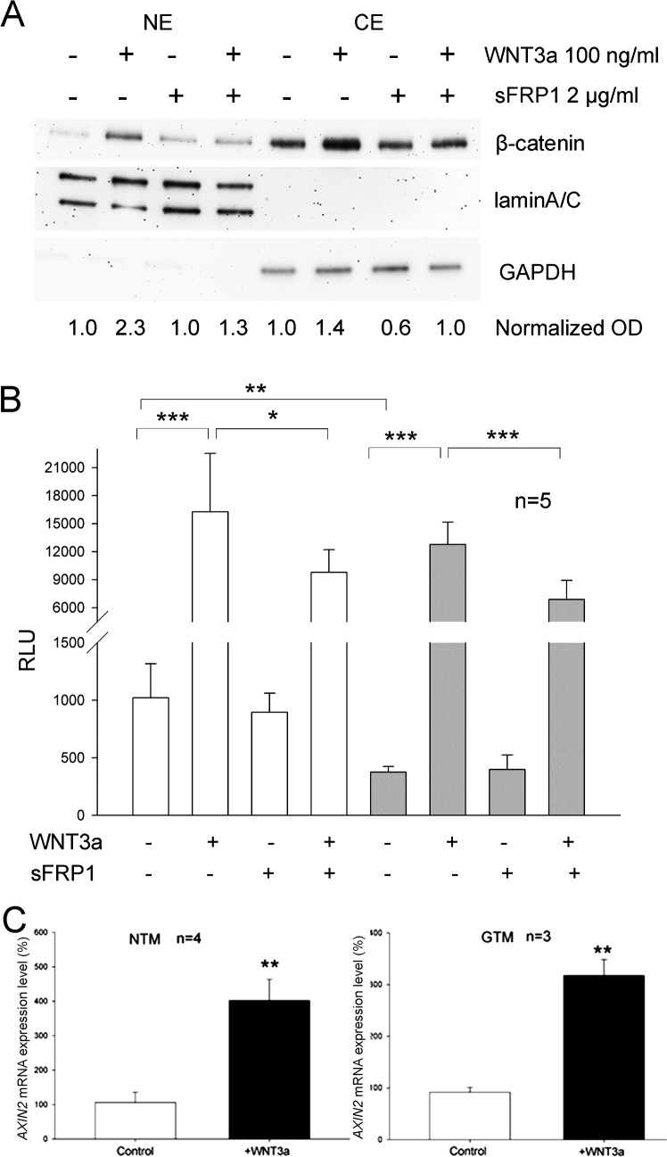
Purpose: We previously discovered elevated levels of secreted frizzled-related protein 1 (sFRP1), the Wnt signaling pathway inhibitor, in the glaucomatous trabecular meshwork (GTM), and found that key canonical Wnt signaling pathway genes are expressed in the trabecular meshwork (TM). The purpose of our study was to determine whether a functional canonical Wnt signaling pathway exists in the human TM (HTM).

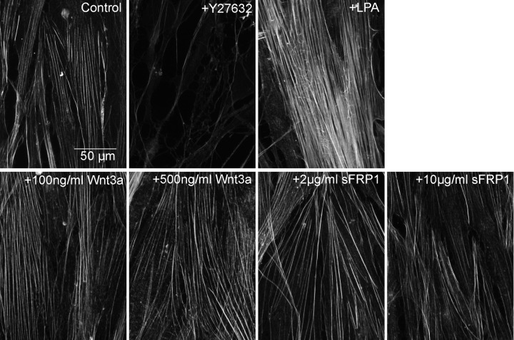
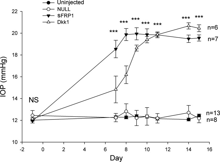
Methods: Western immunoblotting and/or immunofluorescent microscopy were used to study β-catenin translocation as well as the actin cytoskeleton in transformed and primary HTM cells. A TCF/LEF luciferase assay was used to study functional canonical Wnt signaling, which was confirmed further by WNT3a-induced expression of a pathway target gene, AXIN2, via quantitative PCR. Intravitreal injection of an Ad5 adenovirus expressing Dickkopf-related protein-1 (DKK1) was used to study the in vivo effect of canonical Wnt signaling on IOP in mice.

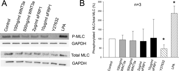
Results: WNT3a induced β-catenin translocation in the HTM, which was blocked by co-treatment with sFRP1. Similarly, WNT3a enhanced luciferase levels in TCF/LEF luciferase assays, which also were blocked by sFRP1. Furthermore, AXIN2 expression was elevated significantly by WNT3a. However, neither WNT3a nor sFRP1 affected actin cytoskeleton organization, which theoretically could be regulated by noncanonical Wnt signaling in HTM cells. Exogenous DKK1, a specific inhibitor for the canonical Wnt signaling pathway, or sFRP1 elevated mouse IOP to equivalent levels.

Conclusions: There is a canonical Wnt signaling pathway in the TM, and this canonical Wnt pathway, but not the noncanonical Wnt signaling pathway, regulates IOP.


Conflict of interest statement

Disclosure: W. Mao, None; J.C. Millar, Alcon (E); W.-H. Wang, Alcon (E); S.M. Silverman, None; Y. Liu, None; R.J. Wordinger, None; J.S. Rubin, None; I.-H. Pang, Alcon (E); A.F. Clark, None

Figures

Figure 1
Figure 1
Immunofluorescent study of WNT3a-induced β-catenin translocation in transformed GTM-3 cells. GTM-3 cell cultures were treated without (A) or with (B) 100 ng/mL WNT3a for 4 hours. (A) β-catenin (green) was expressed at a low level in the control cells, and was located mainly at the submembranous region at the periphery of the cells. (B) WNT3a significantly elevated β-catenin levels in the cytoplasm and nucleus (blue: DAPI staining). Experiments were repeated three times using GTM-3 cells, and representative images are shown.
Figure 2
Figure 2
Existence of a functional canonical WNT signaling pathway in transformed GTM-3 cells. (A) Western blot of nuclear extract proteins (NE) and cytosolic extract proteins (CE) from GTM-3 cells treated with or without WNT3a and/or sFRP1 for 4 hours. LaminA/C served as a nuclear protein loading control, while GAPDH served as a cytosolic protein loading control. The OD of each β-catenin band was normalized and the values are shown on the bottom of the Western blot. (B, C) Plasmid-based luciferase assays of the canonical Wnt signaling pathway in GTM-3 cells. GTM-3 cells were transfected with TCF/LEF luciferase reporter vectors and treated with WNT3a in the absence (B) or presence (C) of sFRP1 at indicated concentrations. Columns represent relative luminescence units (RLU), as a measure of luciferase levels. White columns: control. Black columns: cells treated with WNT3a. Hatched columns: cells treated with WNT3a and sFRP1. Data were analyzed by ANOVA and Bonferroni multiple comparison tests. (D) Comparison of AXIN2 mRNA expression levels of GTM-3 cells treated without (left, white column) or with (right, black column) 100 ng/mL WNT3a for 4 hours. Data were analyzed by Student's t-test. Columns and error bars: mean ± SD. **P < 0.010. ***P < 0.001.
Figure 3
Figure 3
Existence of a functional canonical WNT signaling pathway in primary HTM cells. (A) Western blot of NE proteins and CE proteins from a primary NTM cell strain treated with or without WNT3a (100 ng/mL) and/or sFRP1 (2 μg/mL) for 4 hours. LaminA/C served as a nuclear protein loading control, while GAPDH served as a cytosolic protein loading control. Three different HTM strains were tested and representative data are shown. The OD of each β-catenin band was normalized first to laminA/C (for nuclear β-catenin) or GAPDH (for cytosolic β-catenin). They then were normalized to corresponding control (non-treated, which was set at 1.0), and the values are shown on the bottom of the Western blot. (B) Lentivirus-based luciferase assays of the canonical Wnt signaling pathway in primary NTM (white columns, left panel) and GTM (grey columns,right panel) cells. HTM cells were transfected with the lentiviral TCF/LEF reporter, and treated with or without 100 ng/mL WNT3a and/or 2 μg/mL sFRP1. Columns represent RLU. Data were analyzed by ANOVA and Bonferroni multiple comparison tests except for the comparison between columns 1 and 5 (from left to right), for which Student's t-test was used. Two different HTM strains were tested and representative data are shown. (C) Comparison of AXIN2 mRNA expression levels of primary NTM (white columns) and GTM (black columns) cells treated without or with 100 ng/mL WNT3a for 4 hours. Left panel: NTM cells. Right panel: GTM cells. Data were analyzed by Student's t-test. Columns and error bars: mean ± SD. **P < 0.010. ***P < 0.001.
Figure 4
Figure 4
The organization of actin stress fibers was not affected by WNT3a or sFRP1. Primary GTM cells were treated with WNT3a (100 or 500 ng/mL), sFRP1 (2 or 10 μg/mL), Y27632 (10 μM), or LPA (20 μM) for 24 hours. Actin stress fibers were stained with phalloidin-Alexa-594. Neither WNT3a nor sFRP1 induced significant changes in actin stress fibers. For comparison, the ROCK inhibitor Y27632 disrupted actin stress fiber formation, while the Rho activator LPA enhanced actin stress fiber formation.
Figure 5
Figure 5
MLC phosphorylation was not affected by WNT3a or sFRP1. Primary GTM cells were treated with WNT3a (100 or 500 ng/mL), sFRP1 (2 or 10 μg/mL), Y27632 (10 μM), or LPA (20 μM) for 1 hour. WB of whole cell lysates was used for study MLC phosphorylation, as shown in (A). (B) Quantitative study of MLC phosphorylation. Double phospho-MLC (P-MLC) or total MLC was normalized first to GAPDH. Then, the amount of P-MLC was normalized to total MLC. Finally, the control was set at 100% and the other samples were normalized to the control. The paired Student's t-test was performed. *P < 0.050 compared to control.
Figure 6
Figure 6
DKK1 elevated IOP in mouse eyes. One eye (randomly selected) of each mouse was injected with Ad5-CMV-NULL, Ad5-CMV-DKK1, or Ad5-CMV-sFRP1 vector on day zero. The Ad5-CMV-NULL as well as contralateral uninjected eyes were used as negative controls. Conscious mouse IOPs were measured, and the mean and SEM of each group were plotted over time. The IOP values of DKK1 and sFRP1 transduced eyes were elevated significantly on days 7 to 15. Data were analyzed by ANOVA. ***P < 0.001.

Comment in

References

    1. Resnikoff S, Pascolini D, Etya'ale D, et al. Global data on visual impairment in the year 2002. Bull World Health Organ. 2004; 82: 844– 851. - PMC - PubMed
    1. The Advanced Glaucoma Intervention Study (AGIS): 7. The relationship between control of intraocular pressure and visual field deterioration. The AGIS Investigators. Am J Ophthalmol. 2000; 130: 429– 440. - PubMed
    1. Heijl A, Leske MC, Bengtsson B, Hyman L, Hussein M. Reduction of intraocular pressure and glaucoma progression: results from the Early Manifest Glaucoma Trial. Arch Ophthalmol. 2002; 120: 1268– 1279. - PubMed
    1. Kass MA, Heuer DK, Higginbotham EJ, et al. The Ocular Hypertension Treatment Study: a randomized trial determines that topical ocular hypotensive medication delays or prevents the onset of primary open-angle glaucoma. Arch Ophthalmol. 2002; 120: 701– 713 ; discussion 829–830. - PubMed
    1. Lichter PR. Impact of intraocular pressure reduction on glaucoma progression. JAMA. 2002; 288: 2607– 2608. - PubMed

Publication types

MeSH terms