

Gene expression in human accessory lacrimal glands of Wolfring
- PMID: 22956620
- PMCID: PMC4113189
- DOI: 10.1167/iovs.12-10750
Gene expression in human accessory lacrimal glands of Wolfring
Abstract
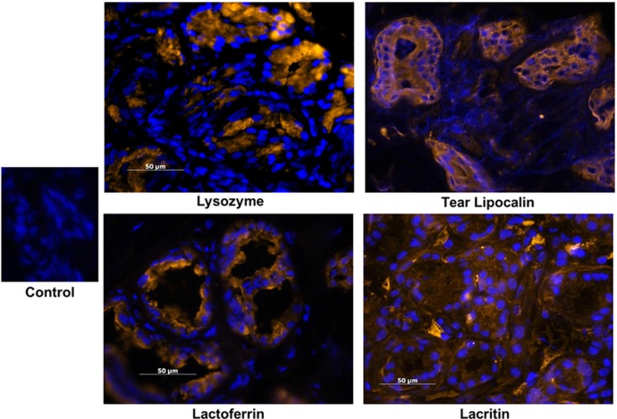
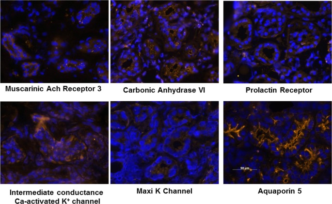
Purpose: The accessory lacrimal glands are assumed to contribute to the production of tear fluid, but little is known about their function. The goal of this study was to conduct an analysis of gene expression by glands of Wolfring that would provide a more complete picture of the function of these glands.
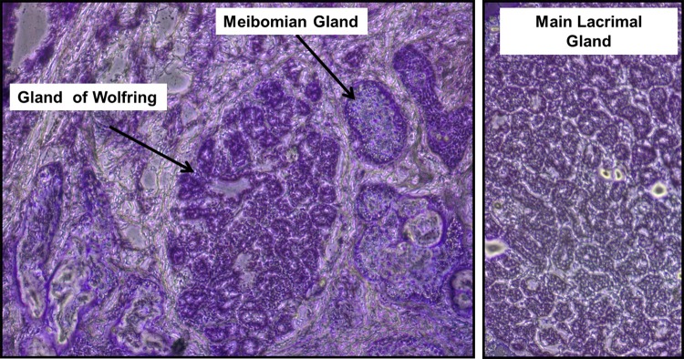
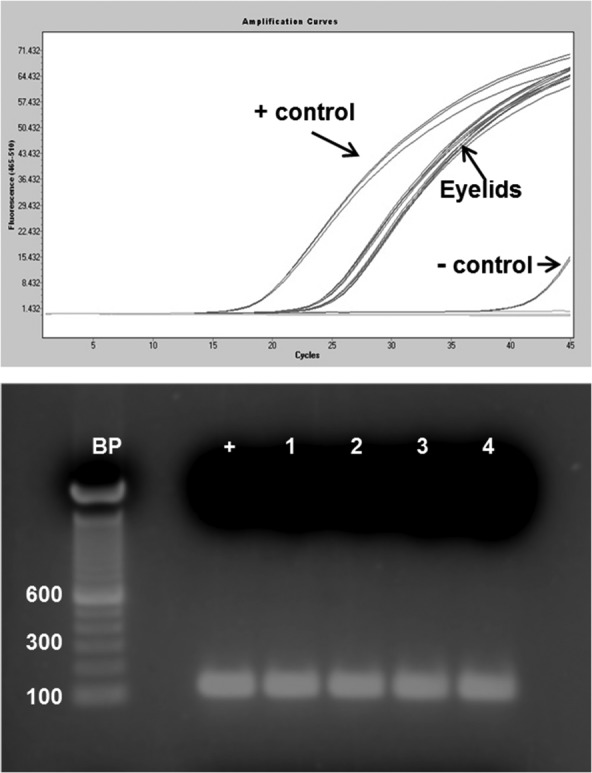
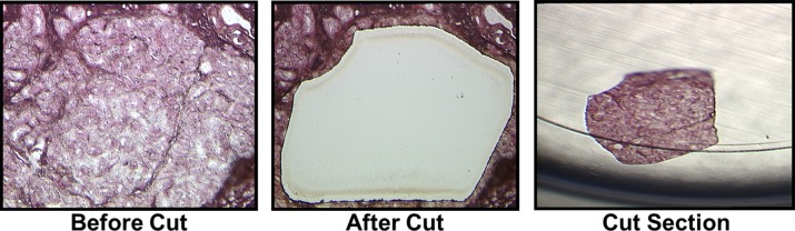
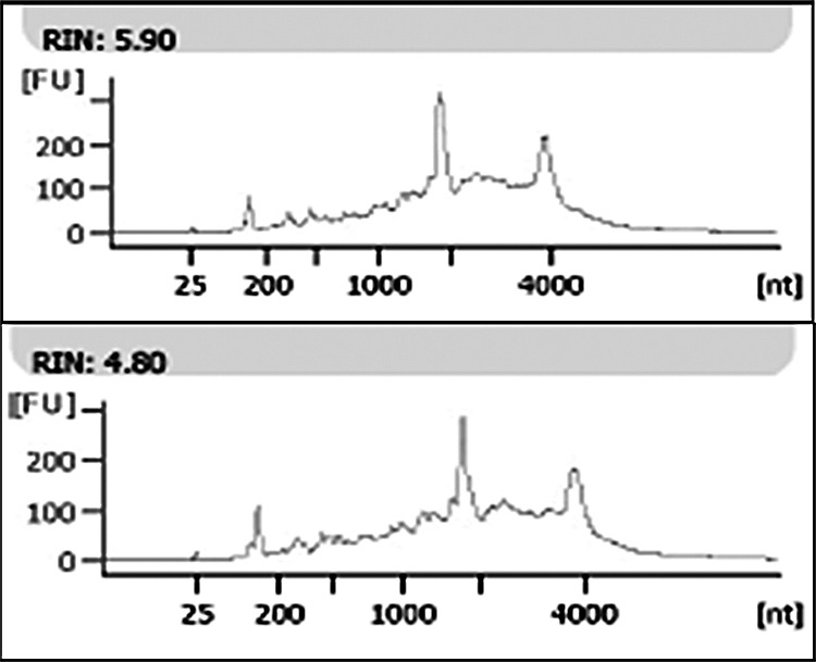
Methods: Glands of Wolfring were isolated from frozen sections of human eyelids by laser microdissection. RNA was extracted from the cells and hybridized to gene expression arrays. The expression of several of the major genes was confirmed by immunohistochemistry.
Results: Of the 24 most highly expressed genes, 9 were of direct relevance to lacrimal function. These included lysozyme, lactoferrin, tear lipocalin, and lacritin. The glands of Wolfring are enriched in genes related to protein synthesis, targeting, and secretion, and a large number of genes for proteins with antimicrobial activity were detected. Ion channels and transporters, carbonic anhydrase, and aquaporins were abundantly expressed. Genes for control of lacrimal function, including cholinergic, adrenergic, vasoactive intestinal polypeptide, purinergic, androgen, and prolactin receptors were also expressed in gland of Wolfring.
Conclusions: The data suggest that the function of glands of Wolfring is similar to that of main lacrimal glands and are consistent with secretion electrolytes, fluid, and protein under nervous and hormonal control. Since these glands secrete directly onto the ocular surface, their location may allow rapid response to exogenous stimuli and makes them readily accessible to topical drugs.
Conflict of interest statement
Disclosure:
Figures
References
-
- Seifert P, Spitznas M, Koch F, Cusumano A. The architecture of human accessory lacrimal glands. Ger J Ophthalmol. 1993;2:444–454 - PubMed
-
- Jordan DR, Anderson RB, Mamalis N. Accessory lacrimal glands. Ophthalmic Surg. 1990;21:146–147 - PubMed
-
- Allansmith MR, Gillette TE. Secretory component in human ocular tissues. Am J Ophthalmol. 1980;89:353–361 - PubMed
-
- Gillette TE, Allansmith MR, Greiner JV, Janusz M. Histologic and immunohistologic comparison of main and accessory lacrimal tissue. Am J Ophthalmol. 1980;89:724–730 - PubMed
-
- Obata H, Horiuchi H, Dobashi Y, Oka T, Sawa M, Machinami R. Immunohistochemical localization of epidermal growth factor in human main and accessory lacrimal glands. Jpn J Ophthalmol. 1993;37:113–121 - PubMed
Publication types
MeSH terms
Substances
Grants and funding
LinkOut - more resources
Full Text Sources
Other Literature Sources
Molecular Biology Databases
Miscellaneous
