. 2012 Nov;23(21):4226-41.
doi: 10.1091/mbc.E12-03-0210. Epub 2012 Sep 5.

BICD2, dynactin, and LIS1 cooperate in regulating dynein recruitment to cellular structures

Affiliations

BICD2, dynactin, and LIS1 cooperate in regulating dynein recruitment to cellular structures

Daniël Splinter et al. Mol Biol Cell. 2012 Nov.

Abstract

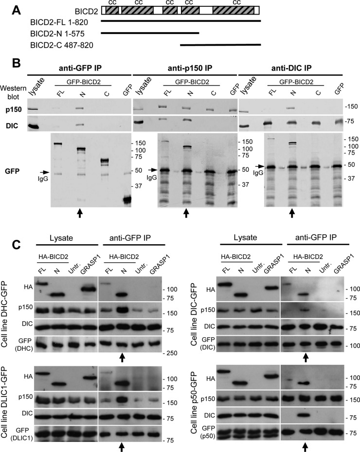
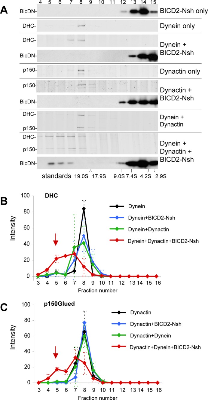
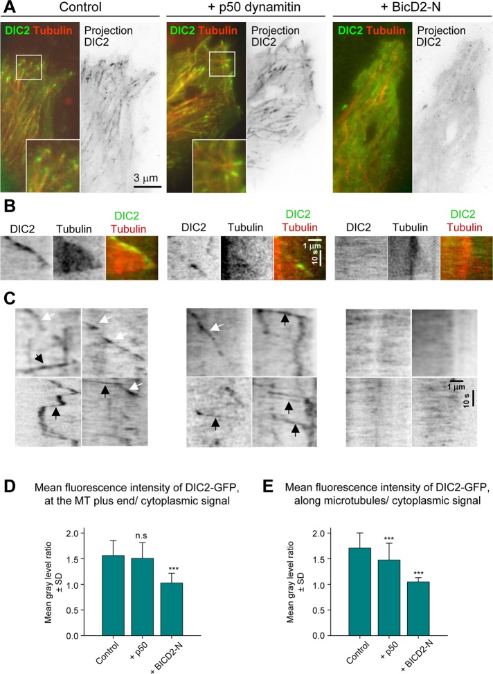
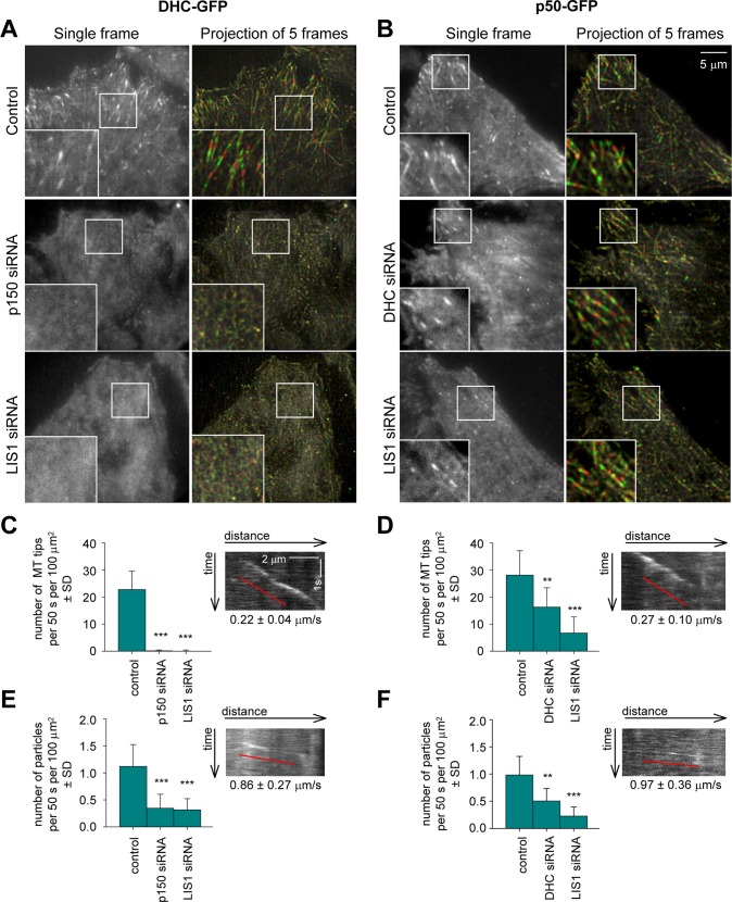
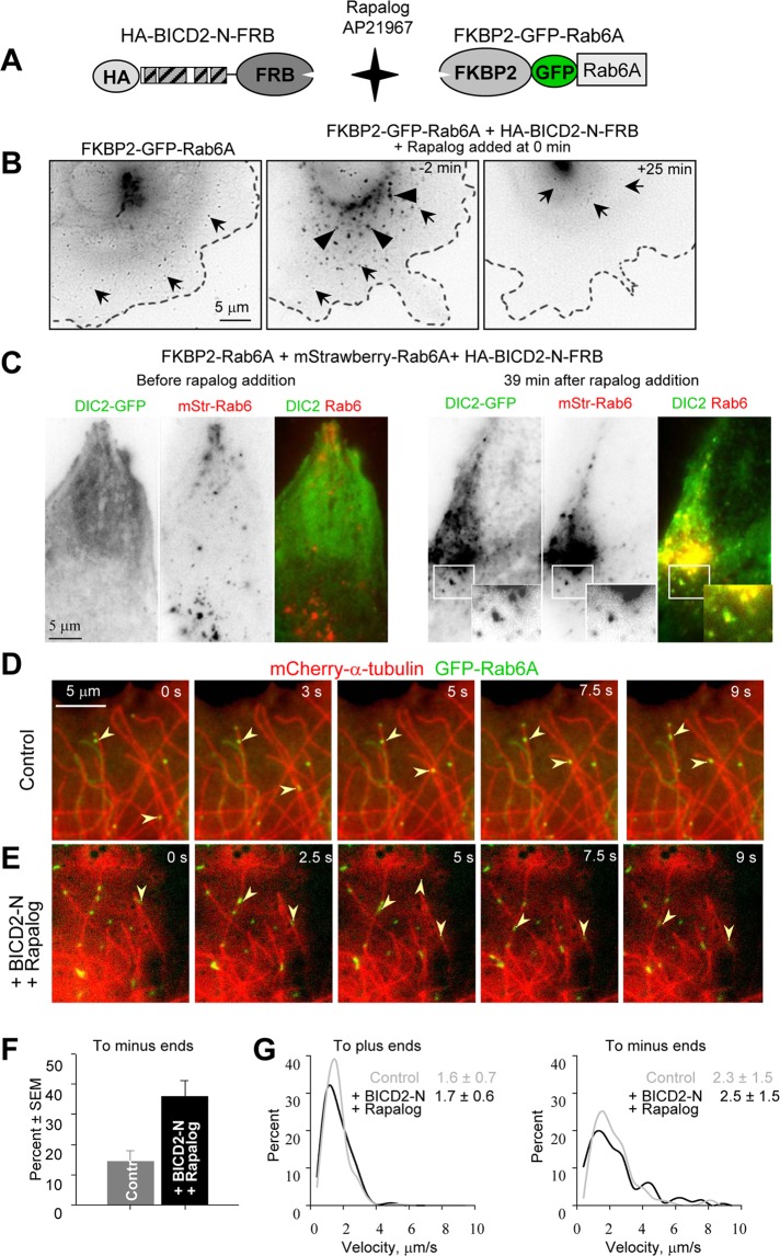
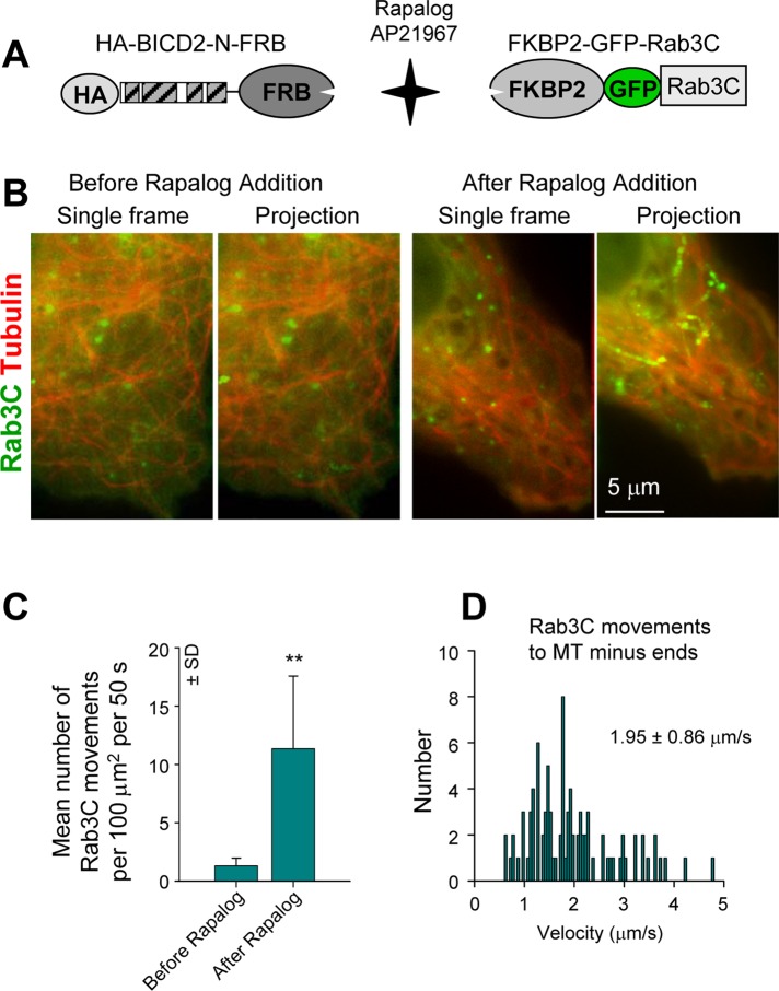
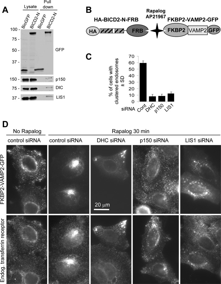
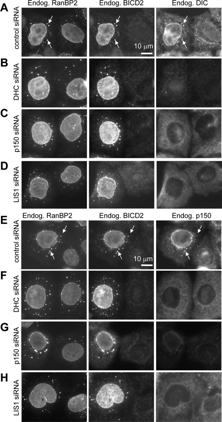
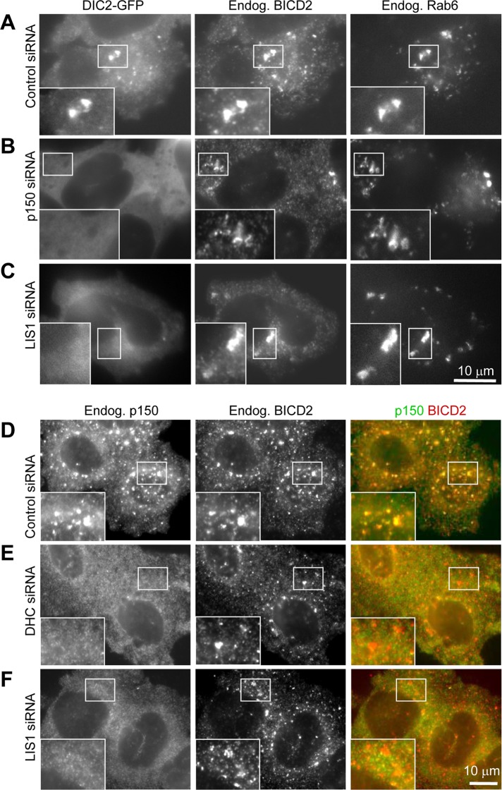
Cytoplasmic dynein is the major microtubule minus-end-directed cellular motor. Most dynein activities require dynactin, but the mechanisms regulating cargo-dependent dynein-dynactin interaction are poorly understood. In this study, we focus on dynein-dynactin recruitment to cargo by the conserved motor adaptor Bicaudal D2 (BICD2). We show that dynein and dynactin depend on each other for BICD2-mediated targeting to cargo and that BICD2 N-terminus (BICD2-N) strongly promotes stable interaction between dynein and dynactin both in vitro and in vivo. Direct visualization of dynein in live cells indicates that by itself the triple BICD2-N-dynein-dynactin complex is unable to interact with either cargo or microtubules. However, tethering of BICD2-N to different membranes promotes their microtubule minus-end-directed motility. We further show that LIS1 is required for dynein-mediated transport induced by membrane tethering of BICD2-N and that LIS1 contributes to dynein accumulation at microtubule plus ends and BICD2-positive cellular structures. Our results demonstrate that dynein recruitment to cargo requires concerted action of multiple dynein cofactors.


Figures

FIGURE 1:
FIGURE 1:
BICD2-N overexpression stabilizes the dynein–dynactin complex in cells. (A) Scheme of BICD2 structure and GFP-BICD2 fusions. (B, C) IP assays with antibodies against GFP, dynactin (p150Glued), and dynein (DIC) were performed with extracts from control HeLa cells transiently overexpressing the indicated GFP-BICD2 fusions or GFP alone (B) or HeLa cells stably expressing GFP-tagged dynein or dynactin subunits either alone (untr.) or in combination with transiently overexpressed, HA-tagged BICD2 or GRASP1 fusions (C). Western blotting was performed with the indicated antibodies. From 1 to 2% of the cell lysate used for the IP and 25% of the IP sample were loaded on gel. In panel B, lanes where GFP-BICD2-N is present show enhanced coprecipitation of dynein and dynactin (vertical arrows below the blots). Bands corresponding to the heavy chain of the antibody used for IP are visible in Western blots with GFP antibodies shown in B (immunoglobulin G, horizontal arrows). In panel C, BICD2-N is coprecipitated with dynein and dynactin and enhances coprecipitation of the two complexes with each other (vertical arrows below the blots). Note that coimmunoprecipitation of dynactin with DIC might be weak because the antibody used, DIC 74.1, can inhibit the interaction between dynein and dynactin.
FIGURE 2:
FIGURE 2:
Purified BICD2-N, dynein, and dynactin form a triple complex in vitro. Dynein, dynactin, or their combinations with or without BICD2-Nsh (as shown on the right) were sedimented on 10–40% linear sucrose gradients. After centrifugation, equal fractions were collected from the bottom of the gradients and subjected to SDS–PAGE (fraction numbers on top). Dynein and dynactin were found in fractions corresponding to ∼20S as determined by silver staining to identify DHC or p150Glued. The position of BICD2-Nsh was determined by Western blotting with anti-BICD2 antibodies. Positions of sucrose density standards are shown at the bottom. Representative gels are shown in A, and quantifications of DHC and p150Glued in different conditions, determined from three independent experiments, are shown in B and C; error bars represent SD. Incubation of dynein and dynactin with the excess of BICD2-Nsh caused a shift of dynein and dynactin and a portion of the BICD2-Nsh protein to denser gradient fractions, indicating that a stable triple complex was present (red arrows in B and C).
FIGURE 3:
FIGURE 3:
BICD2-N overexpression removes cytoplasmic dynein from MTs. HeLa cells stably expressing DIC2-GFP from a GFP-tagged BAC were transfected with either mCherry–α-tubulin alone or in combination with either HA-tagged p50 or BICD2-N, and simultaneous two-color live-cell imaging with 500-ms interval was performed using TIRF microscopy with high penetration depth. Five consecutive frames were averaged. Control stainings showed 100% cotransfection of mCherry–α-tubulin with HA-tagged fusions. (A) Representative images of DIC2-GFP (green) and mCherry–α-tubulin (red) are shown on the left, and projections of sequential frames (181 (control), 221 (p50), and 117 (BicD-N) are shown on the right. Insets show enlargements of the boxed areas. (B, C) Kymographs illustrating DIC2-GFP displacement at MT tips (B) and along MT lattice (C). In C, rapid particle movements along MTs are indicated by black arrows and the slower movements associated with the growing MT tips by white arrows. (D, E) Quantification of the mean DIC2-GFP signal at the MT plus ends or along MTs normalized by the cytoplasmic signal (a value of 1 indicates absence of enrichment along MTs). Error bars indicate SD; ∼30–70 MTs were analyzed in five to seven cells per condition. Values significantly different from control are indicated (Mann–Whitney U test, **p < 0.01, ***p < 0.001).
FIGURE 4:
FIGURE 4:
Dynactin and LIS1 are required for dynein localization to MTs. HeLa cells stably expressing DHC-GFP or p50-GFP from a GFP-tagged BACs were transfected with the indicated siRNAs and used for live-cell imaging with 500-ms interval. Five consecutive frames were averaged. (A, B) Single frames (left) and projections of five consecutive frames (right). Right, odd frames (frames 1, 3 and 5) are shown in green and even frames (frames 2 and 4) are shown in red. (C–F) Analysis of DHC-GFP and p50-GFP dynamics. Quantification of the density of GFP-positive MT ends (C,D) (recognized as comet-like structures with velocity less than 0.5 μm/s) and rapidly moving particles (E, F) (average velocity more than 0.5 μm/s) in different conditions. Plots are represented in the same way as in Figure 3D. Insets show representative kymographs from control cells. Ten cells were analyzed per condition.
FIGURE 5:
FIGURE 5:
Motility of Rab6A vesicles after BICD2-N recruitment. (A) Scheme of the regulated heterodimerization constructs used to attach BICD2-N to Rab6A-positive membranes. (B) Live image of an MRC5-CV cell expressing FKBP2-GFP-Rab6A alone (left) or together with HA–BICD2-N–FRB (middle and right). The cell shown in the middle and right was treated with 1 μM rapalog AP21967; time relative to the moment of drug addition is indicated. Individual Rab6A vesicles are indicated by arrows and dispersed Golgi fragments by arrowheads. Images were processed by applying Unsharp Mask and Blur filters (Photoshop); contrast is inverted. Cell outlines are indicated by stippled lines. (C) HeLa cells stably expressing DIC-GFP were transiently transfected with FKBP2-Rab6A, mStrawberry-Rab6A, and HA–BICD2-N–FRB and imaged using wide-field microscopy with a 500-ms exposure before and after rapalog addition. Contrast is inverted in single-color frames; in the overlay, DIC-GFP is shown in green and mStrawberry-Rab6A in red. Insets show enlargements of the boxed areas. (D, E) Visualization of Rab6A vesicle movement along MTs. (D) Simultaneous live imaging of FKBP2-GFP-Rab6A (green) and mCherry–α-tubulin (red) in a transiently transfected MRC5 cell; time is indicated. (E) The same as in D, but in a cell cotransfected with HAxBICD2-NxFRB, starting at 47.5 s after rapalog addition. Images were processed by applying Blur filter (Photoshop). Arrowheads indicate vesicles moving toward MT plus ends (D) or minus ends (E). (F, G) Analysis of Rab6 vesicle movement within an ∼15-μm-broad area at the cell periphery. Percentage of minus-end–directed movements (F) and averages (in μm/s) and the distributions of movement velocities (G) to MT plus and minus ends in MRC5-CV cells expressing FKBP2-GFP-Rab6A alone or together with HA–BICD2-N–FRB and after rapalog addition. In the latter case, measurements were performed within 25 min after rapalog was added. Approximately 30 cells were analyzed for each condition. The individual distributions and the number of measurements for G are shown in Supplemental Figure S5.
FIGURE 6:
FIGURE 6:
Motility of Rab3C vesicles after BICD2-N recruitment. (A) Scheme of the regulated heterodimerization constructs used to attach BICD2-N to Rab3C-positive membranes. (B) Simultaneous live imaging of FKBP2-GFP-Rab3C (green) and mCherry–α-tubulin (red) in a transiently transfected MRC5-CV cell coexpressing HA–BICD2-N–FRB before and after rapalog addition; single frames are shown on the left, and projections of 40 frames are shown on the right. Imaging was performed with 100-ms interval/exposure using wide-field microscopy. Five consecutive frames were averaged. (C) Quantification of the number of FKBP2-GFP-Rab3C particle movements with length >1 μm. Ten cells were analyzed. (D) Distribution of FKBP2-GFP-Rab3C movement velocities to MT minus ends in MRC5-CV cells coexpressing HA–BICD2-N–FRB after rapalog addition. Approximately 90 events in 10 cells were analyzed.
FIGURE 7:
FIGURE 7:
BICD2-N-dependent motility requires LIS1. (A) Streptavidin pull-down assays with Bio–GFP or Bio–GFP–BICD2-N were analyzed with the indicated antibodies. Two percent of the cell lysate used for the IP and 25% of the IP sample were loaded on gel. (B) Scheme of the regulated heterodimerization constructs used to attach BICD2-N to endosomes. (C, D) HeLa cells were transfected with different siRNAs; 2 d later, cells were cotransfected with HA–BICD2-N–FRB and FKBP2-GFP-VAMP2; after one additional day in culture, cells were treated with rapalog, fixed, and stained for transferrin receptor. (C) Percentage of HA–BICD2-N–FRB– and FKBP2-GFP-VAMP2–coexpressing HeLa cells with endosomes fully clustered in the cell center, 30 min after rapalog addition. Approximately100 cells were analyzed in three independent experiments. (D) Representative images of HA–BICD2-N–FRB– and FKBP2-GFP-VAMP2–coexpressing cells in different conditions.
FIGURE 8:
FIGURE 8:
Dynein and dynactin are mutually dependent on the G2-specific recruitment to the NE and AL. HeLa cells were transfected with the indicated siRNAs; 3 d later, the cells were treated for 5 h with 10 μM nocodazole, fixed with cold methanol, and stained with the antibodies against the nucleoporin RanBP2, BICD2. and DIC (A–D) or RanBP2, BICD2, and p150Glued (E–H). AL (stacks of nuclear pores in the ER membranes localized in the cytoplasm) are indicated by arrows. Note that methanol fixation preferentially preserves the nuclear pore–bound pool of BICD2 present in G2 cells but not the cytosolic and Rab6-bound BICD2 pool in G1 and S cells, as described previously (Splinter et al., 2010).
FIGURE 9:
FIGURE 9:
LIS1 is required for BICD2-dependent recruitment of dynein and dynactin to the NE. (A–C) HeLa cells stably expressing DIC2-GFP (A–C) or control HeLa cells (D, E) were transfected with the indicated siRNAs; 3 d later, cells were treated for 1 h with 10 μM nocodazole, fixed with 4% paraformaldehyde, and stained with the antibodies against BICD2 and Rab6 (A–C) or BICD2 and p150Glued (D–F). Paraformaldehyde fixation preserves the pool of BICD2 associated with Rab6 membranes.

Comment in

References

    1. Aguirre-Chen C, Bulow HE, Kaprielian Z. C. elegans bicd-1, homolog of the Drosophila dynein accessory factor Bicaudal D, regulates the branching of PVD sensory neuron dendrites. Development. 2011;138:507–518. - PMC - PubMed
    1. Bianco A, Dienstbier M, Salter HK, Gatto G, Bullock SL. Bicaudal-D regulates fragile X mental retardation protein levels, motility, and function during neuronal morphogenesis. Curr Biol. 2010;20:1487–1492. - PMC - PubMed
    1. Bingham JB, King SJ, Schroer TA. Purification of dynactin and dynein from brain tissue. Methods Enzymol. 1998;298:171–184. - PubMed
    1. Bolhy S, Bouhlel I, Dultz E, Nayak T, Zuccolo M, Gatti X, Vallee R, Ellenberg J, Doye V. A Nup133-dependent NPC-anchored network tethers centrosomes to the nuclear envelope in prophase. J Cell Biol. 2011;192:855–871. - PMC - PubMed
    1. Bullock SL, Nicol A, Gross SP, Zicha D. Guidance of bidirectional motor complexes by mRNA cargoes through control of dynein number and activity. Curr Biol. 2006;16:1447–1452. - PubMed

Publication types

MeSH terms