. 2012 Sep 5;32(36):12325-36.
doi: 10.1523/JNEUROSCI.2053-12.2012.

Repeated stress dysregulates κ-opioid receptor signaling in the dorsal raphe through a p38α MAPK-dependent mechanism

Affiliations

Repeated stress dysregulates κ-opioid receptor signaling in the dorsal raphe through a p38α MAPK-dependent mechanism

Julia C Lemos et al. J Neurosci. .

Abstract

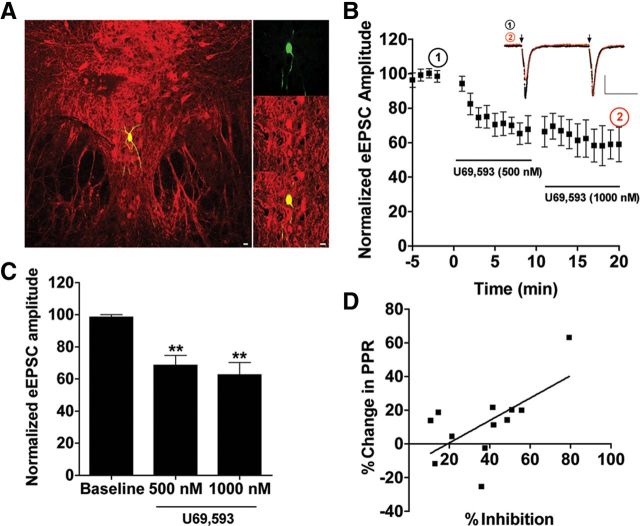
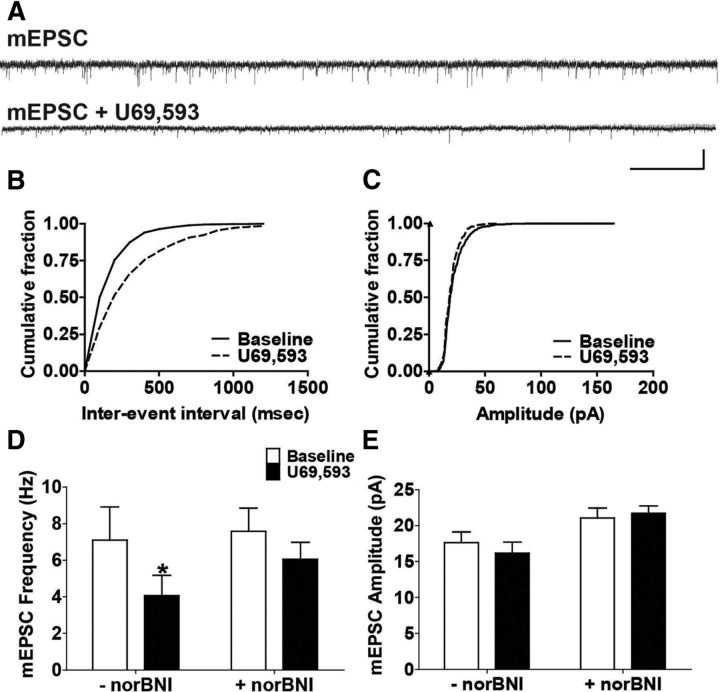
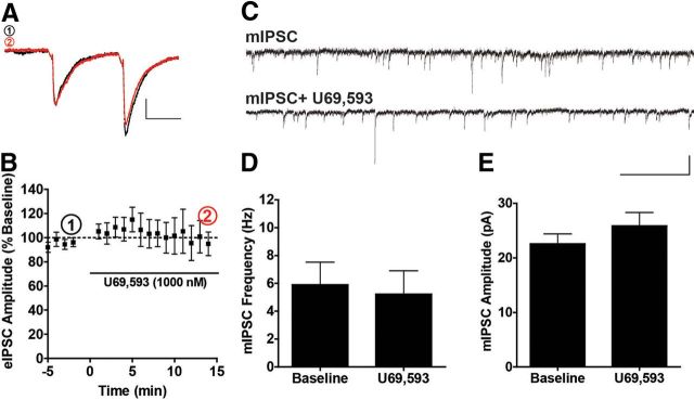
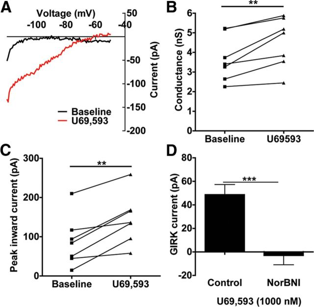
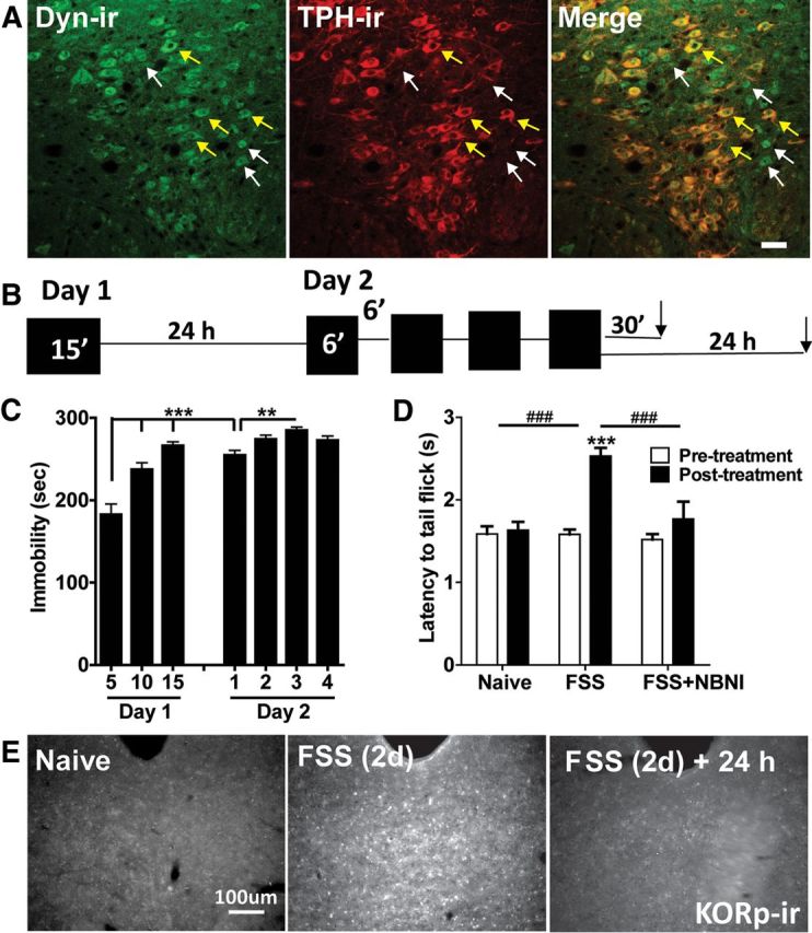
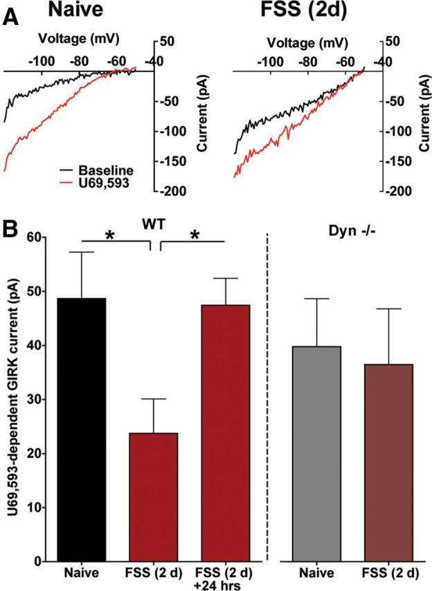
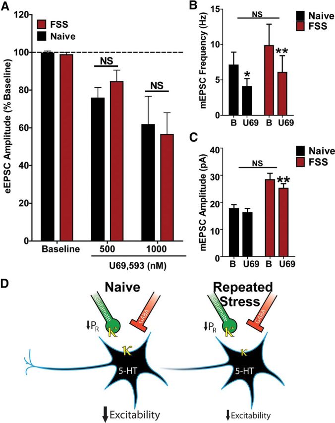
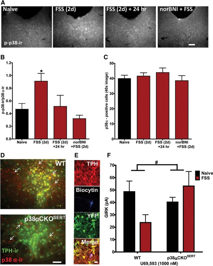
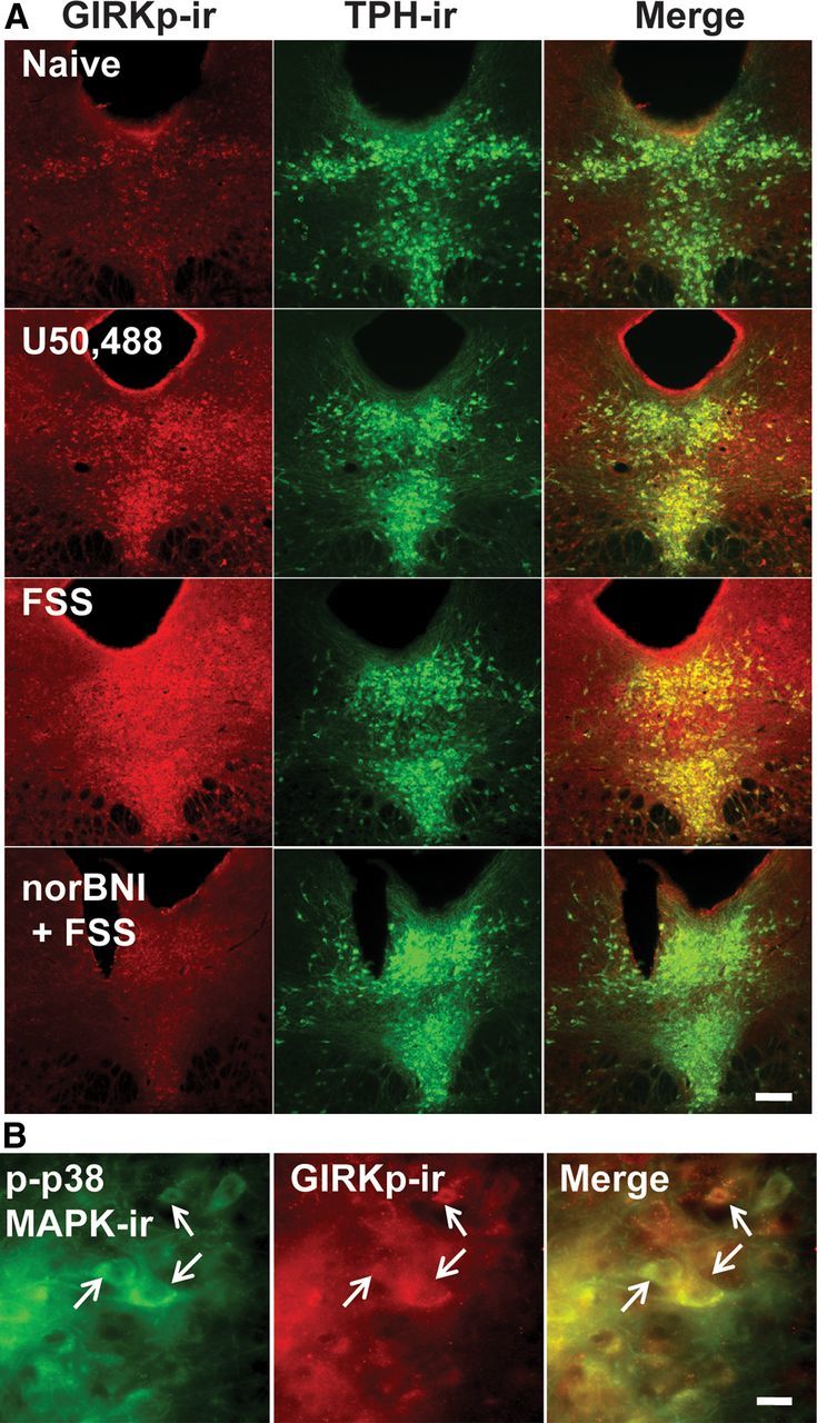
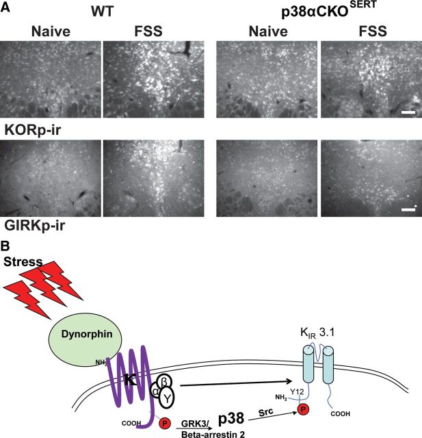
Repeated stress releases dynorphins and causes subsequent activation of κ-opioid receptors (KORs) in limbic brain regions. The serotonergic dorsal raphe nucleus (DRN) has previously been found to be an important site of action for the dysphoric effects of dynorphin-κ-opioid receptor system activation during stress-evoked behaviors, and KOR-induced activation of p38α mitogen-activated protein kinase (MAPK) in serotonergic neurons was found to be a critical mediator of the aversive properties of stress. Yet, how dynorphins and KORs functionally regulate the excitability of serotonergic DRN neurons both in adaptive and pathological stress states is poorly understood. Here we report that acute KOR activation by the selective agonist U69,593 [(+)-(5α,7α,8β)-N-methyl-N-[7-(1-pyrrolidinyl)-1-oxaspiro[4.5]dec-8-yl]benzeneacetamide] inhibits serotonergic neuronal excitability within the DRN through both presynaptic inhibition of excitatory synaptic transmission and postsynaptic activation of G-protein-gated inwardly rectifying potassium channels (GIRKs) electrophysiologically recorded in brain slices. C57BL/6 mice subjected to repeated swim, stress sessions had significantly reduced KOR-mediated GIRK currents recorded in serotonergic neurons in DRN postsynaptically, without significantly affecting presynaptic KOR-mediated regulation of excitatory transmission. This effect was blocked by genetic excision of p38α MAPK selectively from serotonergic neurons. An increase in phospho-immunoreactivity suggests that this functional dysregulation may be a consequence of tyrosine phosphorylation of GIRK (K(IR)3.1) channels. These data elucidate a mechanism for stress-induced dysregulation of the excitability of neurons in the DRN and identify a functional target of stress-induced p38α MAPK activation that may underlie some of the negative effects of pathological stress exposure.


Figures

Figure 1.
Figure 1.
KOR activation by U69,593 depresses evoked glutamatergic EPSCs recorded in 5-HT neurons of the DRN. A, Confocal fluorescent images [20× and 40× (inset)] of a representative recording site within the DRN. Cells were filled with biocytin and postfixed following the conclusion of recording to assess for the presence of TPH. Cells were considered 5-HT-positive if the biocytin-filled cell (green) colocalized with TPH (red). Recordings were confined to the dorsal and ventromedial aspect of the mid to caudal DRN. Scale bar, 20 μm. B, Representative traces (inset; calibration, 25 ms, 100 pA) and time course of normalized evoked EPSC amplitude before and after U69,593 (500 nm, 1000 nm) application. Electrical stimulation (100–1000 μA) was delivered in a pairwise fashion to the slice with an interstimulus interval of 50 ms (arrows). C, Four responses from baseline. U69,593 values at 500 and 1000 nm were averaged to get mean drug responses relative to baseline (**p < 0.01). Both concentrations of significantly decreased the normalized eEPSC amplitude compared with baseline. D, There was a significant correlation between the percentage inhibition of eEPSC amplitude and the percentage increase in paired pulse ratio (r2 = 0.3912, p < 0.05) N = 12 for all graphs.
Figure 2.
Figure 2.
U69,593 produces a norBNI-sensitive decrease in mEPSC frequency. A, Representative traces of pharmacologically isolated mEPSCs recorded from 5-HT DRN neurons before and following U69,593 (1000 nm) application. Calibration, 40 pA, 1 s. B, Cumulative histogram of the interevent interval of baseline mEPSC and that after U69,593 application corresponding to representative traces in A. C, Cumulative histogram of mEPSC amplitudes before and following drug application corresponding to the representative traces in A. D, U69,593 significantly decreased mEPSC frequency by 43% on average that was blunted in slices incubated with norBNI following a stable baseline Bonferroni post hoc, *p < 0.05, N = 7 for both groups). E, U69,593 had no effect on mEPSC amplitude in slices with or without norBNI (*p < 0.05, N = 7 for both groups).
Figure 3.
Figure 3.
KOR activation by U69,593 has no effect of evoked GABAergic IPSCs or mIPSCs. A, Representative traces of eIPSCs before and following U69,593 (1000 nm) application to the slice. Calibration, 50 ms, 200 pA. B, Time course of normalized evoked IPSC amplitude responses to U69,593 (1000 nm) relative to baseline demonstrating no effect of KOR activation on GABAergic synaptic transmission (N = 9). C, Representative traces of mIPSCs before and after U69,593 (1000 nm) bath application (Calibration, 40 pA, 1 s). D, E, U69,593 had no effect of mIPSC frequency or amplitude (N = 6).
Figure 4.
Figure 4.
KOR activation increases GIRK currents postsynaptically. A, Representative traces of baseline inward rectifying currents and KOR-activated (U69,593-induced) currents in which the Ba2+-insensitive current has been subtracted. B, C, U69,593 increases significantly increases the conductance (slope) and the peak inward current (measured at −120 mV) compared with baseline measurements (**p < 0.01, N = 7). D, For both untreated slices and slices preincubated with norBNI (1000 nm), the baseline current was subtracted from the current following U69,593 application to get an absolute change in peak inward current (denoted as GIRK current). U69,593 produced a 48.7 pA GIRK current that was absent in cells that had been preincubated with norBNI (**p < 0.01, N = 7 for both groups).
Figure 5.
Figure 5.
Repeated forced swim stress causes the release of dynorphin and KOR activation. A, 20× fluorescent images of dynorphin and TPH colocalization with the DRN. There was evidence of dynorphin-positive cells in both TPH-negative (white arrows) and TPH-positive cells (yellow arrows) within the DRN. Scale bar, 40 μm. B, Schematic of the 2 d repeated forced swim paradigm used in the study. C, Mice displayed significantly escalating immobility within the first 15 min session on day 1, from day 1 to day 2, and also across sessions on day 2 (**p < 0.01, ***p < 0.001, N = 10). D, Mice showed a significant stress-induced increase in tail-withdrawal latency following exposure to the 2 d swim paradigm that was blocked by norBNI (10 mg/kg) pretreatment (###p < 0.001 interaction; ***p < 0.001 post hoc Bonferroni, N = 7–21). E, Two day repeated swim stress produced an increase in phospho-KOR-ir compared with naive animals that returned to basal levels 24 h after the last swim session. Scale bar, 100 μm.
Figure 6.
Figure 6.
Repeated forced swim stress causes a reduction of KOR-activated GIRK current. A, Representative traces of baseline and KOR-induced GIRK currents in 5-HTcells recorded from the DRN of a naive or stress-exposed mouse. B, In WT animals, repeated swim stress causes a significant decrease in KOR-activated GIRK current that recovered 24 h following the final swim session (*p < 0.05, N = 7–8). In animals lacking the preprodynorphin gene (Dyn−/−), stress exposure did not significantly decrease KOR-mediated GIRK current relative to naive animals (N = 7–9).
Figure 7.
Figure 7.
Repeated stress exposure does not alter KOR-mediated depression of glutamatergic synaptic transmission. A, Repeated swim stress did not alter KOR inhibition of normalized eEPSC amplitude at either 500 or 1000 nm relative to measurements obtained in stress-naive animals (N = 10–12). B, U69,593 (1000 nm) caused a similar inhibition of mEPSC frequency in 5-HT cells recorded from naive versus stress-exposed mice (behavioral treatment by time, F(1,10) = 0.3210, p > 0.05, two-way repeated-measures ANOVA; Naive: 62 ± 7.0% of baseline; FSS: 58 ± 7.8% of baseline, Bonferroni post hoc tests, *p < 0.05, **p < 0.01, N = 5–7). C, 5-HT cells recorded from stress-exposed animals had significantly larger mEPSC amplitudes (two-way repeated-measures ANOVA, main effect of behavioral treatment, F(1,10) = 16.08, p < 0.01). There was not a significant difference in U69,593-induced inhibition of mEPSC amplitude between naive and stress-exposed mice (behavioral by time, F(1,10) = 3.024, p > 0.05, two-way repeated-measures ANOVA; Naive: 92 ± 3.3% of baseline; FSS: 89 ± 2.4% of baseline). There was a trend for a small (8%) decrease in mEPSC amplitude by U69,593 in naive animals, but this was not significant (Bonferroni post hoc test, p > 0.05). U69,593 did produce a small (10%), but significant decrease in mEPSC amplitude in 5-HT cells of stress-exposed animals (Bonferroni post hoc t test, **p < 0.01), N = 5–7. D, Summary of KOR regulation of DRN 5-HT neuronal excitability presynaptically and postsynaptically in stress-naive and stress-exposed mice.
Figure 8.
Figure 8.
p38α MAPK mediates the stress-induced reduction in KOR-activated GIRK current. A, Representative grayscale fluorescent images of phospho-p38 MAPK-ir in the DRN in naive animals, stress-exposed animals, stress-exposed animals allowed to recover for 24 h, and stress-exposed animals pretreated with norBNI. Scale bar, 200 μm. B, Two day swim stress significantly increased the ratio of phospho-p38 MAPK-ir: p38α-ir cells compared with naive animals sampled from a 40× image of the medial DRN. Following 24 h of recovery, this ratio returned to baseline levels. norBNI pretreatment significantly blocked stress-induced activation of phospho-p38MAPK-ir (*p < 0.05, N = 3–5 animals, triplicate sampling per N). C, There was no significant difference in total number of p38α-positive cells across groups (N = 3–5 animals, triplicate sampling per N). D, Representative fluorescent images of p38α-ir (red fluorescence) and TPH-ir (green fluorescence) colocalization in the DRN of WT and p38αCKOSERT animals. Colocalization of both proteins is indicated by the presence of yellow fluorescence above background. p38α-ir was present in both TPH-positive (yellow fluorescence, yellow arrows) and TPH-negative (red fluorescence only, white arrows) cells within the DRN of WT animals. In contrast, p38α-ir was only present above background staining in TPH-negative cells in p38αCKOSERT (red fluorescence only, white arrows). Scale bar, 100 μm. E, Slices obtained from p38αCKOSERT animals used for electrophysiological recording were postfixed following recordings and triple-labeled for biocytin (blue), TPH (red), and YFP (green) to confirm that the recorded cells were both TPH positive and YFP positive, indicating p38α had been excised. Scale bar, 50 μm. F, Stress-induced reduction in KOR-activated GIRK current in 5-HT cells was absent in 5-HT neurons from stress-exposed p38αCKOSERT animals compared with their naive counterparts (#p < 0.05 interaction, N = 7–12).
Figure 9.
Figure 9.
Repeated stressor exposure produces a KOR-dependent increase in phosphorylation of the tyrosine 12 residue of KIR 3.1 in the DRN. A, Fluorescent images of phospho-GIRK-ir and TPH labeling in the DRN of a stress-naive, U50,488 (20 mg/kg)-injected, stress-exposed, and norBNI + stress-exposed animal. Relative to basal levels of phospho-GIRK-ir present in naive animals, U50,488 or repeated swim stress robustly increased GIRKp-ir in TPH-ir cells that was blocked by norBNI pretreatment. Scale bar, 200 μm. B, Fluorescent images (40×) demonstrating colocalization of phospho-p38MAPK-ir and phospho-GIRK-ir. Stress exposure increased phospho-p38MAPK-ir and phospho-GIRK-ir within the same cells in the DRN. Scale bar, 50 μm.
Figure 10.
Figure 10.
Excision of p38α from 5-HT neurons blocks stress-induced phosphorylation of GIRK, but not KOR. A, Top, Repeated swim stress increases phospho-KOR-ir in the DRN compared with naive animals in both WT and p38αCKOSERT animals. Bottom, Excision of p38α reduced stress-induced phospho-GIRK-ir in the DRN compared with WT animals. Scale bar, 100 μm. B, Schematic depicting indirect tyrosine phosphorylation of the GIRK channel by p38α MAPK following stress-induced dynorphin release and subsequent KOR activation.

Similar articles

Cited by

References

    1. Adell A, Casanovas JM, Artigas F. Comparative study in the rat of the actions of different types of stress on the release of 5-HT in raphe nuclei and forebrain areas. Neuropharmacology. 1997;36:735–741. - PubMed
    1. Azmitia EC, Gannon PJ. The primate serotonergic system: a review of human and animal studies and a report on Macaca fascicularis. Adv Neurol. 1986;43:407–468. - PubMed
    1. Bale TL. Stress sensitivity and the development of affective disorders. Horm Behav. 2006;50:529–533. - PubMed
    1. Bausch SB, Esteb TM, Terman GW, Chavkin C. Administered and endogenously released kappa opioids decrease pilocarpine-induced seizures and seizure-induced histopathology. J Pharmacol Exp Ther. 1998;284:1147–1155. - PubMed
    1. Bruchas MR, Macey TA, Lowe JD, Chavkin C. Kappa opioid receptor activation of p38 MAPK is GRK3- and arrestin-dependent in neurons and astrocytes. J Biol Chem. 2006;281:18081–18089. - PMC - PubMed

Publication types

MeSH terms