

Impact of prefrontal cortex in nicotine-induced excitation of ventral tegmental area dopamine neurons in anesthetized rats
- PMID: 22956827
- PMCID: PMC3516404
- DOI: 10.1523/JNEUROSCI.5411-11.2012
Impact of prefrontal cortex in nicotine-induced excitation of ventral tegmental area dopamine neurons in anesthetized rats
Abstract
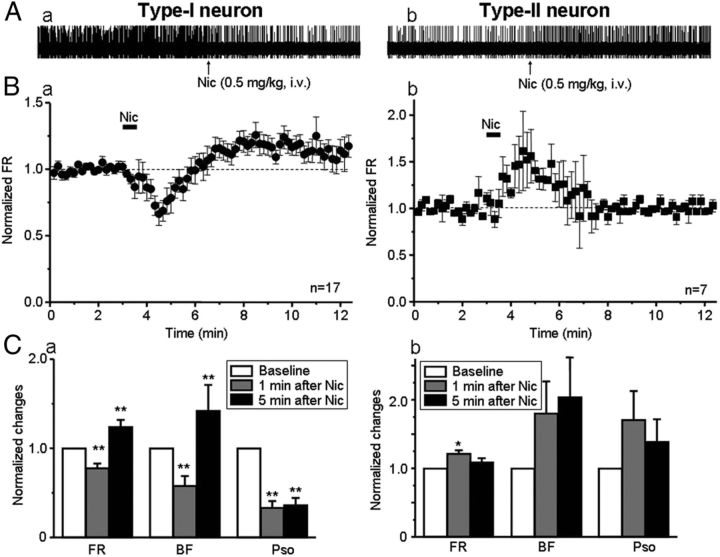
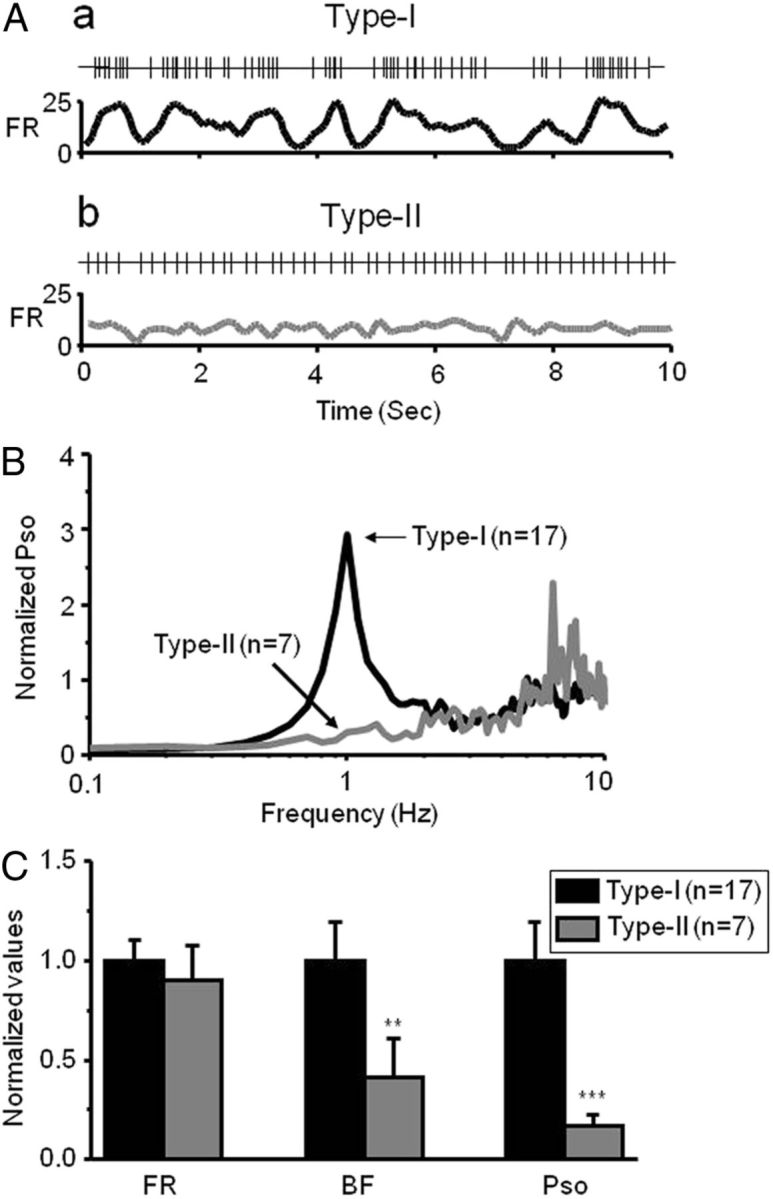
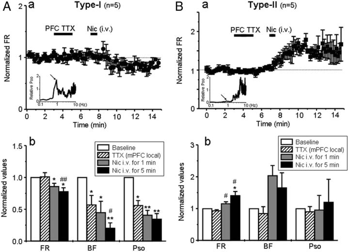
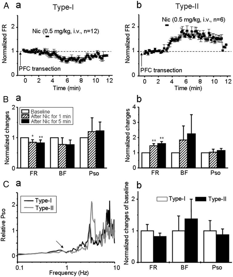
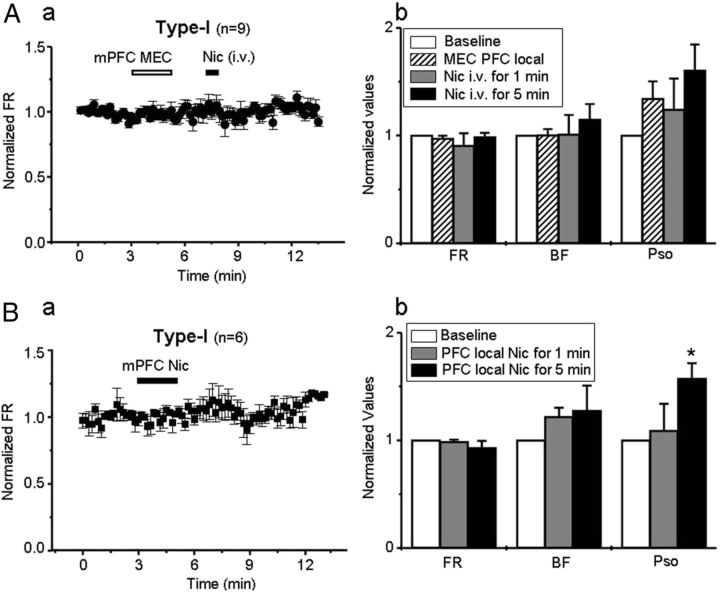
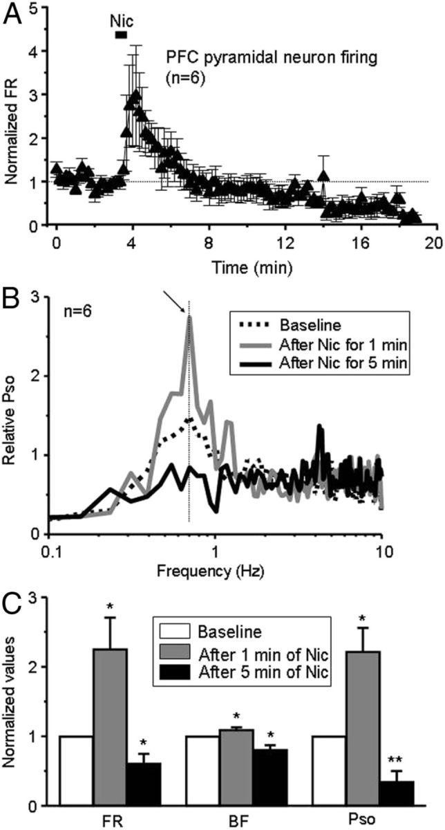
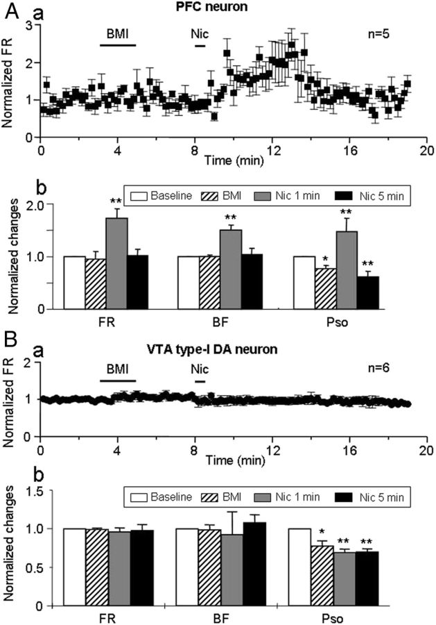
Systemic administration of nicotine increases dopaminergic (DA) neuron firing in the ventral tegmental area (VTA), which is thought to underlie nicotine reward. Here, we report that the medial prefrontal cortex (mPFC) plays a critical role in nicotine-induced excitation of VTA DA neurons. In chloral hydrate-anesthetized rats, extracellular single-unit recordings showed that VTA DA neurons exhibited two types of firing responses to systemic nicotine. After nicotine injection, the neurons with type-I response showed a biphasic early inhibition and later excitation, whereas the neurons with type-II response showed a monophasic excitation. The neurons with type-I, but not type-II, response exhibited pronounced slow oscillations (SOs) in firing. Pharmacological or structural mPFC inactivation abolished SOs and prevented systemic nicotine-induced excitation in the neurons with type-I, but not type-II, response, suggesting that these VTA DA neurons are functionally coupled to the mPFC and nicotine increases firing rate in these neurons in part through the mPFC. Systemic nicotine also increased the firing rate and SOs in mPFC pyramidal neurons. mPFC infusion of a non-α7 nicotinic acetylcholine receptor (nAChR) antagonist mecamylamine blocked the excitatory effect of systemic nicotine on the VTA DA neurons with type-I response, but mPFC infusion of nicotine failed to excite these neurons. These results suggest that nAChR activation in the mPFC is necessary, but not sufficient, for systemic nicotine-induced excitation of VTA neurons. Finally, systemic injection of bicuculline prevented nicotine-induced firing alterations in the neurons with type-I response. We propose that the mPFC plays a critical role in systemic nicotine-induced excitation of VTA DA neurons.
Figures
Similar articles
-
The Nucleus Reuniens of the Midline Thalamus Gates Prefrontal-Hippocampal Modulation of Ventral Tegmental Area Dopamine Neuron Activity.J Neurosci. 2016 Aug 24;36(34):8977-84. doi: 10.1523/JNEUROSCI.1402-16.2016. J Neurosci. 2016. PMID: 27559178 Free PMC article.
-
Prefrontal cortex gates acute morphine action on dopamine neurons in the ventral tegmental area.Neuropharmacology. 2015 Aug;95:299-308. doi: 10.1016/j.neuropharm.2015.03.037. Epub 2015 Apr 14. Neuropharmacology. 2015. PMID: 25882828
-
Prefrontal cortex deletion affects the dopaminergic neural firing complexity in nicotine-treated ventral tegmental area.Annu Int Conf IEEE Eng Med Biol Soc. 2010;2010:4526-9. doi: 10.1109/IEMBS.2010.5626088. Annu Int Conf IEEE Eng Med Biol Soc. 2010. PMID: 21095787
-
Cortical control of VTA function and influence on nicotine reward.Biochem Pharmacol. 2013 Oct 15;86(8):1173-80. doi: 10.1016/j.bcp.2013.07.013. Epub 2013 Aug 7. Biochem Pharmacol. 2013. PMID: 23933294 Review.
-
Tribute to: Self-administered nicotine activates the mesolimbic dopamine system through the ventral tegmental area [William Corrigall, Kathleen Coen and Laurel Adamson, Brain Res. 653 (1994) 278-284].Brain Res. 2016 Aug 15;1645:61-4. doi: 10.1016/j.brainres.2015.12.064. Epub 2016 Feb 8. Brain Res. 2016. PMID: 26867702 Review.
Cited by
-
Smoking, Obesity, and Post-Cessation Weight Gain: Neurobiological Intersection and Treatment Recommendations.J Multidiscip Healthc. 2025 May 24;18:2889-2900. doi: 10.2147/JMDH.S509971. eCollection 2025. J Multidiscip Healthc. 2025. PMID: 40438565 Free PMC article. Review.
-
Role of corticotropin-releasing factor in alcohol and nicotine addiction.Brain Res. 2020 Aug 1;1740:146850. doi: 10.1016/j.brainres.2020.146850. Epub 2020 Apr 21. Brain Res. 2020. PMID: 32330519 Free PMC article. Review.
-
Neural Mechanisms of Neuro-Rehabilitation Using Transcranial Direct Current Stimulation (tDCS) over the Front-Polar Area.Brain Sci. 2023 Nov 18;13(11):1604. doi: 10.3390/brainsci13111604. Brain Sci. 2023. PMID: 38002563 Free PMC article. Review.
-
Choline transporter hemizygosity results in diminished basal extracellular dopamine levels in nucleus accumbens and blunts dopamine elevations following cocaine or nicotine.Biochem Pharmacol. 2013 Oct 15;86(8):1084-8. doi: 10.1016/j.bcp.2013.07.019. Epub 2013 Aug 9. Biochem Pharmacol. 2013. PMID: 23939187 Free PMC article.
-
Cranial radiation disrupts dopaminergic signaling and connectivity in the mammalian brain.Acta Neuropathol Commun. 2025 Mar 13;13(1):59. doi: 10.1186/s40478-025-01976-3. Acta Neuropathol Commun. 2025. PMID: 40083022 Free PMC article.
References
-
- Ahmed SH, Graupner M, Gutkin B. Computational approaches to the neurobiology of drug addiction. Pharmacopsychiatry. 2009;42(Suppl 1):S144–S152. - PubMed
-
- Aracri P, Consonni S, Morini R, Perrella M, Rodighiero S, Amadeo A, Becchetti A. Tonic modulation of GABA release by nicotinic acetylcholine receptors in layer V of the murine prefrontal cortex. Cereb Cortex. 2010;20:1539–1555. - PubMed
-
- Benowitz NL, Kuyt F, Jacob P., 3rd Circadian blood nicotine concentrations during cigarette smoking. Clin Pharmacol Ther. 1982;32:758–764. - PubMed
-
- Bunney BS, Aghajanian GK, Roth RH. Comparison of effects of l-dopa, amphetamine and apomorphine on firing rate of rat dopaminergic neurones. Nat New Biol. 1973;245:123–125. - PubMed
Publication types
MeSH terms
Substances
Grants and funding
LinkOut - more resources
Full Text Sources
Medical