. 2012 Sep 5;32(36):12437-43.
doi: 10.1523/JNEUROSCI.0706-12.2012.

Optogenetic probing of fast glutamatergic transmission from hypocretin/orexin to histamine neurons in situ

Affiliations

Optogenetic probing of fast glutamatergic transmission from hypocretin/orexin to histamine neurons in situ

Cornelia Schöne et al. J Neurosci. .

Abstract

Hypothalamic hypocretin/orexin (hcrt/orx) neurons coordinate sleep-wake cycles, reward seeking, and body energy balance. Neurochemical data suggest that hcrt/orx cells contain several transmitters, but what hcrt/orx cells release onto their projection targets is unknown. A major pathway by which hcrt/orx neurons are thought to promote arousal is through projections to tuberomammillary histamine (HA) neurons. To study the impact of the electrical activity in hcrt/orx cells on HA neurons, we genetically targeted the light-activated excitatory ion channel channelrhodopsin-2 (ChR2) to the plasma membrane of hcrt/orx cells, and performed patch-clamp recordings from HA cells in acute mouse brain slices. Stimulation of ChR2-containing fibers with millisecond flashes of blue light produced fast postsynaptic currents in HA neurons, with a high connection probability (≈60% of HA cells were connected to ≈40% of hcrt/orx cells expressing ChR2). These inputs depended on tetrodotoxin-sensitive action potentials, had kinetics typical of glutamatergic responses mediated by AMPA receptors, were blocked by the AMPA receptor blocker CNQX, and displayed multiple forms of short-term plasticity (depression in ≈70% trials, facilitation in ≈30% trials, both often in the same cell). Furthermore, stimulation of hcrt/orx axons at physiological frequencies rapidly and reversibly increased action potential firing in HA cells, an effect that was abolished by blockade of AMPA receptors. These results provide the first functional evidence that hcrt/orx neurons are capable of fast glutamatergic control of their projection targets, and suggest that variations in electrical activity of hcrt/orx axons can induce rapid changes in long-range signals generated by HA neurons.


Figures
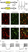
Figure 1.
Figure 1.
Optogenetic control of electrical activity of hcrt/orx cell membrane. A, ChR2-containing construct. When cre is present, the double-floxed inverted open reading frame of ChR2(H134R)-eYFP is flipped, and loxP/lox2272 sites are inactivated. ITR, Inverted terminal repeat; EF1a, elongation factor 1α promoter; WPRE, WHP-post-transcriptional response element; hGH PA, human growth hormone polyadenylation signal. B, Light flashes (black bars) evoke whole-cell currents (bottom trace, holding potential = −60 mV) and action potentials (top trace) (representative example of n = 20 cells). C, Colocalization of orexin-A and cre-recombinase-like immunoreactivities (representative example of n = 3 brains). Scale bar, 50 μm. D, Colocalization of ChR2-eYFP with orexin-A-like immunoreactivities (representative example of n = 4 brains). Scale bar, 50 μm. E, Electrical fingerprints of ChR2-expressing cells showing D and H-type “signatures” of hcrt/orx cells (representative examples of n = 8 cells).
Figure 2.
Figure 2.
Spike-mediated transmitter release from hcrt/orx axons onto HA neurons. A, Left, Coronal schematic at bregma −2.3 mm showing ventral TMN (VTM). f, Fornix; 3V, third ventricle. Right, z-projections from confocal stacks. Top, biocytin (BC)-filled cell, colocalizing with ADA-like immunoreactivity (arrowed). Bottom right, Same cell as in top (red), overlaid with ChR2-eYFP fibers (green). Scale bar, 20 μm. Bottom left, Higher magnification of same cell showing single confocal planes, with fiber–cell appositions arrowed. Representative example of n = 10 cells. B, Typical electrical current-clamp fingerprint of an HA cell (representative example of n = 35 cells). C, Fast PSCs in an HA neuron upon ChR2 stimulation (light flash shown as black bar), and recorded at −60 mV (individual traces in gray, averages in black or blue), with and without tetrodotoxin (TTX). Representative example of n = 8 cells.
Figure 3.
Figure 3.
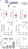
Pharmacological and biophysical properties of fast hcrt/orx → HA inputs. A, Optogenetically induced PSCs (black bar shows light stimulus) with and without CNQX (recordings at −60 mV, individual responses are in gray, averages are in black or blue). Representative example of n = 12 cells. B, Properties of PSCs (black dots are means from individual cells obtained using 0.1 Hz stimulation; n = 15 cells; population means ± SEM are in red). C, Success rate of optogenetic stimulation to evoke PSCs during 10 s stimulus train. D, Total charge transfer in hcrt/orx → HA PSCs during a 10 s stimulus train (sum of charges of PSCs); n = 8–9 cells per condition; **p < 0.01; n.s. = p > 0.05; Student's t test. E, Short-term plasticity in hcrt/orx → HA PSCs. Examples of paired-pulse depression (PPD, top left trace) and paired-pulse facilitation (PPF, top right trace). Bar graph shows mean proportions of different types of responses in 10 cells (percentage of trials that failed to produce PSC on first or second flash are also shown). F, Paired-pulse ratio at different interpulse intervals. *p < 0.05; **p < 0.01; one-way ANOVA with Bonferroni's post-test.
Figure 4.
Figure 4.
Optogenetic stimulation of hcrt/orx cell axons rapidly alters HA cell firing. A, Example membrane potential responses to a 5 ms flash (representative example of n = 8 cells). B, Example changes in HA cell firing induced by optical stimulation in the absence (top trace) and presence (bottom trace) of CNQX (representative example of n = 9 cells). Action potentials are truncated at −15 mV. Bottom graph shows group data for baseline (B), stimulation (S), and recovery (R) at 10 Hz stimulation, expressed as the percentage change in firing, ***p < 0.001, one-way ANOVA followed by Bonferroni's post-test. C, Effects of different stimulation frequencies on firing rate in control and CNQX; n = 4–9 per group. *p < 0.05; ***p < 0.001; n.s. = p > 0.05, two-way ANOVA with Bonferroni's post-test.

References

    1. Adamantidis AR, Zhang F, Aravanis AM, Deisseroth K, de Lecea L. Neural substrates of awakening probed with optogenetic control of hypocretin neurons. Nature. 2007;450:420–424. - PMC - PubMed
    1. Atasoy D, Aponte Y, Su HH, Sternson SM. A FLEX switch targets channelrhodopsin-2 to multiple cell types for imaging and long-range circuit mapping. J Neurosci. 2008;28:7025–7030. - PMC - PubMed
    1. Bernstein JG, Garrity PA, Boyden ES. Optogenetics and thermogenetics: technologies for controlling the activity of targeted cells within intact neural circuits. Curr Opin Neurobiol. 2012;22:61–71. - PMC - PubMed
    1. Carter ME, Adamantidis A, Ohtsu H, Deisseroth K, de Lecea L. Sleep homeostasis modulates hypocretin-mediated sleep-to-wake transitions. J Neurosci. 2009;29:10939–10949. - PMC - PubMed
    1. Chou TC, Lee CE, Lu J, Elmquist JK, Hara J, Willie JT, Beuckmann CT, Chemelli RM, Sakurai T, Yanagisawa M, Saper CB, Scammell TE. Orexin (hypocretin) neurons contain dynorphin. J Neurosci. 2001;21:RC168. - PMC - PubMed

Publication types

MeSH terms

LinkOut - more resources