Randomized Controlled Trial
. 2012 Sep 5;32(36):12499-505.
doi: 10.1523/JNEUROSCI.6508-11.2012.

A neural signature of affiliative emotion in the human septohypothalamic area

Affiliations
Randomized Controlled Trial

A neural signature of affiliative emotion in the human septohypothalamic area

Jorge Moll et al. J Neurosci. .

Abstract

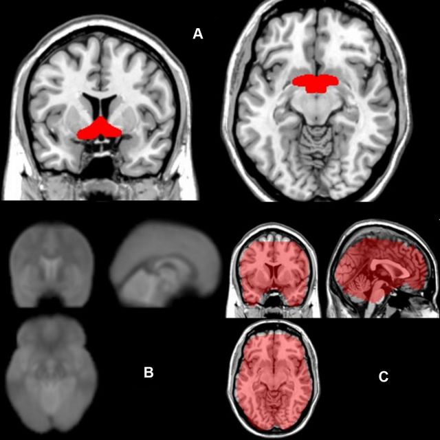
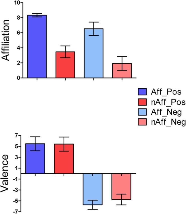
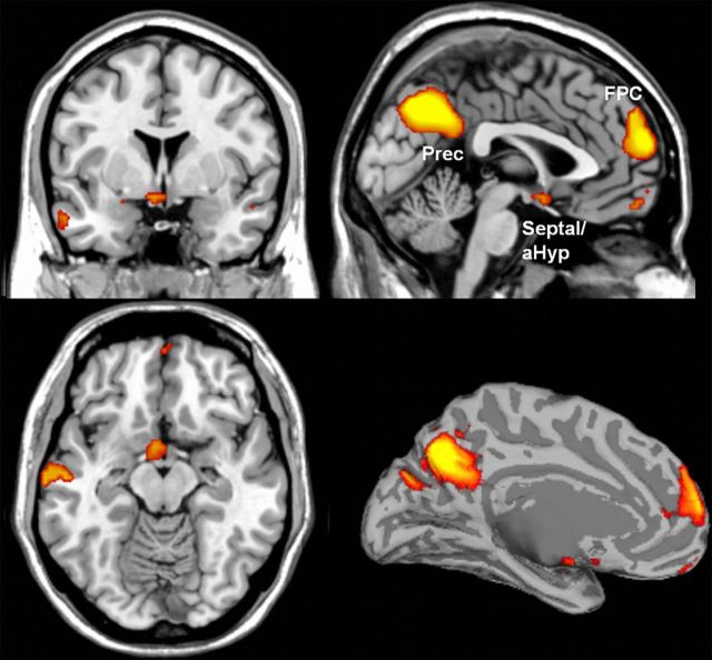
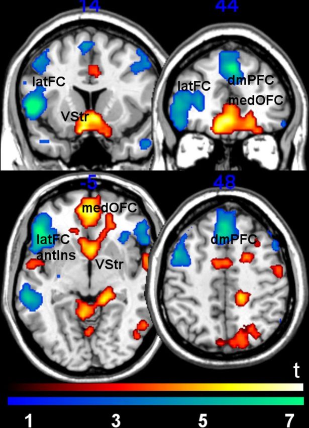
Comparative studies have established that a number of structures within the rostromedial basal forebrain are critical for affiliative behaviors and social attachment. Lesion and neuroimaging studies concur with the importance of these regions for attachment and the experience of affiliation in humans as well. Yet it remains obscure whether the neural bases of affiliative experiences can be differentiated from the emotional valence with which they are inextricably associated at the experiential level. Here we show, using functional MRI, that kinship-related social scenarios evocative of affiliative emotion induce septal-preoptic-anterior hypothalamic activity that cannot be explained by positive or negative emotional valence alone. Our findings suggest that a phylogenetically conserved ensemble of basal forebrain structures, especially the septohypothalamic area, may play a key role in enabling human affiliative emotion. Our finding of a neural signature of human affiliative experience bears direct implications for the neurobiological mechanisms underpinning impaired affiliative experiences and behaviors in neuropsychiatric conditions.


Figures

Figure 1.
Figure 1.
A, Anatomical coverage of the a priori ROI of the septal/preoptic-anterior hypothalamic region, used for small-volume correction for multiple comparisons. B, Average echoplanar images from all subjects show preserved signal at the basal forebrain (i.e., no signal dropouts at the individual and group level, except for a small portion of the ventromedial posterior orbitofrontal cortex). C, Binary mask used by SPM8 in the second-level analysis, overlaid on a T1 anatomical template, shows preserved fMRI signal in basal forebrain and prefrontal regions.
Figure 2.
Figure 2.
Top, Affiliative emotion ratings (care/tenderness) for the affiliative and nonaffiliative main experimental conditions, broken into the four subconditions (Aff_Pos, affiliative-positive; nAff_Pos, nonaffiliative-positive; Aff_Neg, affiliative-negative; nAff_Neg, nonaffiliative-negative). The affiliative condition showed significantly higher care/tenderness ratings than the nonaffiliative condition (n = 80/group; t = 57.57, p < 0.0001). Bottom, Comparison of positive and negative emotional valence ratings between affiliative and nonaffiliative main conditions, broken into the four emotional subconditions. Pairwise comparisons between affiliative versus nonaffiliative main conditions (affiliative-positive vs nonaffiliative-positive and affiliative-negative vs nonaffiliative-negative) did not reveal statistically significant differences (n = 40/group; all p > 0.05, mixed-effects ANOVA, entering subjects as a random variable). Error bars represent SDs.
Figure 3.
Figure 3.
Activation of the septal/preoptic-anterior hypothalamic and medial FPC, predicted a priori, as well as in the left posterior superior temporal sulcus region (data not shown) and precuneus (Prec), observed in the affiliative versus nonaffiliative contrast. To provide a better visualization of the spatial extent of the significantly activated clusters, statistical maps are displayed at uncorrected p = 0.005, minimum cluster size = 5 voxels. These clusters survived FWE correction for multiple comparisons (p < 0.05) either at the whole-brain level or over an a priori anatomical region of interest (Table 2; additional clusters showing in this map did not survive FWE correction). Activation results overlaid on a segmented and inflated brain template are shown at the right inferior quadrant.
Figure 4.
Figure 4.
Brain regions associated with positive versus negative conditions (red-yellow) and negative versus positive contrasts (blue-green). Activation of the ventral striatum (VStr) and medial orbitofrontal cortex (medOFC; BA11/32) was observed in the positive versus negative contrast. For the negative versus positive contrast, activation of dorsomedial prefrontal cortex (dmPFC; BA 8/9) and lateral frontal cortex, including the lateral orbitofrontal cortex and inferior frontal gyrus (latFC), as well as the adjoining anterior insula (antIns) was observed (BA 45/47/48). Only regions that survived whole-brain FWE-correction (voxel-level, p = 0.05) are reported. Statistical images displayed at uncorrected threshold p = 0.005, minimum cluster size = 5 voxels for visualization purposes.

References

    1. Andy OJ, Stephan H. The septum in the human brain. J Comp Neurol. 1968;133:383–410. - PubMed
    1. Aron A, Fisher H, Mashek DJ, Strong G, Li H, Brown LL. Reward, motivation, and emotion systems associated with early-stage intense romantic love. J Neurophysiol. 2005;94:327–337. - PubMed
    1. Bartels A, Zeki S. The neural correlates of maternal and romantic love. Neuroimage. 2004;21:1155–1166. - PubMed
    1. Berridge KC, Robinson TE. Parsing reward. Trends Neurosci. 2003;26:507–513. - PubMed
    1. Bishop MP, Elder ST, Heath RG. Intracranial self-stimulation in man. Science. 1963;140:394–396. - PubMed

Publication types

LinkOut - more resources