. 2012 Sep 5;32(36):12528-42.
doi: 10.1523/JNEUROSCI.1069-12.2012.

Myelin gene regulatory factor is required for maintenance of myelin and mature oligodendrocyte identity in the adult CNS

Affiliations

Myelin gene regulatory factor is required for maintenance of myelin and mature oligodendrocyte identity in the adult CNS

Matthias Koenning et al. J Neurosci. .

Abstract

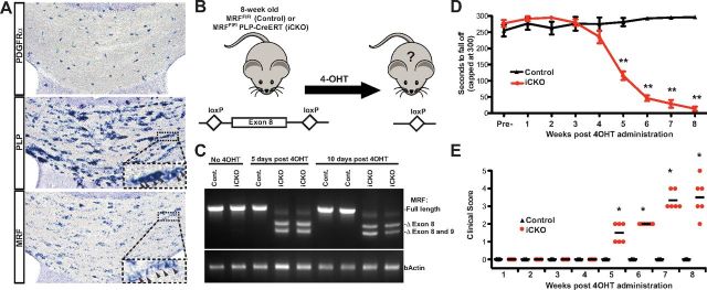
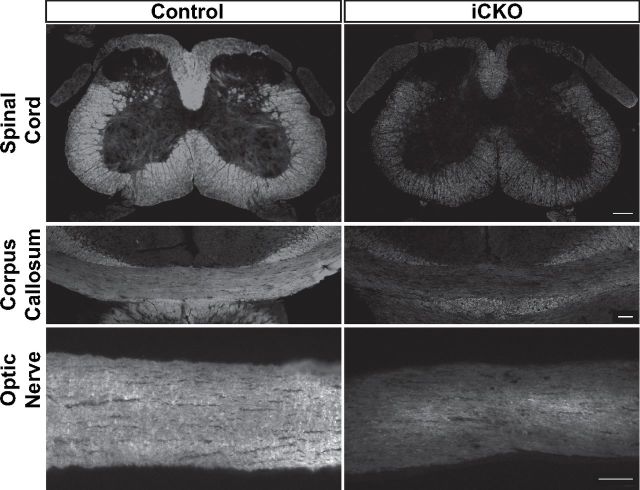
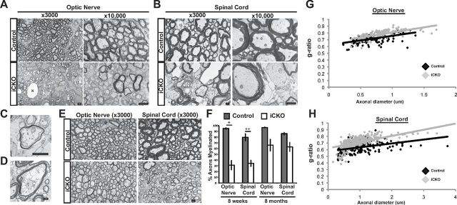
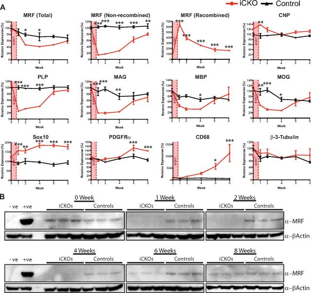
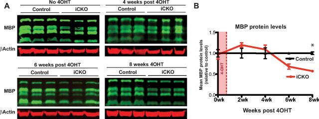
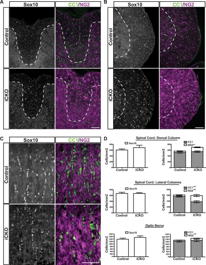
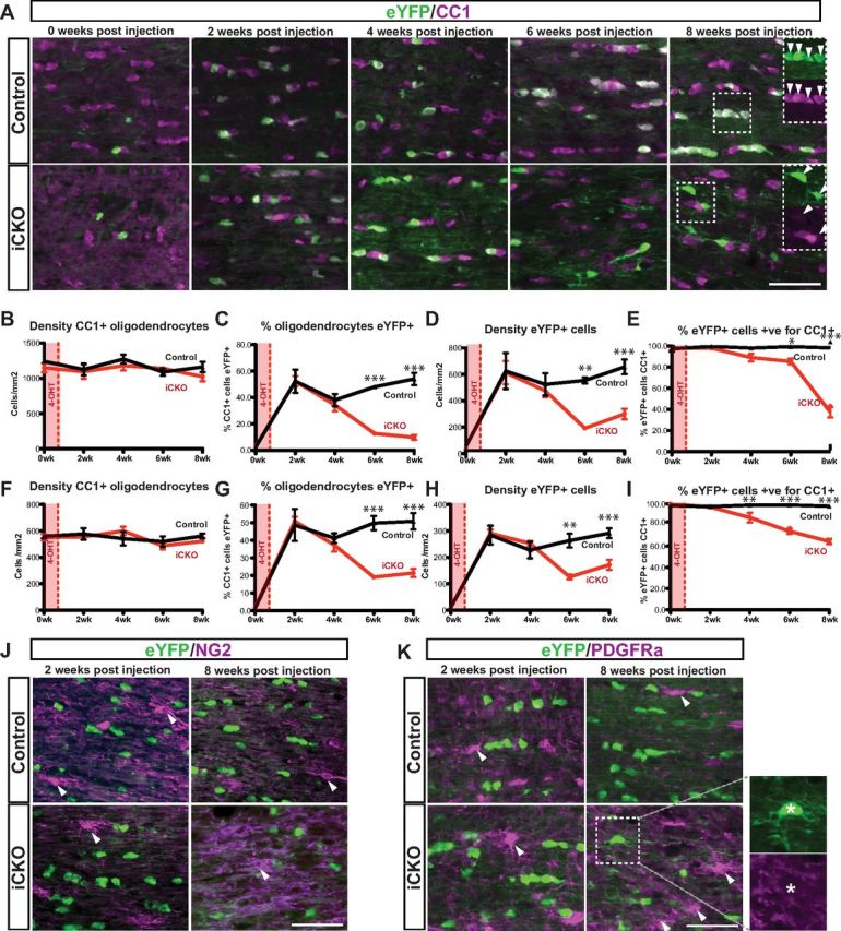
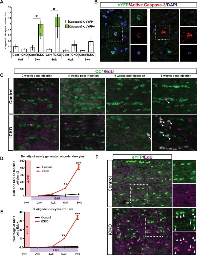
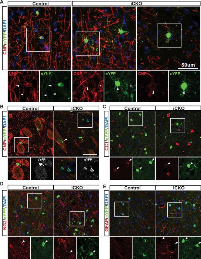
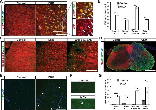
Although the transcription factors required for the generation of oligodendrocytes and CNS myelination during development have been relatively well established, it is not known whether continued expression of the same factors is required for the maintenance of myelin in the adult. Here, we use an inducible conditional knock-out strategy to investigate whether continued oligodendrocyte expression of the recently identified transcription factor myelin gene regulatory factor (MRF) is required to maintain the integrity of myelin in the adult CNS. Genetic ablation of MRF in mature oligodendrocytes within the adult CNS resulted in a delayed but severe CNS demyelination, with clinical symptoms beginning at 5 weeks and peaking at 8 weeks after ablation of MRF. This demyelination was accompanied by microglial/macrophage infiltration and axonal damage. Transcripts for myelin genes, such as proteolipid protein, MAG, MBP, and myelin oligodendrocyte glycoprotein, were rapidly downregulated after ablation of MRF, indicating an ongoing requirement for MRF in the expression of these genes. Subsequently, a proportion of the recombined oligodendrocytes undergo apoptosis over a period of weeks. Surviving oligodendrocytes gradually lose the expression of mature markers such as CC1 antigen and their association with myelin, without reexpressing oligodendrocyte progenitor markers or reentering the cell cycle. These results demonstrate that ongoing expression of MRF within the adult CNS is critical to maintain mature oligodendrocyte identity and the integrity of CNS myelin.


Figures

Figure 1.
Figure 1.
Inducible loss of MRF causes a progressive behavioral deficit. A, In situ hybridization for PDGFRα, PLP, and MRF in the corpus callosum of an 8-week-old mouse. Probes for both PLP and MRF label chains of oligodendrocytes (insets). B, Strategy used to inactivate MRF in myelinating cells in 8-week-old mice. C, RT-PCR analysis of recombination using primers flanking the RNA encoded by the loxP-flanked exon. After 4OHT administration, the full-length transcript is essentially replaced by the truncated transcript lacking exon 8 or exons 8 and 9 in iCKO mice. D, Rotarod analysis of motor function in control or iCKO mice from 0 to 8 weeks after 4OHT. Values are mean ± SEMs, n = 6 animals per genotype. *p < 0.05, **p < 0.01. E, Clinical disease scores for the same cohort shown in D.
Figure 2.
Figure 2.
Representative images of FluoroMyelin staining of key white matter regions (spinal cord, corpus callosum, and optic nerve) of control and iCKO mice at 8 weeks after 4OHT. Widespread demyelination is evident throughout all these regions in the iCKO mice. Scale bars, 100 μm.
Figure 3.
Figure 3.
Ablation of MRF causes severe demyelination. A, Electron microscopy analysis of the optic nerve at 8 weeks after 4OHT reveals substantial demyelination and other signs of myelin pathology, such as vacuolization (*) in the iCKO mice. B, At 8 weeks after 4OHT, the lateral white matter of the spinal cord contains more signs of active demyelination, including substantial vacuolization, delamination, and myelin debris. C, D, Higher-magnification images of a thinly myelinated (presumably remyelinated) axon in the optic nerve (C) and degrading myelin in the spinal cord (D). E, At 8 months after 4OHT, both the optic nerve and spinal cord of the iCKO mice show substantial remyelination as evidenced by numerous thinly myelinated axons. F, Quantification of the proportion of axons myelinated in the optic nerve and lateral white matter of the spinal cord of iCKO mice and controls at 8 weeks and 8 months after 4OHT administration. Data are means ± SEM. *p < 0.05, **p < 0.01. n = 3 animals per condition. Scale bars, 1 μm. G, H, g-ratios of individual axons as a function of axonal diameter in the spinal cord (G) and optic nerve (H) in controls and iCKOs at 8 months after 4OHT.
Figure 4.
Figure 4.
A, qPCR analysis of expression of key genes, including MRF, myelin genes (CNP, MAG, PLP, MBP, and MOG), oligodendrocyte lineage markers (Sox10 and PDGFRα), and markers of microglia/macrophages (CD68) and neurons (β-3-tubulin) after ablation of MRF. Graphs show mean ± SEM expression for each group relative to the mean of the uninjected control (PLP–CreERT+) group. n = 3 animals per condition. *p < 0.05, **p < 0.01, ***p < 0.001. All comparisons between genotypes within time points. B, Western blot analysis of MRF protein expression in the spinal cords of the same animals as in A. Negative and positive controls for MRF expression are lysates from nontransfected and MRF-transfected HEK293T cells, respectively.
Figure 5.
Figure 5.
Quantitative infrared Western blot analysis of MBP protein expression after ablation of MRF. A, Western blots of MBP and β-actin from iCKOs and controls at 0, 4, and 8 weeks after 4OHT. B, Quantification of MBP levels. Graphs show mean ± SEM expression for each group relative to the mean of the control group at each time point. n = 3–4 animals per genotype at each time point. *p < 0.05.
Figure 6.
Figure 6.
Immunohistochemical analysis of the oligodendrocyte lineage at peak clinical severity. A–C, Sox10, CC1, and NG2 staining in the dorsal column of the spinal cord (A), the lateral white matter of the spinal cord (B), and optic nerve (C). D, Quantification of the densities of Sox10, NG2, and CC1 immunopositive cells in control and iCKO mice for each region. The density of Sox10+ cells was not significantly different between genotypes in any region analyzed, although NG2+ numbers were significantly elevated in the iCKO mice in each region. The density of CC1+ cells was reduced in the iCKO animals relative to controls only in the lateral white matter of the spinal cord. **p < 0.01. n = 5 mice per genotype. Scale bars, 100 μm.
Figure 7.
Figure 7.
Ablation of MRF causes a delayed loss of mature phenotype and loss of many of the recombined oligodendrocytes. A, Representative optic nerve sections from MRFWT/FL/PLP–CreERT/Rosa26–eYFP (control) and from MRFFL/FL/PLP–CreERT/Rosa26–eYFP (iCKO) mice either untreated with 4OHT (0 week) or 2, 4, 6, or 8 weeks after 4OHT stained with CC1 and anti-eYFP. Insets in the “8 weeks post injection” images show separated channels for the boxed regions. B–I, Quantification of the density of CC1+ oligodendrocytes, the proportion of CC1+ oligodendrocytes expressing eYFP, the density of eYFP+ cells, and the proportion of eYFP+ cells expressing CC1 for control and iCKO animals at 0–8 weeks after 4OHT in the optic nerve (B–E) and spinal cord (F–I). *p < 0.05, **p < 0.01, ***p < 0.001. n = 3–5 mice per condition. J, K, Optic nerve sections stained with anti-eYFP and the OPC marker NG2 (J) and PDGFRα (K) showing an absence of colocalization. NG2+ and PDGFRα+ cell bodies indicated by arrowheads. Scale bars, 50 μm.
Figure 8.
Figure 8.
Increased oligodendrocyte turnover after ablation of MRF. A, Density of active caspase-3+ cells in the spinal cords of MRFWT/FL/PLP–CreERT/Rosa26–eYFP (control) and MRFFL/FL/PLP–CreERT/Rosa26–eYFP (iCKO) mice after 4OHT administration. These apoptotic cells included both eYFP+ recombined oligodendrocytes and eYFP cells (B); both examples are from iCKO nerve at 4 weeks after 4OHT. n = 5 mice per condition. C, Images showing EdU incorporation and CC1 immunohistochemistry at weeks 2–8 after 4OHT in the optic nerves of control (MRFFL/FL) or iCKO (MRFFL/FL; PLP–CreERT) mice. D, E, Analysis of the density of CC1+/EdU+ double-labeled cells (D) and the proportion of CC1+ oligodendrocytes EdU+ (E) at weeks 2–8 after 4OHT in the optic nerves from C. n = 3–5 mice per condition. F, Double staining for eYFP and EdU incorporation in representative MRFWT/FL/PLP–CreERT/Rosa26–eYFP (control) and MRFFL/FL/PLP–CreERT/Rosa26–eYFP (iCKO) optic nerve sections at 8 weeks after 4OHT. The eYFP+ recombined oligodendrocytes (arrowheads) in both genotypes are overwhelmingly (>99%) EdU. *p < 0.05, **p < 0.01, ***p < 0.001.
Figure 9.
Figure 9.
Surviving eYFP+ iCKO cells lose their mature phenotype. A, Staining for eYFP and CNP in the cerebral cortex of control and iCKO mice at 8 weeks after 4OHT. Control eYFP+ cells are strongly CNP+ and associated with myelin internodes. iCKO eYFP+ cells (arrowheads) are CNP or only weakly positive and display multiple fine processes not associated with myelin internodes. Myelinating oligodendrocytes present in the iCKO cortex are generally eYFP (*). B, A similar loss of CNP expression and association with myelin internodes can be observed in the striatum of iCKO mice, though some eYFP+ cells remain associated with myelin (arrowhead). As in the optic nerve, iCKO eYFP+ cells within the cortex downregulate the expression of CC1 antigen (C) but remain negative for NG2 (D) and GFAP (E).
Figure 10.
Figure 10.
Demyelination caused by ablation of MRF is associated with activated microglia/macrophages and axonal damage. A, Representative sections stained for MBP and CD68 in the lateral columns of the spinal cord (S.C.) in a control and an iCKO animal at 8 weeks after 4OHT. Much of the MBP staining present colocalizes with the CD68+ microglia/macrophages (inset, arrowheads). B, Quantification of the density of CD68+ microglia/macrophages in key white matter regions. C, Anti-CD3 staining in the lateral columns of a control and iCKO animal at 8 weeks after 4OHT and the equivalent region of an EAE mouse. D, Representative image of a control and iCKO spinal cord stained with anti-GFAP. E, F, Representative sections stained for β-APP in the lateral columns of the spinal cord (E) and corpus callosum (F) of a control and iCKO at 8 weeks after 4OHT. Arrowheads indicate β-APP+ axonal terminal bulbs. G, Quantification of the density of β-APP+ axon terminal bulbs in key white matter regions. Graphs show means ± SEMs, n = 5 animals per genotype. *p < 0.05, **p < 0.01. Scale bars, 100 μm

Similar articles

Cited by

References

    1. Aggarwal S, Yurlova L, Simons M. Central nervous system myelin: structure, synthesis and assembly. Trends Cell Biol. 2011;21:585–593. - PubMed
    1. Agrawal D, Hawk R, Avila RL, Inouye H, Kirschner DA. Internodal myelination during development quantitated using X-ray diffraction. J Struct Biol. 2009;168:521–526. - PubMed
    1. Allen Institute for Brain Science. Allen brain atlas. Seattle, WA: Allen Institute for Brain Science; 2009. Available at http://www.brain-map.org.
    1. Bremer M, Fröb F, Kichko T, Reeh P, Tamm ER, Suter U, Wegner M. Sox10 is required for Schwann-cell homeostasis and myelin maintenance in the adult peripheral nerve. Glia. 2011;59:1022–1032. - PubMed
    1. Buch T, Heppner FL, Tertilt C, Heinen TJ, Kremer M, Wunderlich FT, Jung S, Waisman A. A Cre-inducible diphtheria toxin receptor mediates cell lineage ablation after toxin administration. Nat Methods. 2005;2:419–426. - PubMed

Publication types

MeSH terms

Substances