. 2012 Sep 5;32(36):12543-57.
doi: 10.1523/JNEUROSCI.5803-11.2012.

Sox21 promotes hippocampal adult neurogenesis via the transcriptional repression of the Hes5 gene

Affiliations

Sox21 promotes hippocampal adult neurogenesis via the transcriptional repression of the Hes5 gene

Satoru Matsuda et al. J Neurosci. .

Abstract

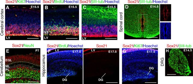
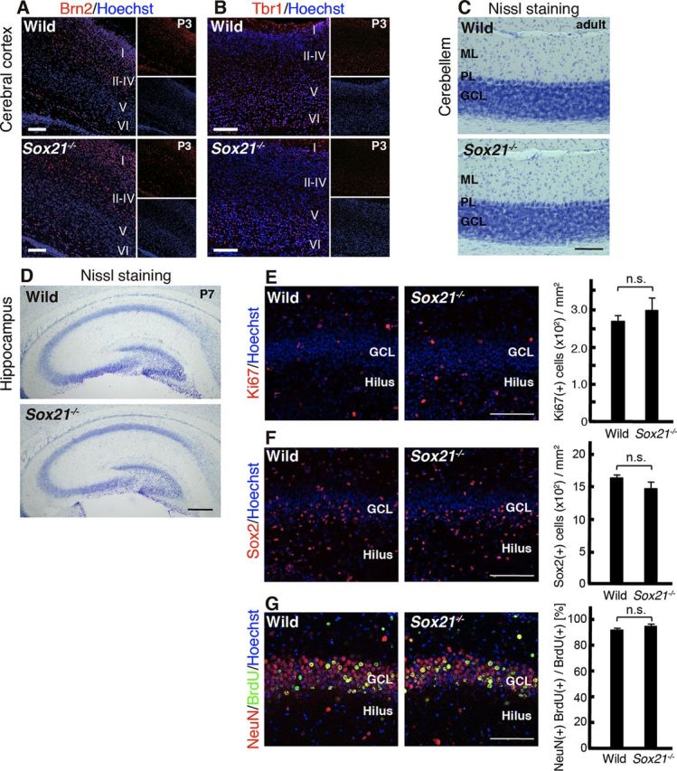
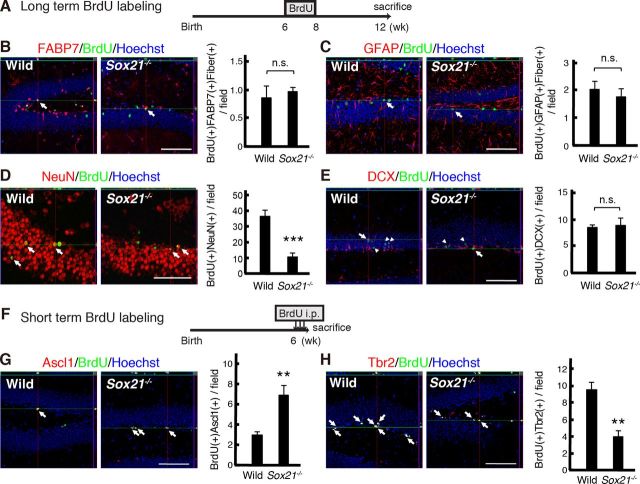
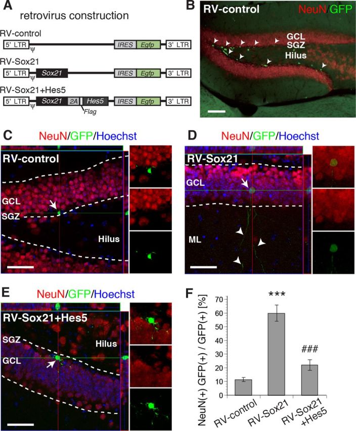
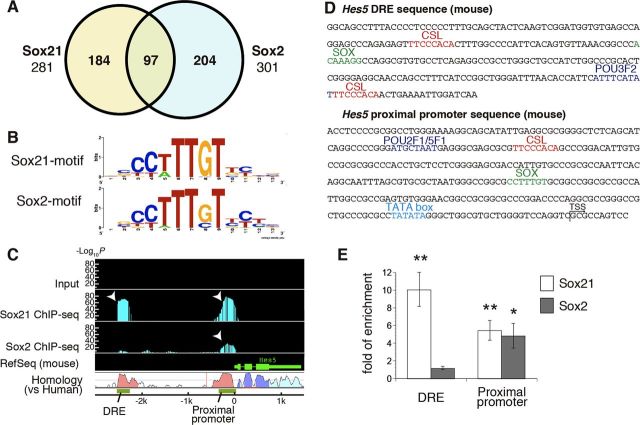
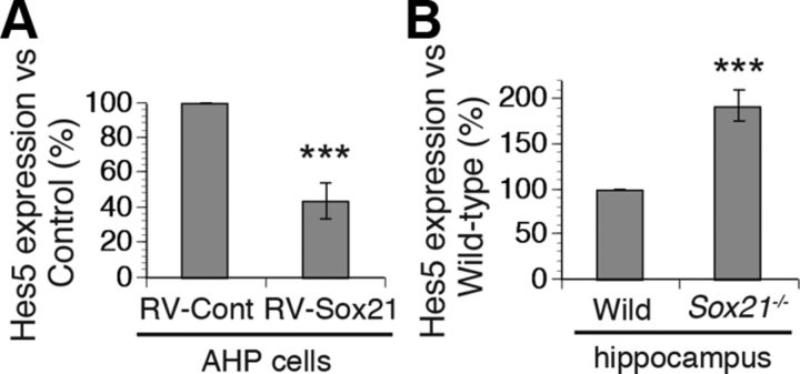
Despite the importance of the production of new neurons in the adult hippocampus, the transcription network governing this process remains poorly understood. The High Mobility Group (HMG)-box transcription factor, Sox2, and the cell surface activated transcriptional regulator, Notch, play important roles in CNS stem cells. Here, we demonstrate that another member of the SoxB (Sox1/Sox2/Sox3) transcription factor family, Sox21, is also a critical regulator of adult neurogenesis in mouse hippocampus. Loss of Sox21 impaired transition of progenitor cells from type 2a to type 2b, thereby reducing subsequent production of new neurons in the adult dentate gyrus. Analysis of the Sox21 binding sites in neural stem/progenitor cells indicated that the Notch-responsive gene, Hes5, was a target of Sox21. Sox21 repressed Hes5 gene expression at the transcriptional level. Simultaneous overexpression of Hes5 and Sox21 revealed that Hes5 was a downstream effector of Sox21 at the point where the Notch and Sox pathways intersect to control the number of neurons in the adult hippocampus. Therefore, Sox21 controls hippocampal adult neurogenesis via transcriptional repression of the Hes5 gene.


Figures

Figure 1.
Figure 1.
Expression of Sox21 in NS/PCs in the developing CNS. Immunohistochemical analyses of Sox21 in the mouse embryonic and postnatal nervous system. A–C, E15.5 cerebral cortex. Sox21 was expressed in Ki67-positive (A) and BrdU-positive (B) proliferative NS/PCs in the VZ; expression was mutually exclusive with that of the neuronal marker βIII-tubulin (C). D, E14.5 spinal cord. Sox21 was not expressed in βIII-tubulin-positive neurons. E, P7 cerebellum. Sox21 was highly expressed in the EGL, and expression was mutually exclusive with that of the neuronal marker, NeuN. F, G, E17.5 hippocampus. BrdU- and Ki67-positive NS/PCs were observed; however, Sox21 was not expressed. H, E14.5 dorsal root ganglia. Sox21 was not expressed in the DRG. Scale bars: A–C, E, H, 100 μm; D, F, G, 200 μm. VZ, Ventricular zone; LV, lateral ventricle; EGL, external granular layer, ML, molecular layer; PL, Purkinje cell layer; IGL, internal granular layer; DRG, dorsal root ganglia; DG, dentate gyrus.
Figure 2.
Figure 2.
Specific expression of Sox21 in NS/PCs in the adult DG. Immunohistochemical analyses of Sox21 in the adult DG. A–D, The areas surrounded by white rectangles in the top panels are shown in the magnified views in the middle three panels. Arrows indicate Sox21 and marker double-positive cells; arrowheads indicate Sox21 single-positive cells. The cells highlighted by yellow arrows are also shown using orthogonal views at higher magnification in the bottom panels (top planes are through the x-z axes, and right planes are through the y-z axes). In the DG, Sox21 expression was restricted to cells in the SGZ. Sox21 was expressed in FABP7-positive (A) and GFAP-positive (B) NS/PCs with horizontally oriented cell bodies typical of type 2a cells and also in GFAP-positive stem-like cells with radial glia-like fibers (B, asterisk) typical of type 1 cells. C, D, The Sox21 expression pattern showed incomplete concordance with the other NS/PC markers, Sox2 (C) and Nestin (D). E, Very few Sox21-positive cells coexpressed PSA-NCAM, a marker for immature neurons. Arrowheads indicate Sox21 single-positive cells. F, The Sox21-positive cell population was mutually exclusive of NeuN-positive neurons in the DG. Arrowheads show Sox21 single-positive cells. G, H, Sox21 was not expressed in S100β-positive astrocytes or GSTπ-positive oligodendrocytes in the DG, although some S100β-positive cells outside the GCL (arrowhead) expressed Sox21. I, A schematic summary of neuronal differentiation in the adult DG and the markers expressed at each stage. Scale bars: A–D, top, 100 μm; middle, 50 μm; bottom, 20 μm; E, F (panels with orthogonal views) G, H, 50 μm; E, F (magnified views), 20 μm.
Figure 3.
Figure 3.
Normal neuronal development in Sox21−/− mice. A, B, Distribution of layer-specific neuronal markers in the cortex of wild-type and Sox21−/− mice at P3. Brn2 (layer II–IV; A) and Tbr1 (layer VI; B) expression patterns in Sox21−/− mice were similar to those in wild-type mice. C, In the adult cerebellum, no impairment was observed in GCL thickness or the cell density in PL and ML. D, The hippocampal structure was normal in P7 Sox21−/− mice. E, F, The numbers of Ki67-positive (F) and Sox2-positive (F) NS/PCs in the hippocampus were not altered in P7 Sox21−/− mice. G, BrdU was administered at E15.5. BrdU-retaining NeuN-positive cells in P7 Sox21−/− mice were comparable with those in wild-type mice. Data represent the mean ± SE. n.s., No significance. Scale bars: A–C, E–G, 100 μm; D, 300 μm. Abbreviations are as in Figure 1.
Figure 4.
Figure 4.
Impaired adult hippocampal neurogenesis in Sox21−/− mice. A, Experimental scheme for long-term BrdU labeling analysis used to detect slowly dividing cells and neurons. B, C, To analyze type 1 cells, marker+/BrdU+/fiber+ cells (arrows) in wild-type and Sox21−/− mice were quantified 4 weeks after the last BrdU administration. The graphs show the numbers of FABP7+/BrdU+/fiber+ cells (B) and GFAP+/BrdU+/fiber+ cells (C) in the DG. D, E, To analyze newly generated mature and immature neurons, the numbers of BrdU+/NeuN+ (D) and BrdU+/DCX+ (E) cells were quantified 4 weeks after the last BrdU administration. F, Experimental scheme for the short-term BrdU labeling analysis used to detect rapidly dividing cells. G, H, The number of BrdU+/Ascl1+ type 2a cells (G, arrows) increased, while the number of BrdU+/Tbr2+ type 2b cells (H, arrows) decreased, in Sox21−/− mice. The top planes are through the x–z axes, and the right planes are through the y-z axes. Data represent the mean ± SE. **p < 0.01; ***p < 0.001 (two-sided t test). n = 3–5. n.s., No significance. Scale bars: 100 μm.
Figure 5.
Figure 5.
Lineage tracing analysis of Sox21-expressing and Sox21-deficient cells in the adult DG. A, B, GFP expression detected in the SGZ. A, GFP was detected in all Sox21-positive cells (arrows) in Sox21+/− mice. B, Sox21 expression was abolished in Sox21−/− mice. C, D, Double staining for GFP and the immature neuronal marker DCX. Top planes are through the x-z axes, and right planes are through the y-z axes. Magnified views are shown in the right panels. Some GFP+ cells (C, arrows) colocalized with DCX+ cells in Sox21+/− mice; however, GFP and DCX expression showed less overlap in Sox21−/− mice (D, arrows). E, The proportion of DCX and GFP double-positive cells within the total GFP-positive cell population in the DG of Sox21+/− and Sox21−/− mice. Scale bars: 50 μm. Data represent the mean ± SE. **p < 0.01 (two-sided t test). n = 3.
Figure 6.
Figure 6.
Sox21 overexpression promoted neuronal differentiation of newborn cells in vivo. A, Schematic showing control-, Sox21-, and Sox21-2A-Hes5-overexpressing retroviral (RV) constructs. ψ depicts a packaging signal used to generate the retrovirus. 2A depicts self-cleaving 2A peptides originating from the foot-and-mouth disease virus. B, Micrographs of GFP-positive cells 3 d after retrovirus infusion into the DG of 6-week-old wild-type mice. The retrovirus infused into the DG delivered its transducing GFP gene selectively into NS/PCs in the SGZ of the hippocampus (arrowheads). C–E, Micrographs showing retrovirus-labeled GFP-positive newborn cells (arrows) 28 d after retrovirus infusion into the DG of 6-week-old wild-type mice in the the controls (C), the Sox21-overexpressing group (D), and the Sox21-2A-Hes5-overexpressing group (E). The GFP-positive infected cell in D represents a mature granule neuron showing NeuN expression and long branches (arrowheads) in the ML of the hippocampus, whereas the majority of GFP-positive cells in the SGZ in C and E lacked NeuN expression and exhibited horizontally expanding branches typical of NS/PCs. F, Quantification of neuronal differentiation in GFP-positive cells. The data represent the proportion of NeuN-positive cells within the total GFP-positive infected cell populations in the SGZ and GCL. Data represent the mean ± SE. ***p < 0.001; ###p < 0.001 (two-sided t test). n = 6–8). Scale bars: B, 100 μm; C–E, 50 μm.
Figure 7.
Figure 7.
Identification of the Hes5 gene as a target of Sox21 using ChIP sequencing analysis. A, Venn diagram showing the number of genomic loci bound by Sox21 and Sox2. Of all the binding sites, 97 common genomic loci were bound by both Sox21 and Sox2. B, Enriched motifs in Sox21- and Sox2-binding sequences identified by de novo computational analysis of ChIP sequencing data. The height of each character represents the relative frequency of the nucleotide appearance in the binding motifs. C, Genomic view of the ChIP sequencing data around the Hes5 gene. Binding by Sox21 and Sox2 is indicated by P values (−log10p) on the y-axis, and the regions in which p values are <10−10 are shown as green bars. Genomic sequence conservation is depicted by the graph showing homology between the mouse and human genomes. Two highly conserved regions upstream of the Hes5 gene corresponded to the positions of the binding sites (arrowheads). D, Genomic sequences of the mouse Hes5 DRE and proximal promoter. Sox21-, Sox2-, CSL-, and POU-binding motifs were present in both regions. E, ChIP–quantitative PCR assay using rat AHP cells. Immunoprecipitated Hes5 DRE or proximal promoter fragments were quantified using the cycle threshold (Ct) values, which were normalized to the Ct of the input DNA. The binding of Sox21 or Sox2 to the DNA was determined by the fold relationship of enrichment of the target fragments in the ChIP DNA over the internal Gapdh gene. Data represent the mean ± SE. *p < 0.05; **p < 0.01 (two-sided t test). n = 3 for Sox21; n = 4 for Sox2.
Figure 8.
Figure 8.
Repression of Hes5 gene promoter activity by Sox21 but not Sox2. AHP cells were transfected with Hes5 promoter–luciferase reporter constructs (1 μg per well) with or without expression vectors for NICD, Sox21, and/or Sox2. The histogram shows the ratio of firefly luciferase to that in cells transfected with the reporter construct and the NICD-expressing vector. The transfection efficiency was normalized to Renilla luciferase activity. A, B, The Hes5 DRE and proximal promoter (−2767 to +73 bp from TSS) and the proximal promoter alone (−687 to +73 bp) were used as reporter constructs in A and B, respectively. The open columns in A show the relative luciferase activities using the intact reporter construct, and the closed columns reflect the construct mutagenized at the Sox-binding sequence of the DRE (ACAAAGG to AagcttG). NICD-expression vector, 0.1 μg per well; Sox21-expression vector, 0.0001, 0.001, and 0.01 μg per well. C, The Hes5 DRE (−2676 to −2244 bp) combined with the β-globin minimal promoter was used. The open columns indicate the intact reporter construct, and the closed columns indicate the mutagenized construct as shown in A. NICD, 1 μg per well; Sox21, 0.0001, 0.001, and 0.01 μg per well. D, E, The Hes5 DRE and proximal promoter were used. D, NICD, 0.1 μg per well, Sox2, 0.0001, 0.001, and 0.01 μg per well. E, NICD, 0.1 μg per well; Sox21, 0.01 μg per well; Sox2, 0.0001, 0.001, and 0.01 μg per well. Data represent the mean ± SE. Statistical significance versus the intact promoter was assessed by two-sided t test (n > 4). **p < 0.01, ***p < 0.001 versus NICD alone; #p < 0.05; ###p < 0.001 versus intact promoter. n.s., No significance versus NICD and Sox21 alone according to the two-sided t test (n > 4). Prox., Proximal promoter.
Figure 9.
Figure 9.
Repressive role of Sox21 in Hes5 expression in vitro and in vivo. A, In vitro analysis of Hes5 expression level confirmed by RT-qPCR. AHP cells infected with a Sox21-expressing retrovirus (RV-Sox21) expressed Hes5 at a lower level than controls (RV-Cont). ***p < 0.001 (two-sided t test). n = 5. B, Endogenous Hes5 expression level in 6-week-old wild-type and Sox21−/− hippocampus determined using RT-qPCR. ***p < 0.001 versus wild-type mice (two-sided t test). n = 4. Data represent the mean ± SE.
Figure 10.
Figure 10.
Colocalization of Sox21 and Hes5 in a small portion of NS/PCs, but not in immature neurons. A, B, Triple immunohistochemical analyses of Hes5 expression in the adult DG using Hes5-NLSlacZ knock-in mice. A, Triple staining for Sox21, Hes5 (lacZ), and the NS/PC marker GFAP. Arrows indicate Sox21+/Hes5+/GFAP+ cells. B, Triple staining for Sox21, Hes5, and the immature neuronal marker PSA-NCAM. Arrowheads indicate Sox21+/Hes5+/PSA-NCAM cells. Scale bars: top row, 100 μm; magnified, 50 μm.
Figure 11.
Figure 11.
Suppression of Hes5 expression promoted neuronal differentiation in the adult DG. A, Western blotting to validate Hes5 shRNA. NIH3T3 cells stably expressing FLAG-Hes5 were infected with a retrovirus encoding control shRNA (RV-shControl) or Hes5 shRNA (RV-shHes5) for 7 d. α-Tubulin expression was used as an internal control. B, C, Micrographs of retrovirus-labeled GFP+ newborn cells (arrows) at 21 d after retrovirus infusion into the DG of 6-week-old wild-type mice. Control shRNA-overexpressing (B) and Hes5 shRNA-overexpressing (C) groups are shown. The GFP-positive infected cells in C demonstrated NeuN expression. D, Quantification of the neuronal differentiation of newborn cells at 21 d after retrovirus injection. Newborn neurons were identified by the presence of NeuN expression, and the data are expressed as the proportion of NeuN+ cells among the total GFP+ infected cell population in the SGZ and GCL. Data represent the mean ± SE. *p < 0.05 (two-sided t test). n = 4. Scale bars: 50 μm.

Similar articles

Cited by

References

    1. Akazawa C, Sasai Y, Nakanishi S, Kageyama R. Molecular characterization of a rat negative regulator with a basic helix-loop-helix structure predominantly expressed in the developing nervous system. J Biol Chem. 1992;267:21879–21885. - PubMed
    1. Bailey TL, Elkan C. Fitting a mixture model by expectation maximization to discover motifs in biopolymers. Proc Int Conf Intell Syst Mol Biol. 1994;2:28–36. - PubMed
    1. Bani-Yaghoub M, Tremblay RG, Lei JX, Zhang D, Zurakowski B, Sandhu JK, Smith B, Ribecco-Lutkiewicz M, Kennedy J, Walker PR, Sikorska M. Role of Sox2 in the development of the mouse neocortex. Dev Biol. 2006;295:52–66. - PubMed
    1. Breunig JJ, Silbereis J, Vaccarino FM, Sestan N, Rakic P. Notch regulates cell fate and dendrite morphology of newborn neurons in the postnatal dentate gyrus. Proc Natl Acad Sci U S A. 2007;104:20558–20563. - PMC - PubMed
    1. Curtis MA, Penney EB, Pearson AG, van Roon-Mom WM, Butterworth NJ, Dragunow M, Connor B, Faull RL. Increased cell proliferation and neurogenesis in the adult human Huntington's disease brain. Proc Natl Acad Sci U S A. 2003;100:9023–9027. - PMC - PubMed

Publication types

MeSH terms

Substances

LinkOut - more resources