. 2012 Sep 5;32(36):12619-29.
doi: 10.1523/JNEUROSCI.1063-12.2012.

Brefeldin A-inhibited guanine exchange factor 2 regulates filamin A phosphorylation and neuronal migration

Affiliations

Brefeldin A-inhibited guanine exchange factor 2 regulates filamin A phosphorylation and neuronal migration

Jingping Zhang et al. J Neurosci. .

Abstract

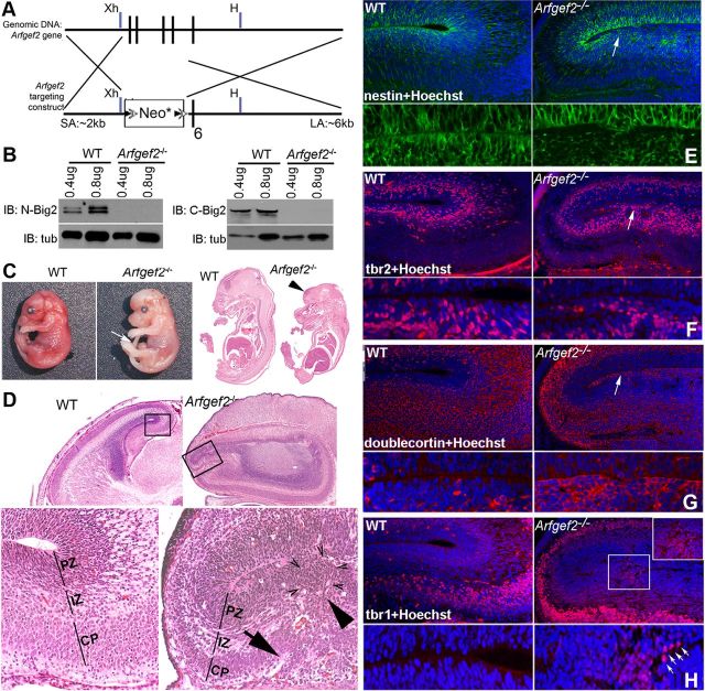
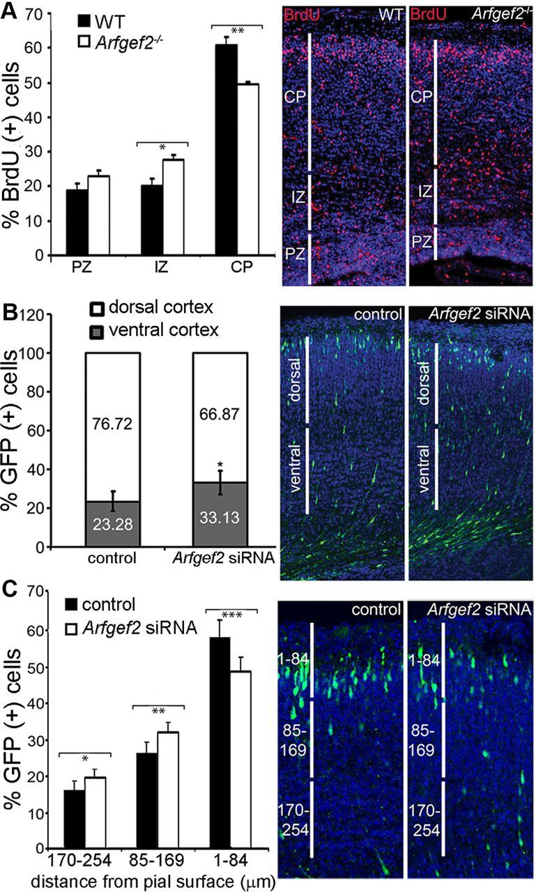
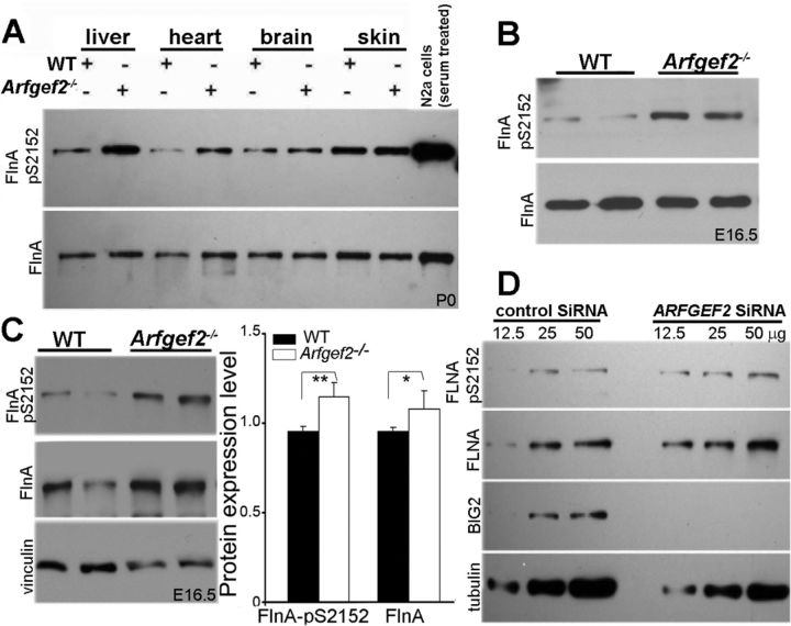
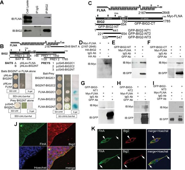
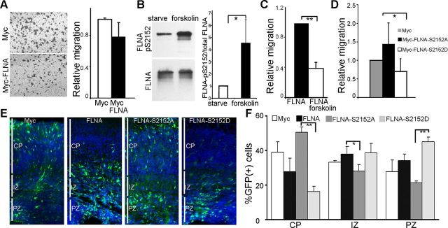
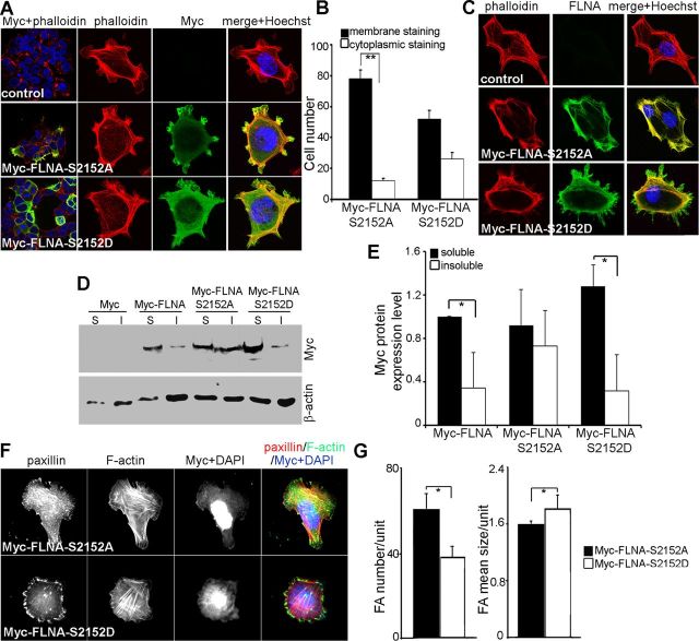
Periventricular heterotopia (PH) is a human malformation of cortical development associated with gene mutations in ADP-ribosylation factor guanine exchange factor 2 (ARFGEF2 encodes for Big2 protein) and Filamin A (FLNA). PH is thought to derive from neuroependymal disruption, but the extent to which neuronal migration contributes to this phenotype is unknown. Here, we show that Arfgef2 null mice develop PH and exhibit impaired neural migration with increased protein expression for both FlnA and phosphoFlnA at Ser2152. Big2 physically interacts with FlnA and overexpression of phosphomimetic Ser2512 FLNA impairs neuronal migration. FlnA phosphorylation directs FlnA localization toward the cell cytoplasm, diminishes its binding affinity to actin skeleton, and alters the number and size of paxillin focal adhesions. Collectively, our results demonstrate a molecular mechanism whereby Big2 inhibition promotes phosphoFlnA (Ser2152) expression, and increased phosphoFlnA impairs its actin binding affinity and the distribution of focal adhesions, thereby disrupting cell intrinsic neuronal migration.


Figures

Figure 1.
Figure 1.
Arfgef2−/− mice develop various cortical malformations, including PH and ectopic distribution of various neural markers in the developing cortex. A, Targeting strategy for generating Arfgef2−/− mice. A LoxP-flanked Neo cassette was inserted downstream of a 2.0 kb fragment containing intron 1 (SA: short arm) and upstream of a 6.0 kb fragment extending from intron 5 to intron 6 (LA: long arm). The targeting vector was designed to replace exons 2–5 of the Arfgef2 gene with a Neo cassette. Xh and H refer to restriction enzyme sites Xho1 and HindIII. B, Immunoblot of total brain lysates with Big2 antibodies against both N- and C-terminal amino acids showed the absence of Big2 expression in Arfgef2−/− mice. Tubulin was used as a loading control. C, Photomicrographs of whole body and H&E-stained sections showed that late gestational (E18.5) Arfgef2−/− embryos could develop exencephaly (arrowhead) and omphalocele (arrow). D, Photomicrograph of sagittal sections of the forebrain in E18.5 WT and Arfgef2−/− mice. Magnified images of the cerebral cortex (shown in boxes) were revealed below. Arfgef2−/− mice displayed PH (arrowhead) and subependymal heterotopia (arrow). E, Nestin (a marker for neural progenitors) positive immunoreactivity (arrow) revealed the presence of ectopic neural progenitor cells within the PH of mutant mice. F, Ectopic localization of the basal progenitor cell marker Tbr2 (arrow) within the PH of Arfgef2−/− mice. G, Immature postmitotic neurons stained by doublecortin within the PH in Arfgef2−/− mice (arrow). H, Tbr1-positive immunoreactivity (inset) revealed the ectopic presence of a few layer six CP neurons within the PH of Arfgef2−/− mice (arrows). For each marker, magnified images were shown below.
Figure 2.
Figure 2.
Loss of Big2 causes defects in neuronal migration in Arfgef2−/− mice. A, The percentage of BrdU-positive cells within the IZ was significantly increased in Arfgef2−/− compared with WT mice (n = 3, *p < 0.01). Conversely, the percentage of BrdU-positive cells within the CP was significantly decreased in Arfgef2−/− mice compared with WT mice (n = 3, **p < 0.02). Images (right) display BrdU immunoreactivity in WT and Arfgef2−/− cortices. Pregnant E14.5 dams were injected with BrdU, and the migratory fates of these BrdU-positive neurons were examined at E18.5. B, Arfgef2 siRNA-mediated knockdown of Big2 resulted in impaired neuronal migration within the CP. The percentage of GFP-positive cells within the ventral half of the CP as opposed to the dorsal half was significantly greater in embryos electroporated with Arfgef2 siRNA compared with control siRNA (n = 3, *p < 0.01). Intraventricular in utero electroporation was performed with coelectroporation of GFP with Arfgef2 siRNA or nontargeting oligonucleotides at E14.5, and mice were examined at E18.5. C, Secondary analyses of the in utero electroporation demonstrated that loss of Big2 function led to progressively fewer progenitors reaching the upper cortical layers (n = 3, *p < 0.05, **p < 0.01, ***p < 0.001). Six or more serial sections were analyzed from each mouse brain.
Figure 3.
Figure 3.
Loss of BIG2 enhances FlnA expression and phosphorylation at Ser2152. A, Western blot displayed levels of phosphorylated FlnA Ser2152 detected in liver, heart, brain, and skin tissues obtained from P0 WT and Arfgef2−/− mice, whereas total levels of FlnA served as loading control. Serum-treated Neuro2a (N2a) cell lysates were used as a control. B, Western blot showed increased level of phosphoFlnA S2152 relative to normalized total FlnA levels in E16.5 mouse whole-brain lysate. C, Increased expression level of both FlnA and phosphoFlnA Ser2152 relative to a housekeeping gene, vinculin, in forebrain tissues from E16.5 WT and Arfgef2−/− embryos. Quantification of FlnA and phosphoFlnA Ser2152 levels in E16.5 Arfgef2−/− and WT mice was shown on the right (n = 4 mice per variable, *p < 0.5, **p < 0.01). D, ARFGEF2 siRNA-mediated knockdown of BIG2 increased FLNA Ser2152 phosphorylation levels by Western blotting analyses. CHP100 cells were transfected with either control nontargeting siRNA or ARFGEF2 siRNA oligonucleotides, and 12.5, 25, or 50 μg of cell lysate was loaded for the detection of FLNA Ser2152 phosphorylation levels. CHP100 cells transfected with ARFGEF2 siRNA oligonucleotides resulted in the loss of BIG2 expression.
Figure 4.
Figure 4.
FLNA interacts and colocalizes with BIG2. A, Endogenous FLNA coimmunoprecipitated with endogenous BIG2 in human CHP100 neuroblastoma cells. B, The receptor binding domain of FLNA (amino acids 2167–2648) interacted with C-terminal (amino acids 786–1452 and 1120–1785) and N-terminal (amino acids 1–654) fragments of BIG2 by directed yeast two-hybrid analysis. Schematic details the regions of FLNA and BIG2 that were used for the baits and preys. Photo images displayed yeast colony growth and β-galactosidase staining. Activation of the LacZ reporter was not observed in yeast expressing BIG2-NT or FLNA (Baits) or BIG2-NT (Prey) alone (bottom left). Expression of BIG2-NT (Bait) with BIG2-C1 or BIG2-C2 (Preys) did not yield growth. Expression of FLNA (Bait) with BIG2-C1, BIG2-C2, or BIG2-NT (Preys) resulted in growth enhancement and color development (bottom right). C, Schematic detailing the extent of the Myc–FLNA and BIG2 constructs used for coimmunoprecipitation. D, Myc–FLNA (amino acids 2167–2648) was detected in HA–BIG2 (full length) immunoprecipitation. Western blot displayed the presence of Myc–FLNA in HA (right) but not IgG (left) immunoprecipitate samples. The Myc–FLNA receptor binding region could be coimmunoprecipitated by both the GFP–BIG2-NT (amino acids 1–654) (E) and GFP–BIG2-CT (amino acids 786–1785) (F) fragments. Myc–FLNA was detected in GFP–BIG2-NT2 (amino acids 221–654) (H) and GFP–BIG2-NT3 (amino acids 329–547) (I) but not in GFP–BIG2-NT1 (amino acids 1–441) (G) immunoprecipitations. This observations suggest that the FLNA receptor binding region binds BIG2 between amino acids 441 and 547. J, Colocalization of FlnA and Big2 along the ventricular lining of the E16.5 mouse cortex. Hoechst staining delineated the nucleus and stained DNA. K, Confocal images displayed the colocalization of FlnA and Big2 within the perinuclear (arrowhead) and peripheral (arrows) domains of a primary neural progenitor cell. Hoechst staining delineated the nucleus and stained DNA. IB, Immunoblot; IP, immunoprecipitation.
Figure 5.
Figure 5.
Increased phosphorylation of FLNA at Ser2152 impairs cell migration. A, Bright-field photomicrographs showed that the cells transfected with either Myc or Myc–FLNA migrated to the lower surface of the transwell membrane. The cells were stained with 0.5% crystal violet for visualization. Quantification of the number of Myc–FLNA-positive cells that had migrated in transwell assay relative to Myc-positive cells alone was shown to the right (n = 3). B, Incubation of serum-starved CHP100 cells with forskolin (10 μm) for 10 min led to an ∼3.5-fold increase in the ratio of phosphorylated FLNA Ser2152/total FLNA (n = 3, *p < 0.05). C, Forskolin treatment significantly reduced the migrated CHP100 cell number to 60% compared with the control group in the transwell assay (n = 4, **p < 0.01). D, Bar graph showed reduced migrating cell numbers in the Myc–FLNA–S2152D (serine 2152 to aspartic acid) group compared with the Myc–FLNA–S2152A (serine 2152 to alanine) group (n = 4, *p < 0.05). All transwell assays were calculated from three or four triplicate individual experiments. E, Representative fluorescent photomicrographs showed GFP-positive cells after in utero electroporation of GFP alone or GFP with FLNA, FLNA–S2152A, and FLNA–S2152D. Hoechst staining was used to define the CP layers. More GFP progenitors were observed near the ventricular lining in mice electroporated with the FLNA–S2152D construct. F, Bar graphs showed the percentage of GFP-positive cells in the CP, IZ, and PZ compared with total GFP cell number 72 h after in utero electroporation (n = 4 per experimental variable; data represents the mean ± SE; *p < 0.5, **p < 0.01).
Figure 6.
Figure 6.
Phosphorylation of FLNA at Ser2152 alters FLNA subcellular distribution, its binding affinity to actin fibers, and focal adhesion structure. A, CHP100 cells transfected with Myc–FLNA–S2152A or FLNA–S2152D were stained with Myc antibody and phalloidin. Myc–FLNA–S2152A accumulated in cell protrusions and colocalized with actin fibers, whereas FLNA–S2152D was found to be diffusely localized throughout the cytoplasm. A low-magnification field of transfected cells with altered cellular distribution was displayed in the left column. B, Statistical analyses of subcellular distribution of Myc–FLNA–S2152A and FLNA–S2152D quantified from a total of 80–100 single cells from three independent experiments (**p < 0.01). C, Similar to the CHP100 studies, FLNA immunostaining of Myc–FLNA–S2152A and FLNA–S2152D in transfected M2 cells (FLNA-depleted melanoma cells). The Myc–FLNA–S2152A appeared colocalized with actin fibers near the cell surface, and FLNA–S2152D was found to be distributed throughout the cytoplasm. D, Western blot analyses demonstrated increased binding affinity of FLNA–S2152A to actin fibers in Triton X-100 insoluble fractions compared with FLNA–S2152D transfected cells. E, Quantification of Western blot experiments in D (n = 3, *p < 0.5). F, Paxillin staining showed increased numbers but reduced size of focal adhesion in Myc–FLNA–S2152A-expressing MEF cells. Myc–FLNA–S2152D-expressing MEFs displayed fewer but larger paxillin-associated focal adhesion. G, Statistical analyses of paxillin staining for focal adhesions in Myc–FLNA–S2152A and Myc–FLNA–S2152D MEFs. The focal adhesion number was quantified relative to total cell surface area using NIH ImageJ software. The focal adhesion size was generated from quantifying the area of cellular fluorescent labeling divided by the number of focal adhesions in the cell (n = 3 independent experiments, 15–20 cells per variable). FA, Focal adhesion (*p < 0.05).

Similar articles

Cited by

References

    1. Achstetter T, Franzusoff A, Field C, Schekman R. SEC7 encodes an unusual, high molecular weight protein required for membrane traffic from the yeast Golgi apparatus. J Biol Chem. 1988;263:11711–11717. - PubMed
    1. Charych EI, Yu W, Miralles CP, Serwanski DR, Li X, Rubio M, De Blas AL. The brefeldin A-inhibited GDP/GTP exchange factor 2, a protein involved in vesicular trafficking, interacts with the beta subunits of the GABA receptors. J Neurochem. 2004;90:173–189. - PubMed
    1. D'Souza-Schorey C, Chavrier P. ARF proteins: roles in membrane traffic and beyond. Nat Rev Mol Cell Biol. 2006;7:347–358. - PubMed
    1. Faux CH, Parnavelas JG. The role of intracellular calcium and RhoA in neuronal migration. Sci STKE. 2007;2007:pe62. - PubMed
    1. Feng Y, Chen MH, Moskowitz IP, Mendonza AM, Vidali L, Nakamura F, Kwiatkowski DJ, Walsh CA. Filamin A (FLNA) is required for cell-cell contact in vascular development and cardiac morphogenesis. Proc Natl Acad Sci U S A. 2006;103:19836–19841. - PMC - PubMed

Publication types

LinkOut - more resources