

Tuning PEG-DA hydrogel properties via solvent-induced phase separation (SIPS)()
- PMID: 22956857
- PMCID: PMC3433064
- DOI: 10.1039/C1JM13943F
Tuning PEG-DA hydrogel properties via solvent-induced phase separation (SIPS)()
Abstract
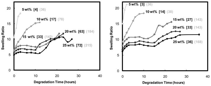
Poly(ethylene glycol) diacrylate (PEG-DA) hydrogels are widely utilized to probe cell-material interactions and ultimately for a material-guided approach to tissue regeneration. In this study, PEG-DA hydrogels were fabricated via solvent-induced phase separation (SIPS) to obtain hydrogels with a broader range of tunable physical properties including morphology (e.g. porosity), swelling and modulus (G'). In contrast to conventional PEG-DA hydrogels prepared from an aqueous precursor solution, the reported SIPS protocol utilized a dichloromethane (DCM) precursor solution which was sequentially photopolymerized, dried and hydrated. Physical properties were further tailored by varying the PEG-DA wt% concentration (5 wt%-25 wt%) and M(n) (3.4k and 6k g mol (-1)). SIPS produced PEG-DA hydrogels with a macroporous morphology as well as increased G' values versus the corresponding conventional PEG-DA hydrogels. Notably, since the total swelling was not significantly changed versus the corresponding conventional PEG-DA hydrogels, pairs or series of hydrogels represent scaffolds in which morphology and hydration or G' and hydration are uncoupled. In addition, PEG-DA hydrogels prepared via SIPS exhibited enhanced degradation rates.
Figures


Similar articles
-
Incorporation of a silicon-based polymer to PEG-DA templated hydrogel scaffolds for bioactivity and osteoinductivity.Acta Biomater. 2019 Nov;99:100-109. doi: 10.1016/j.actbio.2019.09.018. Epub 2019 Sep 16. Acta Biomater. 2019. PMID: 31536841 Free PMC article.
-
PDMS(star)-PEG hydrogels prepared via solvent-induced phase separation (SIPS) and their potential utility as tissue engineering scaffolds.Acta Biomater. 2012 Dec;8(12):4324-33. doi: 10.1016/j.actbio.2012.07.034. Epub 2012 Jul 27. Acta Biomater. 2012. PMID: 22842033 Free PMC article.
-
Enhanced Osteogenic Potential of Phosphonated-Siloxane Hydrogel Scaffolds.Biomacromolecules. 2020 Dec 14;21(12):5189-5199. doi: 10.1021/acs.biomac.0c01293. Epub 2020 Nov 2. Biomacromolecules. 2020. PMID: 33135881
-
Facile fabrication of superporous and biocompatible hydrogel scaffolds for artificial corneal periphery.Colloids Surf B Biointerfaces. 2019 Mar 1;175:26-35. doi: 10.1016/j.colsurfb.2018.11.013. Epub 2018 Nov 8. Colloids Surf B Biointerfaces. 2019. PMID: 30513471
-
Characterization of permeability and network structure of interfacially photopolymerized poly(ethylene glycol) diacrylate hydrogels.Biomaterials. 1998 Jul;19(14):1287-94. doi: 10.1016/s0142-9612(98)00025-8. Biomaterials. 1998. PMID: 9720892
Cited by
-
Incorporation of a silicon-based polymer to PEG-DA templated hydrogel scaffolds for bioactivity and osteoinductivity.Acta Biomater. 2019 Nov;99:100-109. doi: 10.1016/j.actbio.2019.09.018. Epub 2019 Sep 16. Acta Biomater. 2019. PMID: 31536841 Free PMC article.
-
Continuous gradient scaffolds for rapid screening of cell-material interactions and interfacial tissue regeneration.Acta Biomater. 2013 Sep;9(9):8254-61. doi: 10.1016/j.actbio.2013.05.012. Epub 2013 May 22. Acta Biomater. 2013. PMID: 23707502 Free PMC article.
-
Resilin-PEG Hybrid Hydrogels Yield Degradable Elastomeric Scaffolds with Heterogeneous Microstructure.Biomacromolecules. 2016 Jan 11;17(1):128-40. doi: 10.1021/acs.biomac.5b01255. Epub 2015 Dec 22. Biomacromolecules. 2016. PMID: 26646060 Free PMC article.
-
PDMS(star)-PEG hydrogels prepared via solvent-induced phase separation (SIPS) and their potential utility as tissue engineering scaffolds.Acta Biomater. 2012 Dec;8(12):4324-33. doi: 10.1016/j.actbio.2012.07.034. Epub 2012 Jul 27. Acta Biomater. 2012. PMID: 22842033 Free PMC article.
-
Polymerization- and solvent-induced phase separation in hydrophilic-rich dentin adhesive mimic.Acta Biomater. 2014 Jul;10(7):3038-47. doi: 10.1016/j.actbio.2014.03.001. Epub 2014 Mar 12. Acta Biomater. 2014. PMID: 24631658 Free PMC article.
References
-
- Dutta RC, Dutta AK. Biotechnol Adv. 2009;27:334–339. - PubMed
-
- Kleinman HK, Philp D, Hoffman MP. Curr Opin Biotechnol. 2003;14:526–532. - PubMed
-
- Lutolf MP, Hubbell JA. Nat Biotechnol. 2005;23:47–55. - PubMed
-
- Brandl F, Sommer F, Goepferich A. Biomaterials. 2007;28:134–136. - PubMed
-
- Pennesi C, Scaglione S, Gionnoni P, Quarto R. Curr Pharm Biotechnol. 2011;12:151–159. - PubMed
Grants and funding
LinkOut - more resources
Full Text Sources