. 2012;8(8):e1002896.
doi: 10.1371/journal.ppat.1002896. Epub 2012 Aug 30.

CPSF6 defines a conserved capsid interface that modulates HIV-1 replication

Affiliations

CPSF6 defines a conserved capsid interface that modulates HIV-1 replication

Amanda J Price et al. PLoS Pathog. 2012.

Abstract

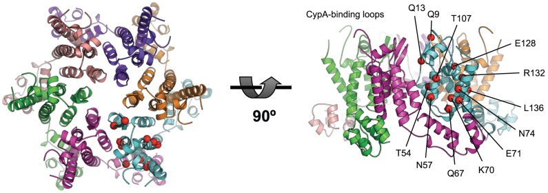
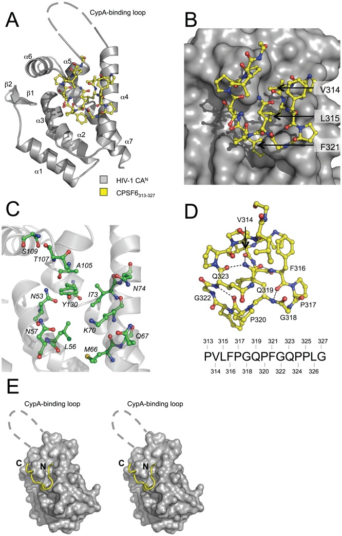
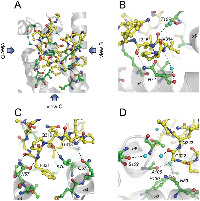
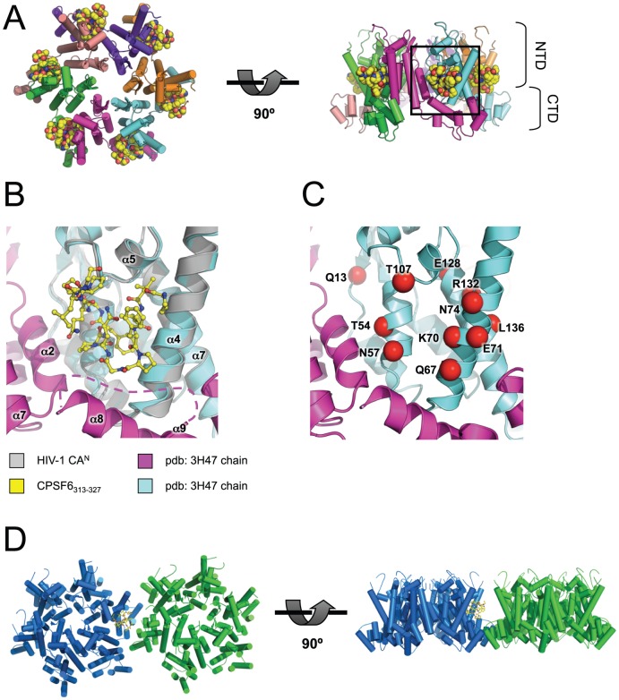
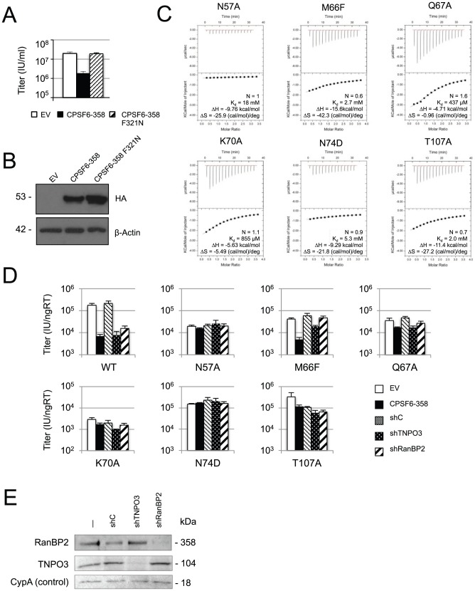
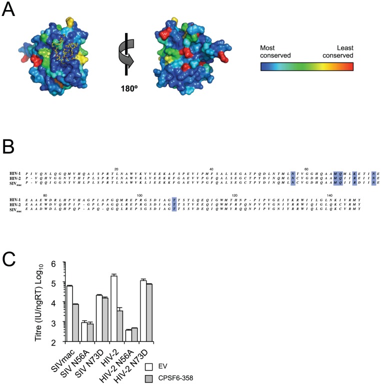
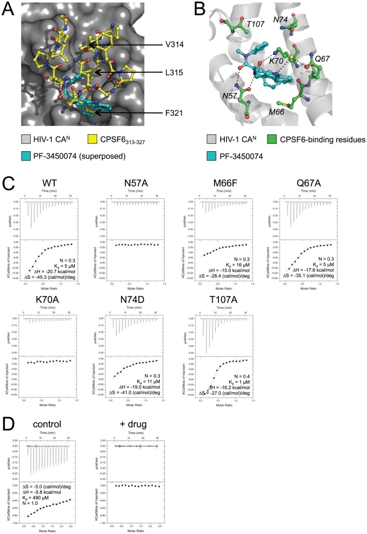
The HIV-1 genome enters cells inside a shell comprised of capsid (CA) protein. Variation in CA sequence alters HIV-1 infectivity and escape from host restriction factors. However, apart from the Cyclophilin A-binding loop, CA has no known interfaces with which to interact with cellular cofactors. Here we describe a novel protein-protein interface in the N-terminal domain of HIV-1 CA, determined by X-ray crystallography, which mediates both viral restriction and host cofactor dependence. The interface is highly conserved across lentiviruses and is accessible in the context of a hexameric lattice. Mutation of the interface prevents binding to and restriction by CPSF6-358, a truncated cytosolic form of the RNA processing factor, cleavage and polyadenylation specific factor 6 (CPSF6). Furthermore, mutations that prevent CPSF6 binding also relieve dependence on nuclear entry cofactors TNPO3 and RanBP2. These results suggest that the HIV-1 capsid mediates direct host cofactor interactions to facilitate viral infection.


Conflict of interest statement

The authors have declared that no competing interests exist.

Figures

Figure 1
Figure 1. Exposed CA mutations affecting HIV-1 infectivity.
Location of CA mutations that are solvent-exposed in the hexameric CA structure and which are found to result in decreased HIV-1 infectivity . CA mutations are labeled and represented as red spheres. The CA hexamer structure was derived from pdb: 3H47 by generating symmetry-related copies in PyMOL. Left: view looking down onto the hexamer, right: view from the side, with CypA-binding loops at the top.
Figure 2
Figure 2. CPSF6 binds diverse lentiviral capsids.
Isothermal titration calorimetry (ITC) of CPSF6313–327 against CAN domains of lentiviral capsids. CPSF6313–327 binding to HIV-1 is specific (A), being abolished by CA mutation N74D (B). CPSF6313–327 also binds to diverse primate lentiviral capsids, HIV-2, FIV and SIVmac (C). The stoichiometry (N), affinity (Kd), enthalpy (ΔH) and entropy (ΔS) of interaction are shown.
Figure 3
Figure 3. Crystal structure of HIV-1 CAN in complex with CPSF6313–327.
(A) Crystal structure of the HIV-1 CAN:CPSF6313–327 complex. HIV-1 CAN is shown as cartoon representation and CPSF6 as sticks. Secondary structure elements in HIV-1 CAN are labeled. The electron density for the CypA-binding loop was poor, so this region is represented by a dashed line. (B) Close-up view of the HIV-1 CAN:CPSF6313–327 interface, showing HIV-1 CAN as surface representation. The three CPSF6 residues that fill the centre of the channel in HIV-1 are indicated. (C) The CPSF6-binding interface on HIV-1 CAN. Residues involved in binding to CPSF6313–327 are labeled and shown in green. (D) Intramolecular interactions in CPSF6313–327. Residues involved in intramolecular hydrogen bonding interactions are labeled and the interactions shown as dashed lines. The sequence of CPSF6313–327 is shown for reference. (E) Stereo figure showing overview of the HIV-1 CAN:CPSF6313–327 interaction. The N- and C-termini of CPSF6313–327 (labeled) project out of the binding channel in HIV-1 CAN.
Figure 4
Figure 4. Interactions in the HIV-1 CAN:CPSF6313–327 complex.
(A–D) Detailed views of the HIV-1 CAN:CPSF6313–327 interface. HIV-1 CAN is shown as grey cartoon, HIV-1 residues that bind CPSF6 are in green and CPSF6313–327 in yellow. Water molecules involved in water-mediated interactions in the complex are shown as cyan spheres. (A) Overview of the HIV-1 CAN:CPSF6313–327 interface, showing all interacting residues and intermolecular hydrogen-bonding interactions. Views of the close-ups shown in (B), (C) and (D) are indicated. (B–D) HIV-1 CAN α-helices are labeled to aid orientation. Interacting residues are labeled (CPSF6 in normal font; HIV-1 in italics).
Figure 5
Figure 5. The CPSF6-binding interface is accessible and highly conserved in HIV-1 virions.
(A) Model of CPSF6313–327 binding to HIV-1 CA hexamer. The hexamer structure was derived from pdb: 3H47 by generating symmetry-related copies in PyMOL. The model was composed by superposition of CAN chains from HIV-1 CAN:CPSF6313–327 on the hexamer using secondary structure matching. HIV-1 CA helices are shown as cylinders and CPSF6313–327 as spheres. Left: top view, right: side view. N-terminal and C-terminal CA domains (NTD and CTD) are labeled. (B) Close-up of boxed region in (A). CAs are shown as cartoons and CPSF6313–327 as sticks. The region between CTD positions 175 and 188 was disordered and so is represented by a dashed line. (C) Same view as in (B), showing exposed CA mutations (labelled and shown as red spheres) that result in decreased HIV-1 infectivity (see Figure 1 ). (D) Model of CPSF6313–327 binding at a hexamer-hexamer interface. Neighbouring hexamers were derived from pdb: 3H47 by generating extended symmetry-related copies in PyMOL. HIV-1 CAN:CPSF6313–327 was superposed on the hexamer using secondary structure matching. The model shows that CPSF6313–327 binding is likely to be accommodated at neighbouring CTD-mediated hexamer-hexamer junctions.
Figure 6
Figure 6. The CPSF6-binding interface determines HIV-1 nuclear entry requirements.
(A–B) CPSF6 residue F321 is critical for interaction with HIV-1 CAN. (A) HeLa cells expressing empty vector (white bar), HA-tagged CPSF6-358 (black bar) or CPSF6-358 bearing mutation F321N (striped bar) were infected with GFP-encoding VSV-G pseudotyped HIV-1 vector. F321N abolished restriction by CPSF6-358, confirming the importance of this residue in the HIV-1 CAN:CPSF6 interaction. (B) Western blot to show CPSF6-358 and CPSF6-358 F321N expression levels, with actin as loading control. (C) ITC of CPSF6313–327 against mutant HIV-1 CANs. All mutations at the CPSF6-binding interface resulted in reduced affinity to CPSF6313–327. The stoichiometry (N), affinity (Kd), enthalpy (ΔH) and entropy (ΔS) of interaction are shown. (D) Titres of VSV-G pseudotyped GFP-encoding HIV-1 vectors bearing wild type or mutant CA on HeLa cells expressing empty vector (EV), CPSF6-358, control knockdown cells (shC) and cells depleted for TNPO3 (shTNPO3) or RanBP2 (shRanBP2). The data are representative of two independent experiments, each using three different virus doses. Mutation of HIV-1 CAN residues involved in binding to CPSF6 resulted in the loss of dependence on TNPO3 and RanBP2, suggesting a link between CPSF6 binding and normal nuclear import of HIV-1. (E) Western blot to show knockdown of TNPO3 and RanBP2, with cyclophilin A (CypA) as loading control.
Figure 7
Figure 7. The CPSF6 interface is conserved in HIV-1, HIV-2, SIVmac CAN.
(A) The CPSF6-binding interface is highly conserved within HIV-1 viruses. The ConSurf Server , was used to map ∼100 unique HIV-1 CAN sequences onto the HIV-1 CAN:CPSF6313–327 structure. HIV-1 CAN is shown as surface representation and CPSF6313–327 as yellow sticks. The level of conservation at each position in CAN is shown by the colour; dark blue = most conserved, red = least conserved. Residues at the CPSF6-binding interface are among the most highly conserved of all, suggesting that this is a functionally important interface. The figure was generated using the PyMOL script output by ConSurf, with conservation grades replacing the B-factor column. (B) Sequence alignment of HIV-1, HIV-2, SIVmac CAN. (C) Titres of VSV-G pseudotyped GFP-encoding HIV-2 and SIVmac vectors bearing wild type or mutant CA on HeLa cells expressing empty vector (EV) or CPSF6-358.
Figure 8
Figure 8. Drug PF-3450074 binds to part of the CPSF6-binding interface in HIV-1 CAN.
(A) Superposition of HIV-1 CAN:PF-3450074 structure (pdb: 2XDE) on HIV-1 CAN:CPSF6313–327 using secondary structure matching of the HIV-1 CAN domains. Drug PF-3450074 is shown in cyan, CPSF6313–327 in yellow. The three CPSF6 residues that fill the centre of the channel in HIV-1 are indicated. One of the phenyl rings in PF-3450074 superposes almost exactly with the phenyl ring of F321 in CPSF6313–327. (B) Crystal structure of HIV-1 CAN:PF-3450074 (pdb: 2XDE) showing residues involved in binding to CPSF6 (green sticks). The yellow sphere represents a water molecule involved in a water-mediated interaction in the complex. (C) ITC of PF-3450074 against wild type and mutant HIV-1 CANs. (D) Titration of 1 mM CPSF6313–327 into 80 µM HIV-1 CAN was carried out in the absence or presence of 100 µM PF-3450074 (‘control’ and ‘+ drug’ respectively). PF-3450074 completely inhibits binding of CPSF6313–327 to HIV-1 CAN.

References

    1. Fassati A, Goff SP (2001) Characterization of intracellular reverse transcription complexes of human immunodeficiency virus type 1. J Virol 75: 3626–3635. - PMC - PubMed
    1. Miller MD, Farnet CM, Bushman FD (1997) Human immunodeficiency virus type 1 preintegration complexes: studies of organization and composition. J Virol 71: 5382–5390. - PMC - PubMed
    1. Hulme AE, Perez O, Hope TJ (2011) Complementary assays reveal a relationship between HIV-1 uncoating and reverse transcription. Proc Natl Acad Sci U S A 108: 9975–80. - PMC - PubMed
    1. Arhel NJ, Souquere-Besse S, Munier S, Souque P, Guadagnini S, et al. (2007) HIV-1 DNA Flap formation promotes uncoating of the pre-integration complex at the nuclear pore. EMBO J 26: 3025–3037. - PMC - PubMed
    1. Arhel N (2010) Revisiting HIV-1 uncoating. Retrovirology 7: 96. - PMC - PubMed

Publication types

MeSH terms

Associated data