

doi: 10.1007/s13225-012-0152-2.
The Longibrachiatum Clade of Trichoderma: a revision with new species
Affiliations
- PMID: 22956918
- PMCID: PMC3432902
- DOI: 10.1007/s13225-012-0152-2
Item in Clipboard
The Longibrachiatum Clade of Trichoderma: a revision with new species
Fungal Divers.
.
Abstract
The Longibrachiatum Clade of Trichoderma is revised. Eight new species are described (T. aethiopicum, T. capillare, T. flagellatum, T. gillesii, T. gracile, T. pinnatum, T. saturnisporopsis, T. solani). The twenty-one species known to belong to the Longibrachiatum Clade are included in a synoptic key. Trichoderma parareesei and T. effusum are redescribed based on new collections or additional observations. Hypocrea teleomorphs are reported for T. gillesii and T. pinnatum. Previously described species are annotated.
Figures
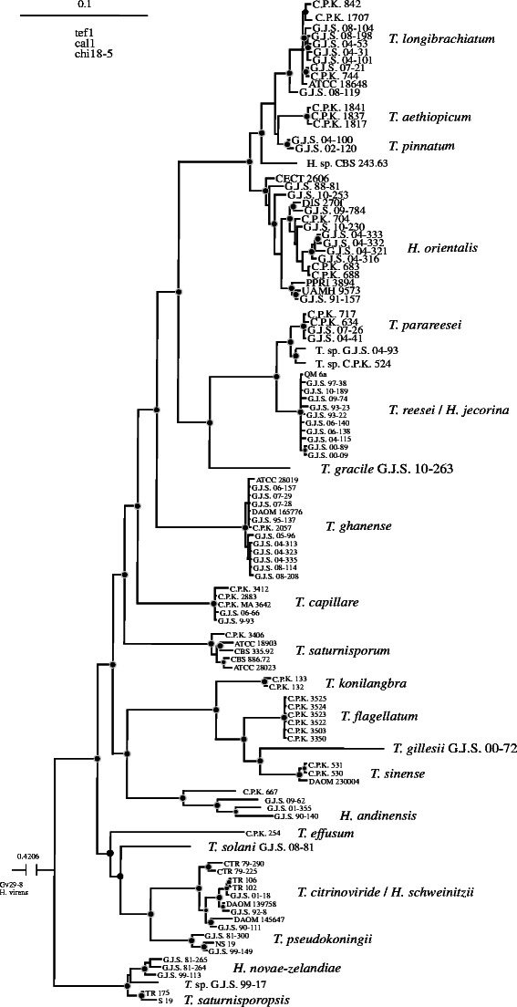
Bayesian phylogram obtained from the concatenated alignment of tef1, cal1 and chi18-5 loci. See Druzhinina et al. (2012) for details
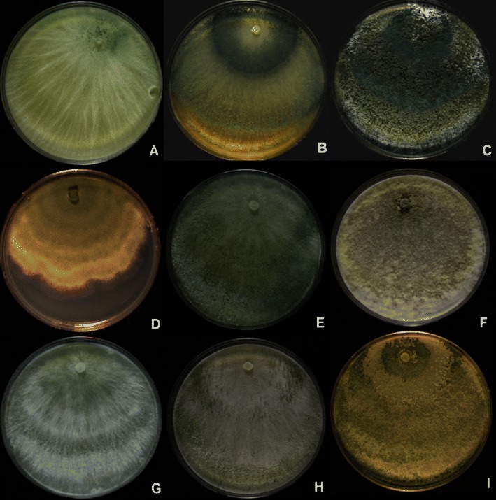
Longibrachiatum Clade. Cultures grown on PDA. a, b
T. aethiopicum, G.J.S. 10–165. c
T. capillare, G.J.S. 10–170. d
T. effusum, DAOM 230007. e, f
T. flagellatum, G.J.S. 10–162. g, h
T. gracile, G.J.S. 10–263, just beginning to sporulate. i. G.J.S. 99–17. All grown 1 week at 25°C under light, except b, e, h, which were grown 1 week at 35°C in darkness with intermittent light. Note the increased sporulation in colonies grown at 35°C when compared to the same strain grown at 25°C (b vs. a, e vs. f)
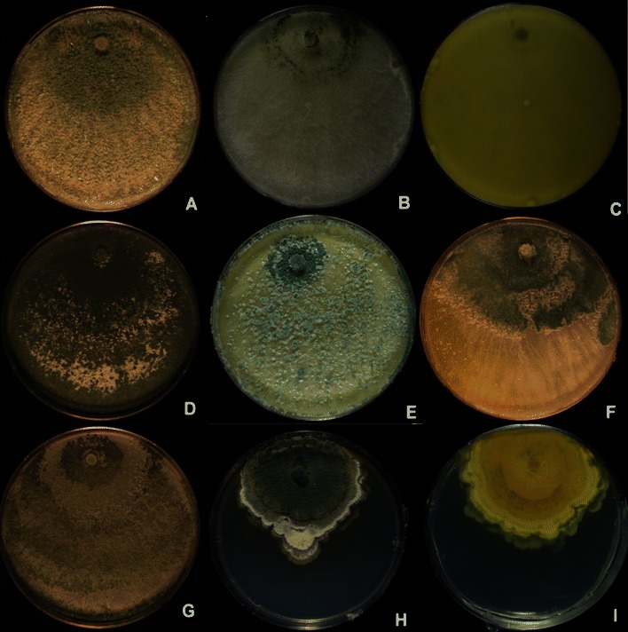
Longibrachiatum Clade. Cultures grown on PDA. a–c
Hypocrea orientalis (a G.J.S. 06–317, b G.J.S. 04–321, c G.J.S. 04–316, reverse showing diffusing yellow pigment). d
T. parareesei G.J.S. 04–41. e, f
T. pinnatum (e G.J.S. 04–100, f G.J.S. 02–120). g
T. saturnisporopsis Tr 175. h, i
T. solani G.J.S. 08–81 (h colony from above, i colony reverse)
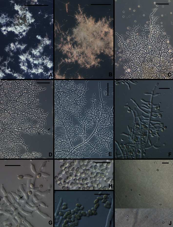
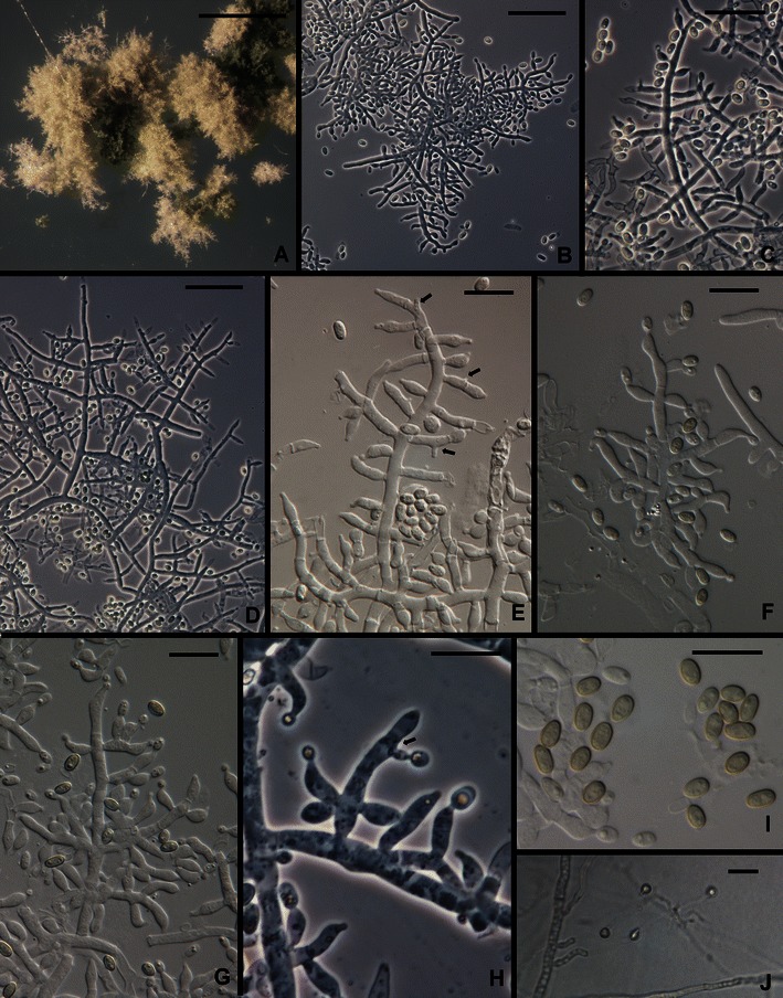
Trichoderma aethiopicum. a, b Pustules on SNA. c–g Conidiophores from SNA (Arrows in d, g show intercalary phialides). h, i Conidia. j Chlamydospores. All from SNA. a, b, d, e, h, j from G.J.S. 10–167; c, g from 10 to 166; f, i from G.J.S. 10–165. Scale bars: a = 0.5 mm, b = 100 μm, c–e, j = 20 μm, f–i = 10 μm
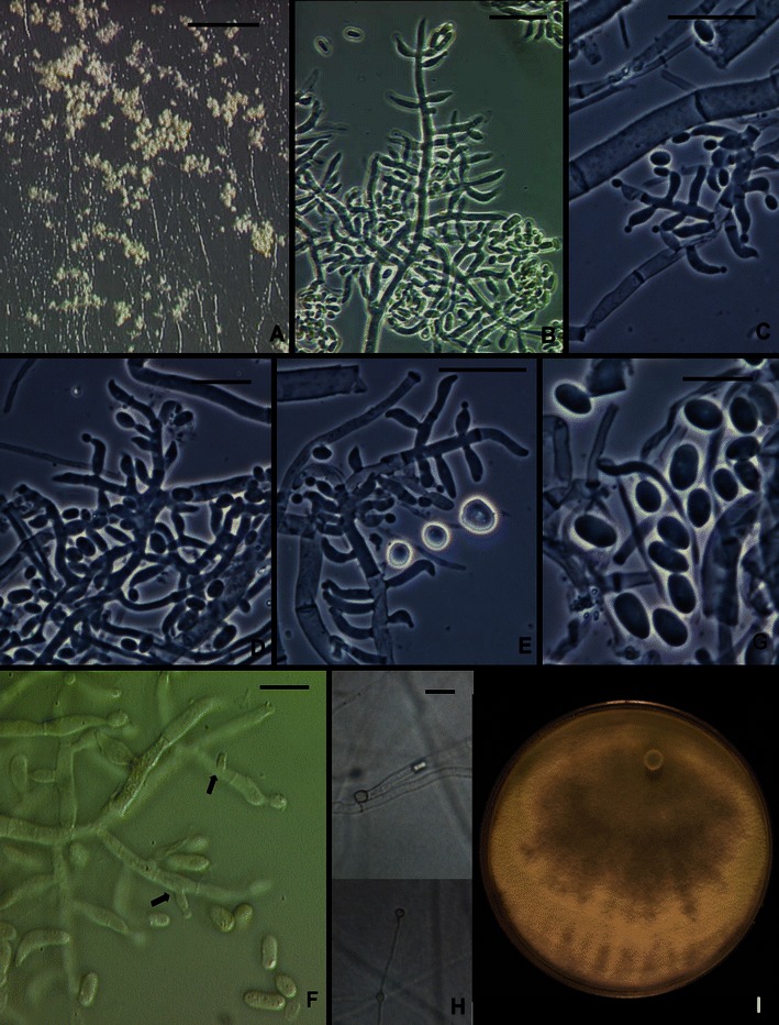
Trichoderma sp. CBS 243.63. a Pustules from CMD. b–e, f Conidiophores and phialides. f, g Conidia. Intercalary phialides indicated by arrows. h. Chlamydospores. i. Colony 1 week on PDA under light just beginning to sporulate. b, f from CMD; b–e, g, h from SNA. Scale bars: a = 2 mm, b–e, h = 20 μm. g = 10 μm
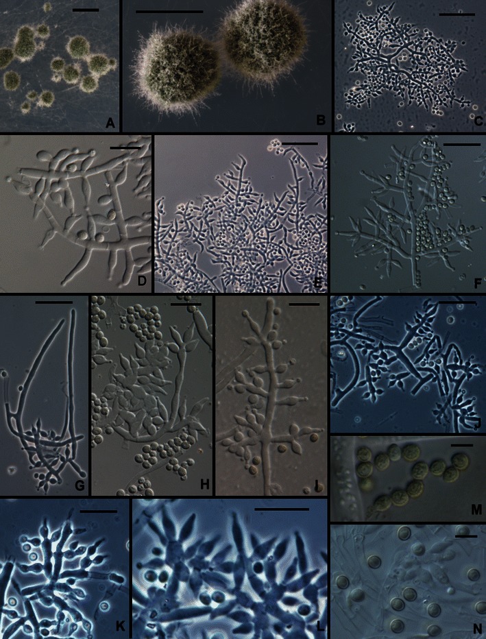
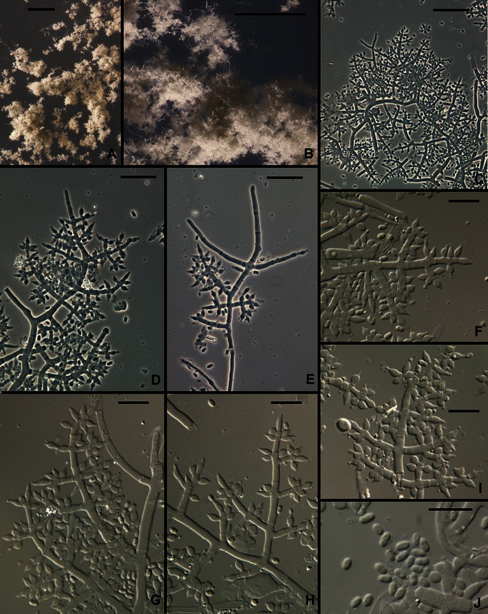
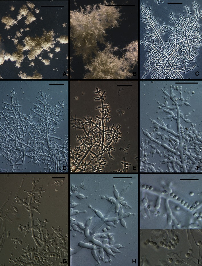
Trichoderma capillare. a, b Pustules (Hairs seen in b). c–l Conidiophores (Hairs seen in g, m). n Conidia. All from SNA except M, which is from CMD. a–c, g–i from G.J.S. 10–170; d, e from G.J.S. 06–66; f, j–l, n from G.J.S. 10–169; m from ATCC 20898. Scale bars: a, b = 0.5 mm; c, e–f, j, k = 20 μm; d, h, i, l–n = 10 μm
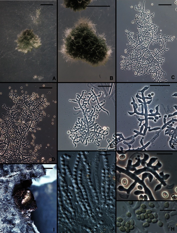
Trichoderma effusum. a–i Conidiophores. j Phialides and aphanophialides in immersed hyphae. k Conidia. All from SNA. All from DAOM 230007. Scale bars: a = 0.5 mm; b–e, g–i, k = 10 μm; f, j = 20 μm
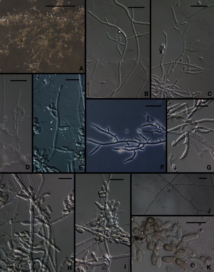
Trichoderma flagellatum.
a, b. Pustules. c–h Conidiophores. Hairs visible in c–e, g, i Phialides (Arrow shows an intercalary phialide). j Conidia. k Chlamydospores. All from SNA. a from G.J.S. 10–156; b from G.J.S. 10–163; c, e, f, g, j from G.J.S. 10–164; d from G.J.S. 10–162; h, i, k from G.J.S. 10–161. Scale bars: a, b = 0.5 mm; c–h = 20 μm; I, J = 10 μm
Trichoderma gillesii anamorph. a, b Pustules. c–i Conidiophores (Hairs visible in e). j Conidia. All from SNA. All from G.J.S. 00–72. Scale bars: a = 1 mm, b = 0.25 mm; c–e = 20 μm; f–i = 10 μm
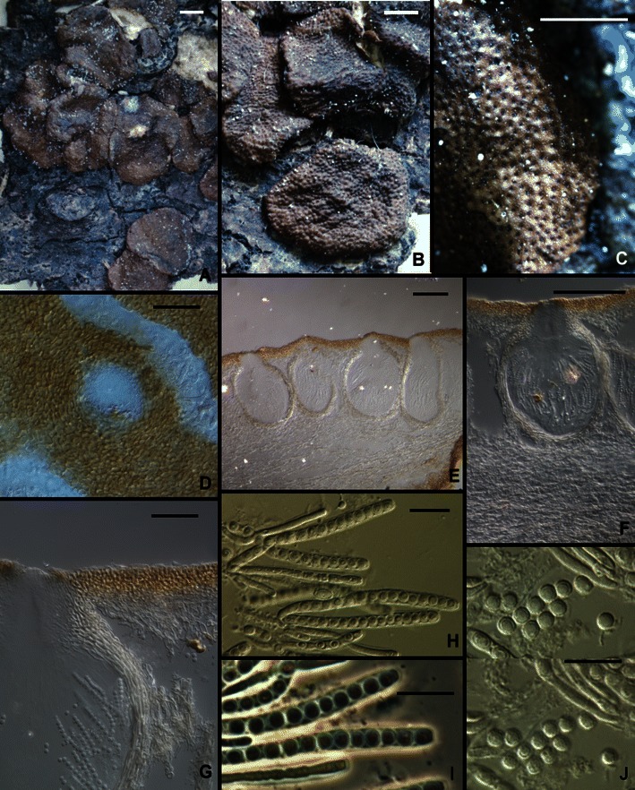
Trichoderma gillesii, Hypocrea teleomorph. a, b Stroma morphology. c Stroma surface, macro view. d Stroma surface, micro view. e–g Perithecia, median longitudinal sections showing surface region and internal tissue of stroma. h, i Asci. j Part-ascospores. Note the subglobose part-ascospores in Figs. i and j All from G.J.S. 00–72. Scale bars: a, b = 1 mm; c = 0.5 mm; d, g = 20 μm; e = 50 μm, f = 100 μm; h–j = 10 μm
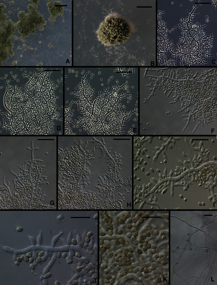
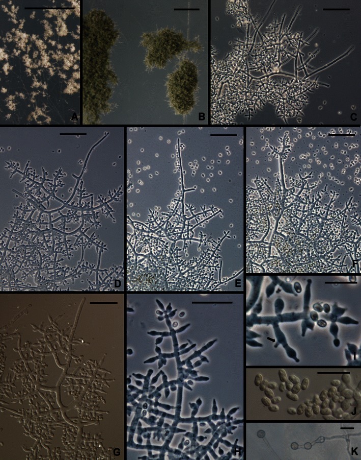
Trichoderma gracile. a, b. Pustules. c–j. Conidiophores (Arrows in e, j show intercalary phialides). k Conidia. l Chlamydospores. All from SNA. All from G.J.S. 10–263. Scale bars: a = 1 mm, b = 0.5 mm; c–h, l = 20 μm; i–k = 10 μm
Hypocrea orientalis. a–c Pustules. d–f Conidiophores. g Phialides. arrows show intercalary phialides. h Conidia. i Part-ascospores; note the globose to subglobose shape. j, k Stromata. a–g from SNA. a, c, g from G.J.S. 04–316; b, d–f, h from DIS 270f; i–k from WU 31609. Scale bars
a = 0. 5 mm; b, c = 250 μm; d–f = 20 μm; g–i = 10 μm; j, k = 1 mm
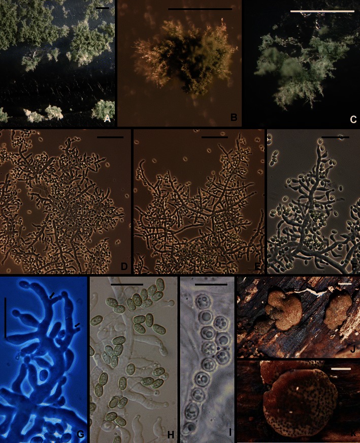
Trichoderma parareesei. a Pustules. b–h Conidiophores and phialides (Arrows in e, h show intercalary phialides). i. Conidia.. j. Chlamydospores. All from SNA. a, d, e from G.J.S. 10–168; b, f, g, i from G.J.S. 07–26; c, from G.J.S. 04–41; h, j from G.J.S. 04–250. Scale bars: a = 0.5 mm; b–d, j = 20 μm; e–i = 10 μm
Trichoderma pinnatum. a, b Pustules. c–g Conidiophores. h Conidia. i Overmature stroma. J. Asci with subglobose part ascospores. a–h From SNA. a, c, e–j from G.J.S. 02–120; b, d from G.J.S. 04–100. Scale bars: a, b = 0.5 mm; c–f = 20 μm; g, h, j = 10 μm; i = 1 mm
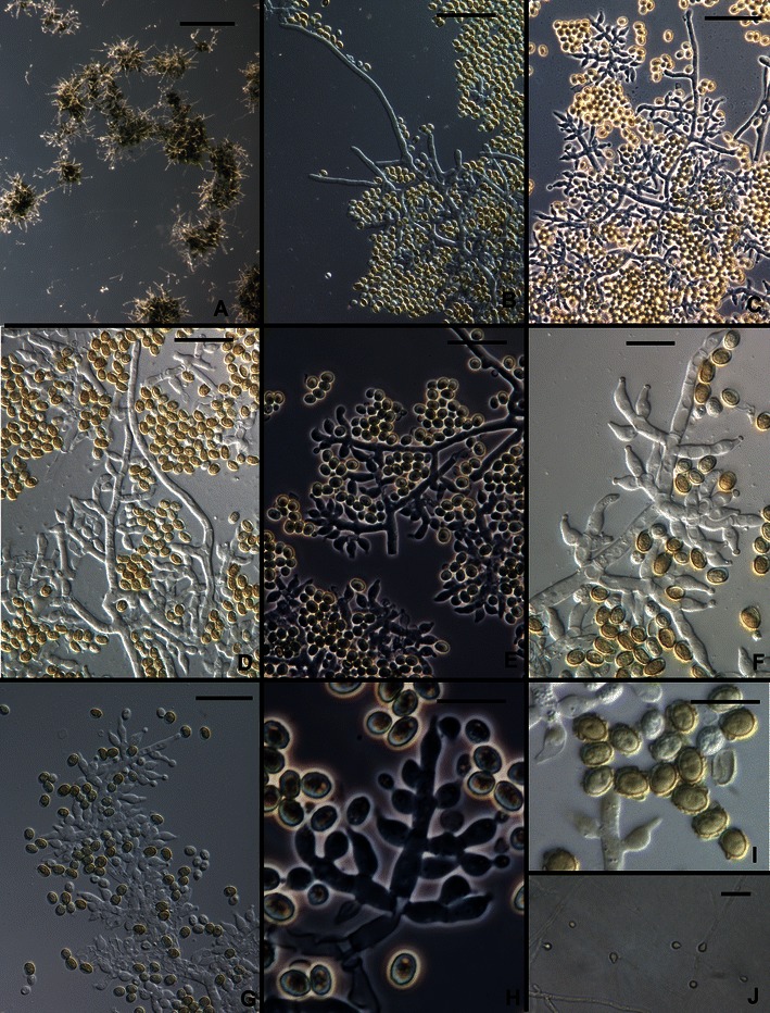
Trichoderma saturnisporopsis. a Pustules. b–h Conidiophores (hairs seen in b–d). i Conidia. j Chlamydospores. All from SNA. a–d, f, i from Tr 175; e, g, h, j from Jaklitsch S 19. Scale bars: a = 0.5 mm; b–e, g, j = 20 μm; f, h, i = 10 μm
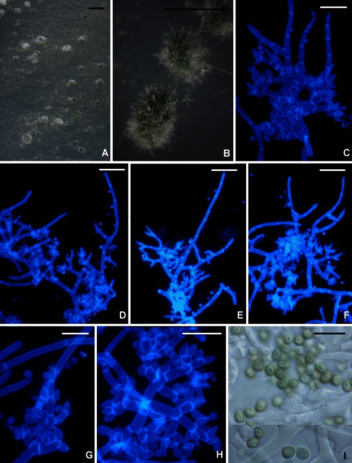
Trichoderma sp. G.J.S. 99–17. a, b Pustules. c–h Conidiophores. i Conidia. All from CMD. c–h fluorescence microscopy in calcofluor (hairs visible in b–f). Scale bars: a = 1 mm, b = 0.5 mm; c–h = 20 μm; i = 10 μm
Trichoderma solani.
a, b Young pustules, conidia just beginning to turn green. c–h Conidiophores. i Conidia. All from G.J.S. 88–81. Scale bars: a = 1 mm, b =250 μm, c–f = 20 μm, g–i = 10 μm
References
-
- Bisby GR. Trichoderma viride Pers. ex Fries, and notes on Hypocrea. Trans Br Mycol Soc. 1939;23:149–168. doi: 10.1016/S0007-1536(39)80020-1. - DOI
-
- Bissett J. A revision of the genus Trichoderma. I. Section Longibrachiatum sect. nov. Can J Bot. 1984;62:924–931. doi: 10.1139/b84-131. - DOI
-
- Bissett J. A revision of the genus Trichoderma. II. Infrageneric classification. Can J Bot. 1991;69:2357–2372. doi: 10.1139/b91-297. - DOI
Grants and funding
LinkOut - more resources
Full Text Sources