

Spinal cord compression secondary to intrathecal catheter-induced granuloma: a report of four cases
- PMID: 22956937
- PMCID: PMC3427967
- DOI: 10.1055/s-0030-1267087
Spinal cord compression secondary to intrathecal catheter-induced granuloma: a report of four cases
Abstract
Objective: The management of nonmalignant pain by morphine pump implantation has become an effective and increasingly frequent strategy of care. We report a rare complication of intrathecal granuloma formation adjacent to the intrathecal catheter tip resulting in spinal cord compression in four patients undergoing intrathecal treatment for chronic pain.
Methods: Four patients presented with chronic back pain and lower extremity pain and weakness and were treated with morphine pump implantation (Fig 1). Each patient developed a mass at the level of the intrathecal catheter tip resulting in increased back pain and diminished neurological function. Following clinical examination and x-ray workup, the patients underwent surgical resection of the mass and removal of the intrathecal catheter. One patient received conservative saline therapy first, and another patient had granuloma resection first and removal of the intrathecal catheter at a later date. Pathological analysis showed granulation tissue with extensive necrosis and chronic inflammation, with negative culture results. No evidence of neoplasm was found.
Results: Patients showed varying degrees of improvement following removal of the intrathecal mass. Two patients had moderate pain reduction following resection of the granuloma; a third had minimal pain improvement; and a fourth had significant pain improvement but continued lower extremity weakness.
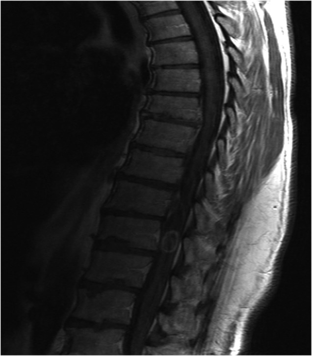
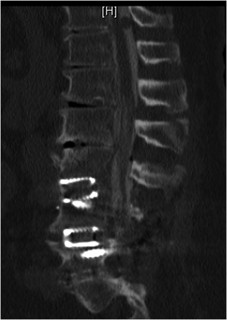
Conclusions: The formation of granulomas caused by intrathecal catheter implantation is a rare but serious complication. Imaging studies including magnetic resonance imaging with contrast and computed tomography with myelogram should be used to follow up a neurological examination consistent with spinal cord compression. Timely surgical intervention may result in marked improvement of symptoms.
Conflict of interest statement
No funding was received for this work. No investigational device was used. The authors report no conflict of interest.
Figures
Similar articles
-
Spinal cord compression by catheter granulomas in high-dose intrathecal morphine therapy: case report.Neurosurgery. 1998 May;42(5):1176-80; discussion 1180-1. doi: 10.1097/00006123-199805000-00142. Neurosurgery. 1998. PMID: 9588567 Review.
-
A Patient with an Intradural Tumor: An Unexpected Finding.Cureus. 2020 Mar 23;12(3):e7376. doi: 10.7759/cureus.7376. Cureus. 2020. PMID: 32226698 Free PMC article.
-
The Management of Unresectable Intrathecal Catheter-Tip-Associated Granuloma Using Morphine Therapy Cessation and Spinal Cord Stimulation.Cureus. 2020 Aug 31;12(8):e10160. doi: 10.7759/cureus.10160. Cureus. 2020. PMID: 33014655 Free PMC article.
-
Formation of two consecutive intrathecal catheter tip granulomas within nine months.Cent Eur Neurosurg. 2010 Feb;71(1):39-42. doi: 10.1055/s-0029-1202359. Cent Eur Neurosurg. 2010. PMID: 20201126
-
Spinal cord compression from intrathecal catheter-tip inflammatory mass: case report and a review of etiology.Reg Anesth Pain Med. 2004 May-Jun;29(3):237-42. doi: 10.1016/j.rapm.2004.01.012. Reg Anesth Pain Med. 2004. PMID: 15138910 Review.
Cited by
-
Blame it on the pump.Clin Case Rep. 2021 Jun 9;9(6):e04195. doi: 10.1002/ccr3.4195. eCollection 2021 Jun. Clin Case Rep. 2021. PMID: 34136231 Free PMC article.
-
Intrathecal pump catheter-tip granuloma recurrence with associated myelomalacia - How safe is intrathecal analgesic infusion therapy? A case report.Surg Neurol Int. 2019 Apr 24;10:62. doi: 10.25259/SNI-33-2019. eCollection 2019. Surg Neurol Int. 2019. PMID: 31528400 Free PMC article.
-
Scar Tissue Catheter Tip Occlusion From an Intrathecal Baclofen Delivery: A Case Report and Review of the Literature.Cureus. 2024 Oct 2;16(10):e70720. doi: 10.7759/cureus.70720. eCollection 2024 Oct. Cureus. 2024. PMID: 39493071 Free PMC article.
References
-
- Magill S T, Wang P, Eller J L. et al.Differentiating intrathecal catheter tip granulomas from normal magnetic resonance image distortion caused by metallic catheter tips. Neurosurgery. 2008;62(1):242–248. - PubMed
-
- Medel R, Pouratian N, Elias W J. Catheter-tip mass mimicking a spinal epidural hematoma: case report. J Neurosurg Spine. 2010;12(1):66–71. - PubMed
-
- Schuchard M, Krames E S, Lanning R M. Intraspinal analgesia for nonmalignant pain: a retrospective analysis for efficacy, safety, and feasibility in 50 patients. Neuromodulation. 1998;1:46–56. - PubMed
-
- Winkelmüller M, Winkelmüller W. Long-term effects of continuous intrathecal opioid treatment in chronic pain of nonmalignant etiology. J Neurosurg. 1996;85(3):458–467. - PubMed
-
- Miele V J, Price K O, Bloomfield S. et al.A review of intrathecal morphine therapy related granulomas. Eur J Pain. 2006;10(3):251–261. - PubMed
LinkOut - more resources
Full Text Sources
