. 2012;1(1):71-87.
doi: 10.3233/JHD-2012-120016.

Deletion of the huntingtin proline-rich region does not significantly affect normal huntingtin function in mice

Affiliations

Deletion of the huntingtin proline-rich region does not significantly affect normal huntingtin function in mice

Michelle Neveklovska et al. J Huntingtons Dis. 2012.

Abstract

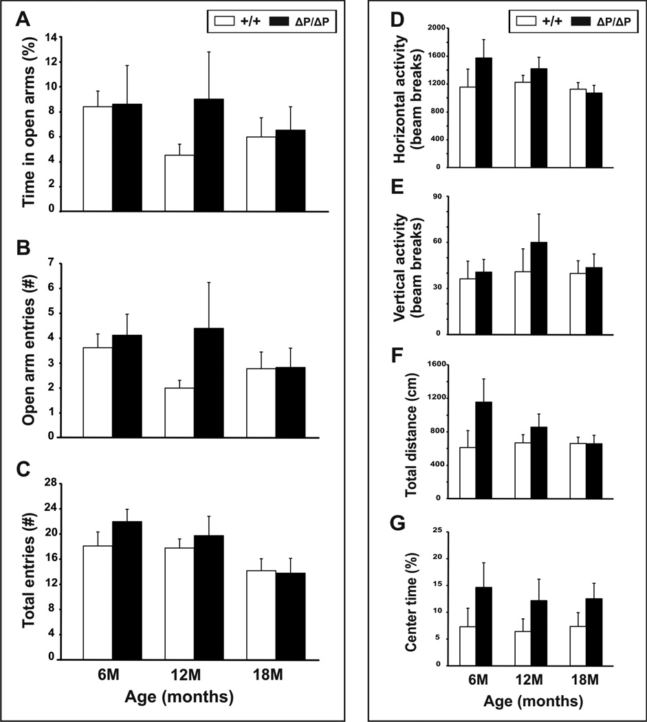
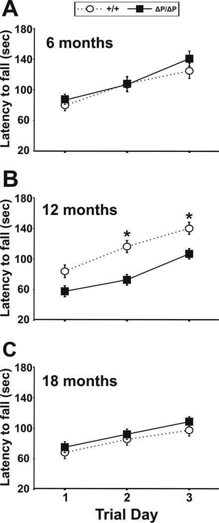
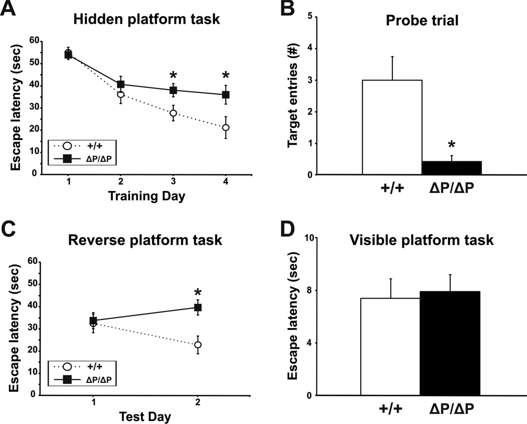
The N-terminus of Huntingtin, the protein encoded by the Huntington's disease gene, contains a stretch of polyglutamine residues that is expanded in Huntington's disease. The polyglutamine stretch is flanked by two conserved protein domains in vertebrates: an N1-17 domain, and a proline-rich region (PRR). The PRR can modulate the structure of the adjacent polyglutamine stretch, and is a binding site for several interacting proteins. To determine the role of the PRR in Huntingtin function, we have generated a knock-in allele of the mouse Huntington's disease gene homolog that expresses full-length normal huntingtin lacking the PRR. Mice that are homozygous for the huntingtin PRR deletion are born at the normal Mendelian frequency, suggesting that the PRR is not required for essential huntingtin functions during embryonic development. Moreover, adult homozygous mutants did not exhibit any significant differences from wild-type controls in general motor function and motor learning. However, 18 month-old male, but not female, homozygous PRR deletion mutants exhibited deficits in the Morris water task, suggesting that age-dependent spatial learning and memory may be affected in a sex-specific fashion by the huntingtin PRR deletion.


Conflict of interest statement

Conflict of interest statement

The authors have no conflicts of interest arising from the publication of this paper.

Figures

Figure 1
Figure 1. Generation of the HdhΔPRR allele
(A) Schematic of the wild-type Hdh allele surrounding the first exon is shown (Hdh) along with the gene targeting construct (T) lacking the PRR, and the targeted locus following recombination (HdhΔP). The “~” in the targeting vector represents plasmid sequence, and the “II” indicates the restriction site used to linearize the targeting vector prior to ES cell electroporation. The location of the neomycin phosphotransferase cassette (pgkneo, box) flanked by loxP sites (black arrowheads) that was used for positive selection of the transfected ES cells is shown. The transcriptional orientations of the Hdh and pgkneo genes are indicated with arrows. The small gray arrows indicate the location of the forward and reverse oligonucleotide primers used for PCR genotyping. The sizes of the wild-type and targeted NcoI-digested genomic DNA fragments recognized by the 3´flanking probe (small black rectangle) are shown above (Hdh) and below (HdhΔP), respectively. Restriction enzyme sites are NotI (Not), NcoI (N), HindIII (H), XmnI (X) and KpnI (K). The schematic is not drawn to scale. (B) Exon-1 of the wild-type (Hdh) mouse htt gene was modified by gene targeting to generate a deletion of the sequence encoding the mouse PRR (HdhΔP). The XmnI and KpnI restriction sites used for the modifications are shown. (C) DNA from tail biopsies was used to genotype progeny by PCR with primers that were designed to discriminate between the wild-type (Hdh) and modified (HdhΔP) alleles: Lane 1- Hdh+/+; Lane 2 - HdhΔPRRPRR; Lane 3 - HdhΔPRR/+; M – 100bp DNA molecular weight marker. (D) Nucleotide and encoded amino acid sequence of the Hdh+ (top) and HdhΔPRR (bottom) exon-1 XmnI–KpnI restriction fragment. Intron-1 sequence is presented in lower case, and the first and last codons of the PRR are indicated in larger font
Figure 2
Figure 2. Deletion of the PRR does not affect htt expression levels
(A) Densitometry analysis of wild-type and ΔPRR-htt levels calculated from western blots of whole brain protein extracts obtained from wild-type (+/+) and HdhΔPRRPRR (ΔP/ΔP) mice. (B) Whole brain protein extracts (100µg) from HdhΔPRRPRR (ΔP/ΔP), HdhΔPRR/+ (ΔP/+), and Hdh+/+ (+/+) mice were fractionated on 5% SDS-PAGE, and analyzed by western blotting using an antibody that recognizes both wild-type and ΔPRR-htt (MAB2166). The position of a 250 kD protein standard is indicated on the left. (C) Immunohistochemical analyses of coronal brain sections obtained from 20 month-old wild-type (+/+) and HdhΔPRRPRR (ΔP/ΔP) mice using an anti-htt antibody (BML-PW0595, epitope: htt N2-17) recognizing both wild-type and ΔPRR-htt. Nuclei were stained with To-Pro-3 iodide. Scale bars = 100µm.
Figure 3
Figure 3. Htt subcellular localization and fractionation are not affected by the PRR deletion
(A) Immunocytochemical analyses of PMEFs obtained from wild-type (+/+) and HdhΔPRRPRR (ΔP/ΔP) embryos using an anti-htt antibody (BML-PW0595), as well as Mitotracker Red CMXRos, a red-fluorescent dye that stains mitochondria. Nuclei were stained with To-Pro-3 iodide. Scale bars = 50µm. (B) Nuclear (Nuc) and cytoplasmic (Cyto) protein fractions (40µg) isolated from HdhΔPRR/+ (ΔP/+) P3 PMEFs were fractionated on 9% SDS-PAGE, and analyzed by western blotting using antibodies recognizing htt (MAB2166), PSD95 and tubulin (proteins preferentially enriched in the cytoplasmic fraction), and CREB (a protein that preferentially associates with the nuclear fraction). (C) Whole brain protein extracts from 5 month-old wild-type, HdhΔPRR/+ and HdhΔPRRPRR mice were fractionated on 5% SDS-PAGE, and analyzed by western blotting using an anti-htt phospho-S13 antibody (S13-P), and MAB2166. A non-specific protein recognized by the phospho-S13 antibody in both wild-type and HdhΔPRRPRR protein samples is indicated with an “*”. The sizes (in kD) of protein standards are indicated on the left.
Figure 4
Figure 4. Deletion of the htt PRR does not affect anxiety and overall activity levels in HdhΔPRRPRR mice
(A–C) The elevated plus maze was used to evaluate anxiety-like traits. Time spent in the open arms, number of open arm entries, and total number of entries were recorded for male mice at 6, 12, and 18 months of age. (D–G) General activity levels were collected using an automated activity cage. Horizontal activity, vertical activity, total distance, and percent time spent in the center were measured in male mice at 6, 12, and 18 months of age. All data are expressed as mean ± SEM.
Figure 5
Figure 5. Rotarod testing of motor coordination and balance in HdhΔPRRPRR and control mice
(A–C) Motor coordination and balance, as well as motor learning were tested on an accelerating rotarod. Male mice were tested at 6, 12 and 18 months of age. All data are expressed as mean ± SEM.
Figure 6
Figure 6. Spatial learning and memory in the Morris water maze task is affected in older male HdhΔPRRPRR mice
Escape latencies for the hidden platform (A), reversal (C) and visible platform tasks (D), and number of target entries during the probe trial (B) were recorded for male mice at 18 months of age. All data are expressed as mean ± SEM.

Similar articles

Cited by

References

    1. Pecheux C, Gall AL, Kaplan JC, Dode C. Sequence analysis of the CAG triplet repeats region in the Huntington disease gene (IT15) in several mammalian species. Ann Genet. 1996;39(2):81–86. - PubMed
    1. Karlovich CA, John RM, Ramirez L, Stainier DY, Myers RM. Characterization of the Huntington's disease (HD) gene homologue in the zebrafish Danio rerio. Gene. 1998 Sep 14;217(1–2):117–125. - PubMed
    1. Baxendale S, Abdulla S, Elgar G, Buck D, Berks M, Micklem G, Durbin R, Bates G, Brenner S, Beck S. Comparative sequence analysis of the human and pufferfish Huntington's disease genes. Nat Genet. 1995 May;10(1):67–76. - PubMed
    1. Li Z, Karlovich CA, Fish MP, Scott MP, Myers RM. A putative Drosophila homolog of the Huntington's disease gene. Hum Mol Genet. 1999 Sep;8(9):1807–1815. - PubMed
    1. Gissi C, Pesole G, Cattaneo E, Tartari M. Huntingtin gene evolution in Chordata and its peculiar features in the ascidian Ciona genus. BMC Genomics. 2006;7:288. - PMC - PubMed

Publication types

MeSH terms

LinkOut - more resources