. 2012;7(9):e44038.
doi: 10.1371/journal.pone.0044038. Epub 2012 Sep 5.

Depletion of the C. elegans NAC engages the unfolded protein response, resulting in increased chaperone expression and apoptosis

Affiliations

Depletion of the C. elegans NAC engages the unfolded protein response, resulting in increased chaperone expression and apoptosis

Paul T Arsenovic et al. PLoS One. 2012.

Abstract

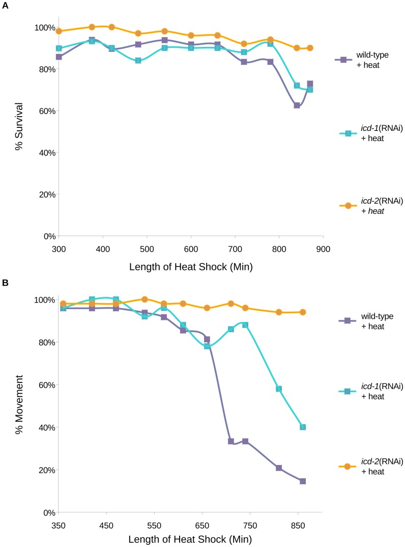
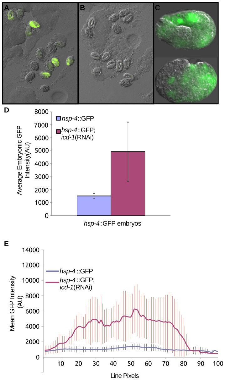
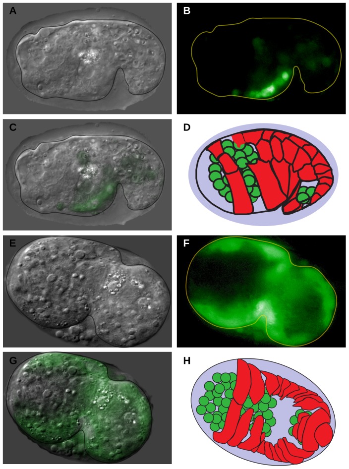
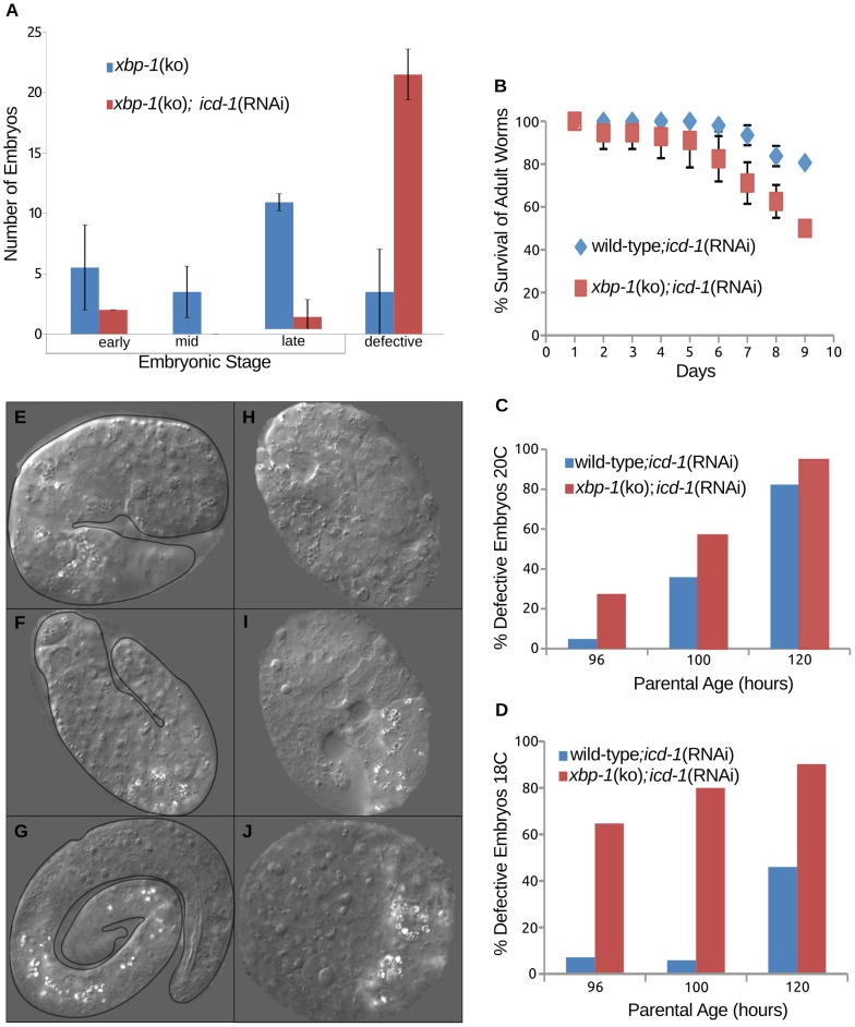
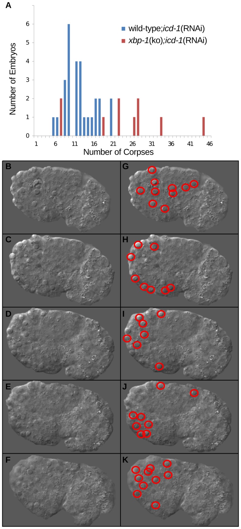
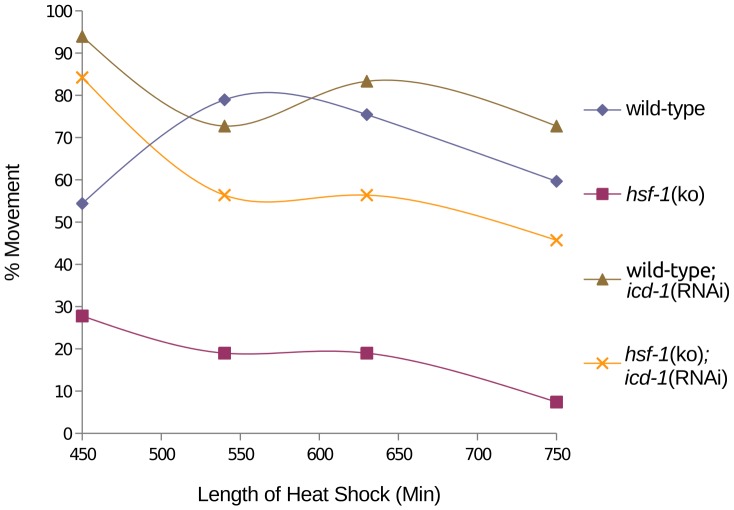
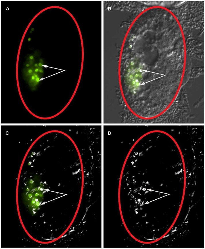
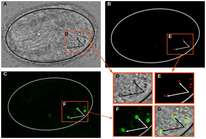
The nascent polypeptide-associated complex (NAC) is a highly conserved heterodimer important for metazoan development, but its molecular function is not well understood. Recent evidence suggests the NAC is a component of the cytosolic chaperone network that interacts with ribosomal complexes and their emerging nascent peptides, such that the loss of the NAC in chaperone-depleted cells results in an increase in misfolded protein stress. We tested whether the NAC functions similarly in Caeonorhabditis (C.) elegans and found that its homologous NAC subunits, i.e. ICD-1 and -2, have chaperone-like characteristics. Loss of the NAC appears to induce misfolded protein stress in the ER triggering the unfolded protein response (UPR). Depletion of the NAC altered the response to heat stress, and led to an up-regulation of hsp-4, a homologue of the human chaperone and ER stress sensor GRP78/BiP. Worms lacking both ICD-1 and the UPR transcription factor XBP-1 generated a higher proportion of defective embryos, showed increased embryonic apoptosis and had a diminished survival rate relative to ICD-1-depleted animals with an intact UPR. Up-regulation of hsp-4 in NAC-depleted animals was specific to certain regions of the embryo; in embryos lacking ICD-1, the posterior region of the embryo showed strong up-regulation of hsp-4, while the anterior region did not. Furthermore, loss of ICD-1 produced prominent lysosomes in the gut region of adults and embryos putatively containing lipofuscins, lipid/protein aggregates associated with cellular aging. These results are the first set of evidence consistent with a role for C. elegans NAC in protein folding and localization during translation. Further, these findings confirm C. elegans as a valuable model for studying organismal and cell-type specific responses to misfolded protein stress.


Conflict of interest statement

Competing Interests: Tim A. Bloss is an author on patent# 20060199781 “Assays based on BTF3 activity”.

Figures

Figure 1
Figure 1. Mortality and movement of ICD-1 or ICD-2-depleted animals during heat stress.
Developmentally synchronized wild-type (N2) C. elegans larvae were fed icd-1 or icd-2(RNAi)-specific bacteria for 30 hours and exposed to continuous heat stress at 36°C for 850 minutes. A) Survival of animals was measured by periodic assessment of pharyngeal pumping. B) Movement was measured by periodic assessment of escape reaction to physical prodding. N = 50 for each of the three populations tested per experiment. Data presented in A) and B) represent the results of two independent trials for each experiment. The three experimental populations were also exposed to no heat (20°C) as a measure of basal rates of mortality and effects on movement, and showed no inherent lethality or immobility (N = 50 per population) (data not shown).
Figure 2
Figure 2. Embryonic expression of hsp-4 in ICD-1-depleted animals.
Animals containing an hsp-4::GFP expression vector were fed (A) icd-1(RNAi)-specific bacteria or (B) OP50 (E. coli) bacteria expressing no double stranded RNA for 36 hours and their progeny embryos were randomly assessed for expression of GFP. C) Two examples of embryos selected from the icd-1(RNAi) population. Both embryos were viable and morphologically wild-type. D) Average GFP signal of hsp-4::GFP-containing embryos treated with icd-1(RNAi) compared with GFP signal generated in control hsp-4::GFP-containing embryos. Populations of embryos were chosen at random, and embryos within a given field of observation were assessed and averaged regardless of GFP status. Up-regulation of hsp-4::GFP was observed in three independent experiments, and up-regulation was quantified in two of these experiments. Error bars represent the standard deviation of the mean GFP intensity of the embryos assessed in these two experiments (two-tailed t-test, P-value = 0.01). E) Averaged line scans measuring pixel intensity across all assessed icd-1(RNAi) embryos compared with control embryos; vertical bars represent the standard deviation of line pixels.
Figure 3
Figure 3. Localization of hsp-4 expression in embryos depleted of ICD-1.
Animals containing an hsp-4::GFP expression vector were treated with icd-1(RNAi) for 48–56 hours, and their embryos were assessed for GFP expression. Localization of hsp-4::GFP signal was determined in three independent experiments, totaling 30 GFP-positive embryos assessed. All embryos showed similar localization patterns. In this time period of the RNAi treatment, a majority of embryos are morphologically defective with high levels of cell death. A–C) DIC, GFP and merged images, respectively, of a representative icd-1(RNAi) embryo ∼470 minutes post fertilization. D) Diagram of embryo 470 minutes post fertilization. E–G) DIC, GFP and merged images, respectively, of a representative icd-1(RNAi) embryo ∼360 minutes post-fertilization. H) Diagram of embryo 360 minutes post fertilization. In both D) and H) green cells represent location of neurons, red cells represent location of developing epithelial layer. Diagrams in D) and H) adapted from Chin-sang and Chisholm, 2000 .
Figure 4
Figure 4. Increased sensitivity of xbp-1(ko) animals to depletion of ICD-1.
[hsp-4::GFP] xbp-1(ko) worms were treated with icd-1(RNAi) and characterized for effects on mortality and embryonic development. A) Profile of embryonic development in [hsp-4::GFP] xbp-1(ko) animals untreated or treated with icd-1(RNAi). Embryos were scored as either phenotypically wild-type or defective. The stage of wild-type embryos was determined primarily by the extent of the vermiform structure, e.g. early embryos showed no tube-like formation, while late embryos contained fully formed worms. Defective embryos were morphologically abnormal with high levels of cell death. Error bars represent one standard deviation from the mean frequency of embryos per developmental stage over two independent experiments. B) Survival curve of [hsp-4::GFP] xbp-1(ko) and wild-type animals during icd-1(RNAi) treatment over a ten day period; survival was determined by the presence of pharyngeal pumping. Error bars represent the standard deviation from the mean of two independent experiments. C,D) Percentage of embryos characterized as defective in wild-type and [hsp-4::GFP] xbp-1(ko) animals treated with icd-1(RNAi) for 48–56 hours grown at 20°C (C) or 18°C (D) over two independent experiments. E–G) Representative defective embryos from wild-type adults treated with icd-1(RNAi) for 48 hours. All three embryos displayed moderate morphological defects with increased levels of cell death, but also display vermiform structure to some extent. H–J) Representative defective embryos from [hsp-4::GFP] xbp-1(ko) embryos treated with icd- 1(RNAi) for 48 hours. All three embryos showed severe morphological defects and significant cell death, with no detectable tube-like formations.
Figure 5
Figure 5. Embryonic apoptosis in xbp-1(ko) animals depleted of ICD-1.
[hsp-4::GFP] xbp-1(ko) animals were treated with icd-1(RNAi), and their comma-stage embryos were scored for apoptotic cell corpses. Developmental apoptosis occurs normally throughout C. elegans embryogenesis, and the comma-stage of development generates a high number of apoptotic cell corpses relative to other stages. A) The number of comma-stage embryos containing a specific number of cell corpses in wild-type (n = 28) and [hsp-4::GFP] xbp-1(ko) (n = 11) embryos treated with icd-1(RNAi). B–F) Multiple focal planes of a comma-stage embryo produced by an [hsp-4::GFP] xbp-1(ko) worm treated with icd-1(RNAi). Cell corpses are distinguishable from surrounding cells by their raised-up, button-like structures. G–K) Duplicate images of A–E highlighting each of the 43 cell corpses observed in this embryo.
Figure 6
Figure 6. Effects of ICD-1 depletion on movement of hsf-1(ko) animals exposed to heat stress.
hsf-1(ko) animals were fed icd-1(RNAi)-expressing bacteria at time 0 and exposed to heat stress at 36°C for 750 minutes. Movement of worms was first measured at 450 minutes of heat stress by escape response to prodding, and compared to wild-type;icd-1(RNAi) animals under heat stress (n = 400 for each population). All experimental populations showed 100% movement at the beginning of the time course (data not shown). As a control, both strains of worms were assessed for movement at 20°C (n = 400) (data not shown).
Figure 7
Figure 7. Generation of GLO-1-positive lysosomal structures in icd-1(RNAi) embryos.
Animals containing a glo-1::GFP expression vector were treated with icd-1(RNAi) for 48–56 hours and the resulting embryos were scored for the presence of large lysosomal structures in their gut cells. GLO-1 is a marker for C. elegans lysosomes that are also distinguishable by bio-fluorescence under polarized light. A majority of the embryos (n = 20) displayed glo-1::GFP signal coincident with lysosomal structures. A–D) A representative [glo-1::GFP] icd-1(RNAi) embryo visualized by A) fluorescent light; B) fluorescent light and DIC; C) fluorescent light and polarized DIC and D) polarized DIC. White arrows indicate lysosomes visualized by DIC with overlapping glo-1::GFP signal, red ovals outline the shell of the embryo.
Figure 8
Figure 8. Identification of lipofuscin granules in icd-1(RNAi) embryos.
Wild-type embryos were treated with icd-1(RNAi) and scored for the presence of lipofuscin granules using two-photon microscopy. A–C) a representative icd-1(RNAi) embryo visualized by A) transmission; B) thresholded transmission and; C) two-photon auto-fluorescence. Transmission microscopy was used to visualize large lysosomal structures in the gut cells, thresholded transmission used to visualize saturated pixels that correlated with lysosomal structures and two-photon excitation used to visualize specifically lipofuscin granules using a 500 nm bandwidth filter. D–F) magnified regions of A–C respectively, as demarcated by a red border. G) An overlay of images D–F. Arrows point to auto-flourescent lipofuscin granules.

Similar articles

Cited by

References

    1. Hartl FU, Bracher A, Hayer-Hartl M (2011) Molecular chaperones in protein folding and proteostasis. Nature 475: 324–332. - PubMed
    1. Voisine C, Pedersen JS, Morimoto RI (2010) Chaperone networks: tipping the balance in protein folding diseases. Neurobiology of Disease 40: 12–20. - PMC - PubMed
    1. Waldmeier PC (2003) Prospects for antiapoptotic drug therapy of neurodegenerative diseases. Prog Neuropsychopharmacol Biol Psychiatry 27: 303–321. - PubMed
    1. Hetz C (2012) The unfolded protein response: controlling cell fate decisions under ER stress and beyond. Nat Rev Mol Cell Biol 13: 89–102. - PubMed
    1. Vabulas RM, Raychaudhuri S, Hayer-Hartl M, Hartl FU (2010) Protein folding in the cytoplasm and the heat shock response. Cold Spring Harb Perspect Biol 2: 1–18. - PMC - PubMed

Publication types

Substances