

Computational studies on the substrate interactions of influenza A virus PB2 subunit
- PMID: 22957044
- PMCID: PMC3434214
- DOI: 10.1371/journal.pone.0044079
Computational studies on the substrate interactions of influenza A virus PB2 subunit
Abstract
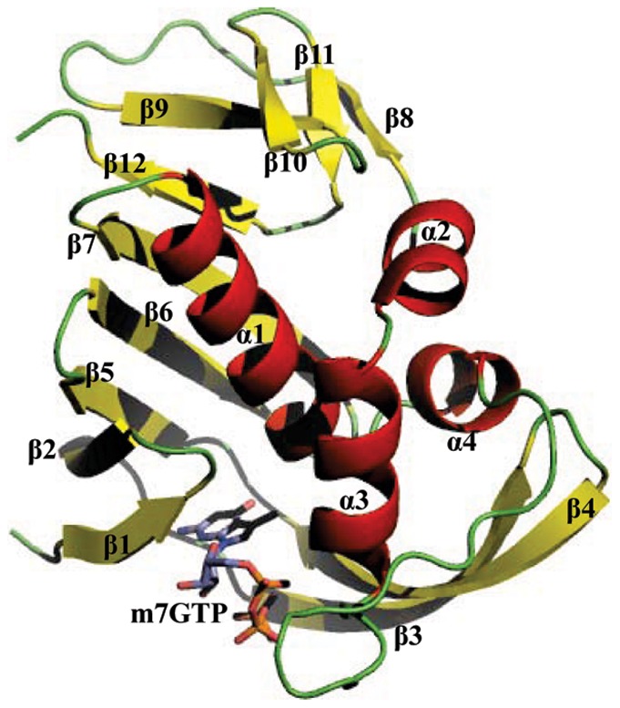
Influenza virus, which spreads around the world in seasonal epidemics and leads to large numbers of deaths every year, has several ribonucleoproteins in the central core of the viral particle. These viral ribonucleoproteins can specifically bind the conserved 3' and 5' caps of the viral RNAs with responsibility for replication and transcription of the viral RNA in the nucleus of infected cells. A fundamental question of most importance is that how the cap-binding proteins in the influenza virus discriminates between capped RNAs and non-capped ones. To get an answer, we performed molecular dynamics simulations and free energy calculations on the influenza A virus PB2 subunit, an important component of the RNP complexes, with a cap analog m7GTP. Our calculations showed that some key residues in the active site, such as Arg355, His357, Glu361 as well as Gln406, could offer significant hydrogen bonding and hydrophobic interactions with the guanine ring of the cap analog m7GTP to form an aromatic sandwich mechanism for the cap recognition and positioning in the active site. Subsequently, we applied this idea to a virtual screening procedure and identified 5 potential candidates that might be inhibitors against the PB2 subunit. Interestingly, 2 candidates Cpd1 and Cpd2 have been already reported to have inhibitory activities to the influenza virus cap-binding proteins. Further calculation also showed that they had comparatively higher binding affinities to the PB2 subunit than that of m7GTP. We believed that our findings could give an atomic insight into the deeper understanding of the cap recognition and binding mechanism, providing useful information for searching or designing novel drugs against influenza viruses.
Conflict of interest statement
Figures


Similar articles
-
A molecular modelling approach to understand the effect of co-evolutionary mutations (V344M, I354L) identified in the PB2 subunit of influenza A 2009 pandemic H1N1 virus on m7GTP ligand binding.J Gen Virol. 2016 Aug;97(8):1785-1796. doi: 10.1099/jgv.0.000500. Epub 2016 May 6. J Gen Virol. 2016. PMID: 27154164
-
The structural basis for cap binding by influenza virus polymerase subunit PB2.Nat Struct Mol Biol. 2008 May;15(5):500-6. doi: 10.1038/nsmb.1421. Epub 2008 May 4. Nat Struct Mol Biol. 2008. PMID: 18454157
-
Structural and functional characterization of K339T substitution identified in the PB2 subunit cap-binding pocket of influenza A virus.J Biol Chem. 2013 Apr 19;288(16):11013-23. doi: 10.1074/jbc.M112.392878. Epub 2013 Feb 22. J Biol Chem. 2013. PMID: 23436652 Free PMC article.
-
[Structural and Biochemical Analyses on the RNA-dependent RNA Polymerase of Influenza Virus for Development of Novel Anti-influenza Agents].Yakugaku Zasshi. 2017;137(2):205-214. doi: 10.1248/yakushi.16-00195. Yakugaku Zasshi. 2017. PMID: 28154333 Review. Japanese.
-
The Influenza Virus Polymerase Complex: An Update on Its Structure, Functions, and Significance for Antiviral Drug Design.Med Res Rev. 2016 Nov;36(6):1127-1173. doi: 10.1002/med.21401. Epub 2016 Aug 29. Med Res Rev. 2016. PMID: 27569399 Free PMC article. Review.
Cited by
-
Identification of a Novel Anti-cancer Protein, FIP-bbo, from Botryobasidium botryosum and Protein Structure Analysis using Molecular Dynamic Simulation.Sci Rep. 2019 Apr 9;9(1):5818. doi: 10.1038/s41598-019-42104-1. Sci Rep. 2019. PMID: 30967569 Free PMC article.
-
Scaffold-based pan-agonist design for the PPARα, PPARβ and PPARγ receptors.PLoS One. 2012;7(10):e48453. doi: 10.1371/journal.pone.0048453. Epub 2012 Oct 31. PLoS One. 2012. PMID: 23119024 Free PMC article.
-
Identification of novel multitargeted PPARα/γ/δ pan agonists by core hopping of rosiglitazone.Drug Des Devel Ther. 2014 Nov 7;8:2255-62. doi: 10.2147/DDDT.S70383. eCollection 2014. Drug Des Devel Ther. 2014. PMID: 25422585 Free PMC article.
References
-
- Lee RV (2007) Transmission of influenza in human beings. Lancet Infect Dis 7: 759–760. - PubMed
-
- Gambotto A, Barratt-Boyes SM, de Jong MD, Neumann G, Kawaoka Y (2008) Human infection with highly pathogenic H5N1 influenza virus. Lancet 371: 1464–1475. - PubMed
-
- Olsen B, Munster VJ, Wallensten A, Waldenström J, Osterhaus AD, et al. (2006) Global patterns of influenza A virus in wild birds. Science 312: 384–388. - PubMed
Publication types
MeSH terms
Substances
LinkOut - more resources
Full Text Sources
Miscellaneous
