. 2012;7(9):e44476.
doi: 10.1371/journal.pone.0044476. Epub 2012 Sep 5.

Cilostazol prevents endothelin-induced smooth muscle constriction and proliferation

Affiliations

Cilostazol prevents endothelin-induced smooth muscle constriction and proliferation

Yoshifumi Kawanabe et al. PLoS One. 2012.

Abstract

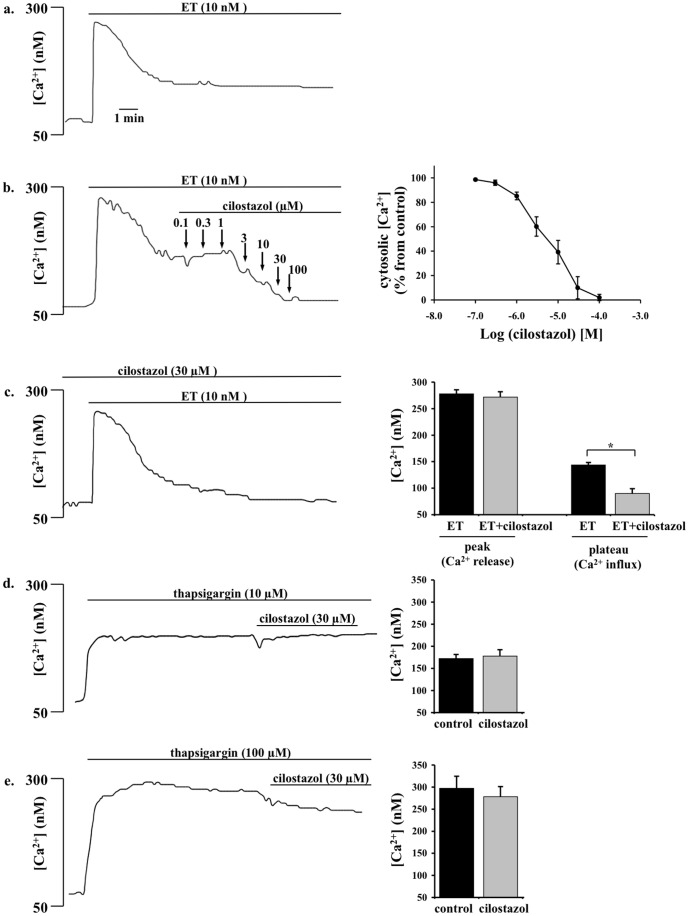
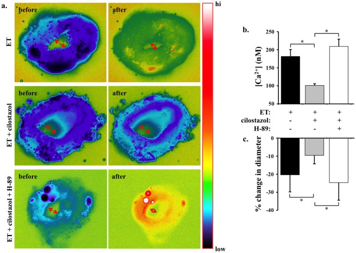
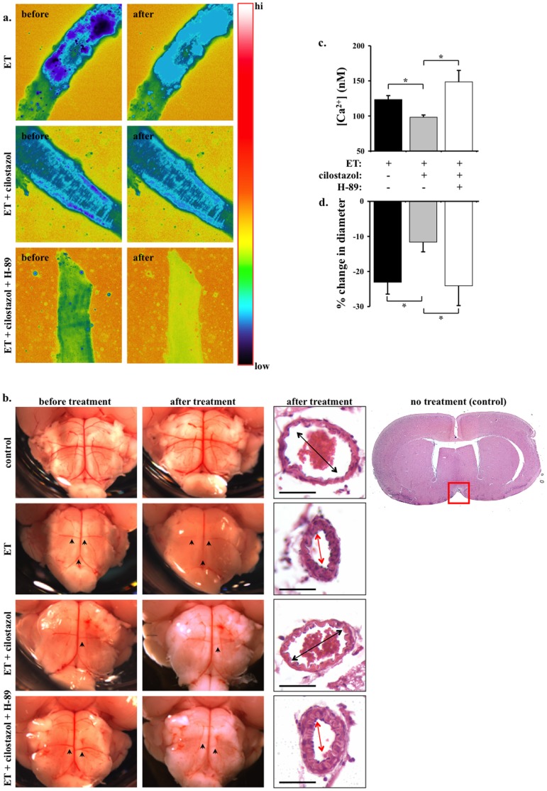
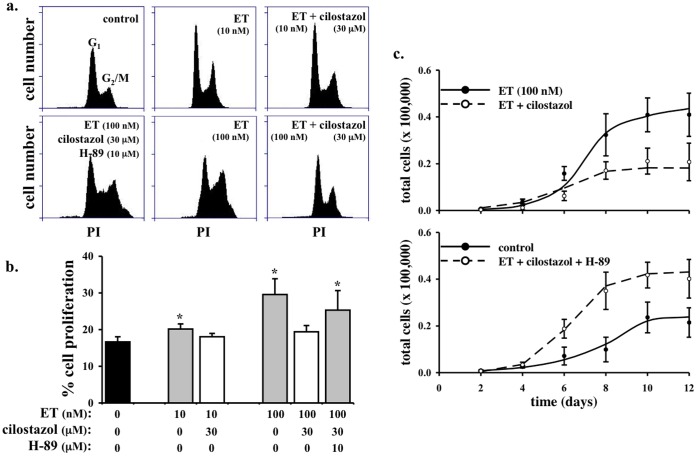
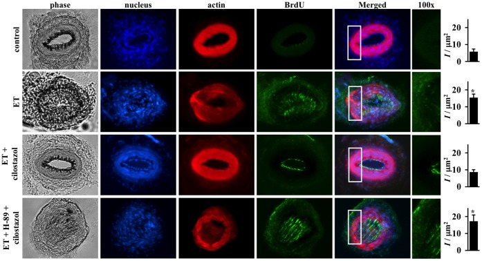
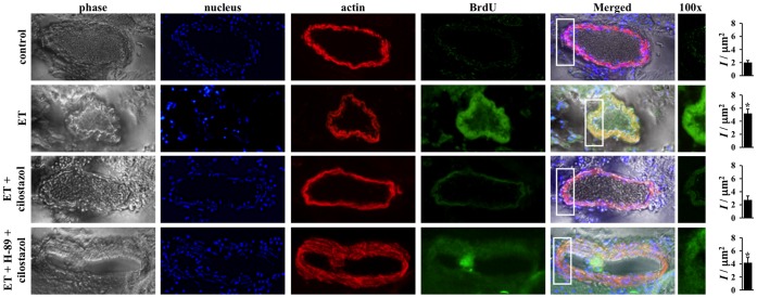
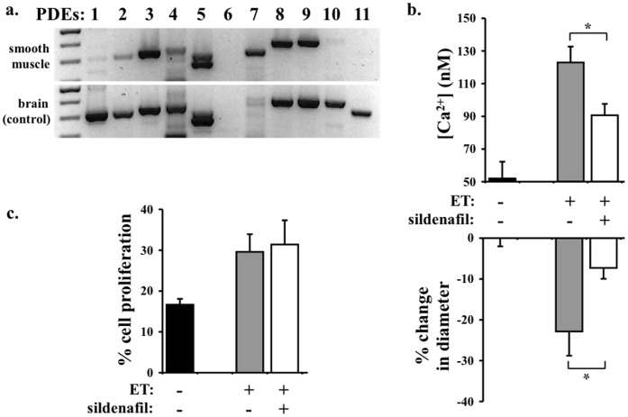
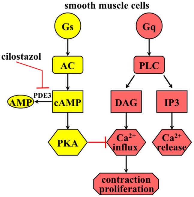
Cilostazol is a phosphodiesterase inhibitor that has been shown to inhibit platelet activation. Endothelin is known to be the most potent endogenous growth promoting and vasoactive peptide. In patients and animal models with stroke, the level of circulating endothelin increases and complicates the recovery progress contributed by vascular constriction (an immediate pathology) and vascular proliferation (a long-term pathology). However, the effects of cilostazol on endothelin have not been explored. To demonstrate the dual-antagonizing effects of cilostazol on vasoconstriction and cell proliferation induced by endothelin, we used primary culture of mouse vascular smooth muscle cells in vitro, mouse femoral artery ex vivo, and intracranial basilar artery ex vivo. We show that the dual-inhibition effects of cilostazol are mediated by blocking endothelin-induced extracellular calcium influx. Although cilostazol does not inhibit endothelin-induced intraorganellar calcium release, blockade of extracellular calcium influx is sufficient to blunt endothelin-induced vasoconstriction. We also show that cilostazol inhibits endothelin-induced cellular proliferation by blocking extracellular calcium influx. Inhibition of cAMP-dependent protein kinase (PKA) can block anti-proliferation activity of cilostazol, confirming the downstream role of PKA in cellular proliferation. To further demonstrate the selectivity of the dual-antagonizing effects of cilostazol, we used a different phosphodiesterase inhibitor. Interestingly, sildenafil inhibits endothelin-induced vasoconstriction but not cellular proliferation in smooth muscle cells. For the first time, we show selective dual-antagonizing effects of cilostazol on endothelin. We propose that cilostazol is an excellent candidate to treat endothelin-associated diseases, such as stroke.


Conflict of interest statement

Competing Interests: The authors have declared that no competing interests exist.

Figures

Figure 1
Figure 1. Cilostazol inhibits endothelin-induced extracellular calcium influx in a dose-dependent manner. a.
Endothelin (ET) increases intracellular free calcium in primary culture of mouse femoral smooth muscle cells in a biphasic manner. ET induces intraorganellar calcium release (initial peak) and extracellular calcium influx (plateau phase). b. After the intracellular calcium increase is stabilized, dose-dependent effects of cilostazol is examined by applying various concentrations of cilostazol to the cells. Percent changes in cytosolic calcium is presented on the right panel (N = 4). c. Pretreatment with sub-optimal cilostazol concentration blocks endothelin-induced calcium influx but not calcium release. Effects of endothelin-induced calcium influx and release are quantified in the presence or absence of cilostazol pretreatment (N = 5). d. 10 µM of thapsigargin promotes release of intraorganellar calcium release. After the intracellular calcium increase is stabilized, sub-optimal concentration of cilostazol does not show a substantial effect on calcium release (N = 4). e. Cilostazol has no significant role in 100 µM thapsigargin-induced calcium release (N = 5).
Figure 2
Figure 2. Cilostazol inhibits endothelin-induced cytosolic calcium increase and vasoconstiction in mouse femoral arteries. a.
Cytosolic calcium (pseudocolor) and inner diameter (arrows) of femoral arteries are studied ex vivo. Segment of artery is imaged and recorded before and after endothelin (ET) treatment in the presence or absence of cilostazol and/or H-89. Artery segment is pseudocolored to denote changes in cytosolic free calcium level from low to high (hi) as indicated in the color bar. b. Cilostazol significantly decreases endothelin-induced cytosolic calcium increase. c. Changes in diameter denoting vascular constriction are expressed in percentage relative to baseline diameter prior to endothelin treatment. N = 5.
Figure 3
Figure 3. Cilostazol inhibits endothelin-induced calcium increase and vasoconstriction in mouse intracranial arteries. a.
Cytosolic calcium of basilar arteries are studied ex vivo. Segment of artery is imaged and recorded before and after endothelin (ET) treatment in the presence or absence of cilostazol. Artery segment is pseudocolored to denote changes in cytosolic free calcium level from low to high (hi) as indicated in the color bar. b. Micrographs show the pons region of the whole brain before and after endothelin treatment in the presence or absence of cilostazol and/or H-89. The vascular reflection line denoting vascular tone (constriction) of intracranial arteries is adequately visible. Arrowheads point at the anterior inferior cerebellar arteries or basilar arteries to compare vascular tone before and after treatment. Sagittal sections of mouse brain are obtained at the pons, near the interpeduncular fossa boundary. A standard H&E staining is shown in the far right. Red box indicates the area of interest, where basilar artery is located. Arrows denote basilar artery constriction. Bar = 50 µm c. Cilostazol significantly decreases endothelin-induced cytosolic calcium increase. Administration of H-89 blocks the effect of cilostazol d. Changes in diameter denoting vascular constriction are expressed in percentage relative to baseline diameter prior to endothelin treatment. N = 5.
Figure 4
Figure 4. Cilostazol inhibits endothelin-induced cell proliferation in vascular smooth muscle cells. a.
Endothelin (ET) at 10 and 100 nM increases cell proliferation in primary culture of smooth muscle cells as indicated by flow cytometry study. Cell proliferation is studied with nuclear marker, propodium iodide (PI). b. Cilostazol significantly decreases endothelin-induced cellular division. Effect of cilostazol can be blocked by H-89 (N = 3). c. Cell counting experiments were performed in the present and absent of ET, cilostazol and/or H-89. Cilostazol significantly decreases endothelin-induced cell growth. H-89 attenuates the effect of cilostazol in endothelin-induced cell growth (N = 4).
Figure 5
Figure 5. Cilostazol inhibits endothelin-induced cell proliferation in mouse femoral arteries.
Effects of endothelin (ET) in the presence or absence of cilostazol and PKA inhibitor (H-89) on cell proliferation are studied with immunolabeling in femoral arteries ex vivo. Segment of artery is imaged and recorded for phase contrast to indicate the morphology of the artery, dapi to visualize cell nucleus, α-actin to outline contractile smooth muscle cells and BrdU to determine cellular proliferation property of the vascular cells. Endothelin increases cell proliferation as indicated by BrdU staining and cell growth into the lumen of the artery. Cilostazol blocks cell proliferation induced by endothelin, and H-89 inhibits efficacy of cilostazol. White box denotes a larger magnification. Asterisk indicates significant difference in fluorescence intensity per area within the smooth muscle cells, compared to control groups. N = 4.
Figure 6
Figure 6. Cilostazol inhibits endothelin-induced cell proliferation in mouse basilar arteries.
Effects of endothelin (ET) in the presence or absence of cilostazol and PKA inhibitor (H-89) on cell proliferation are studied with immunolabeling in basilar arteries ex vivo. Segment of artery is imaged and recorded for phase contrast to indicate the morphology of the artery, dapi to visualize cell nucleus, α-actin to outline contractile smooth muscle cells and BrdU to determine cellular proliferation property of the vascular cells. Endothelin increases cell proliferation as indicated by BrdU staining. Cilostazol blocks cell proliferation induced by endothelin, and H-89 inhibits efficacy of cilostazol. White box denotes a larger magnification. Asterisk indicates significant difference in fluorescence intensity per area within the smooth muscle cells, compared to control groups. N = 3.
Figure 7
Figure 7. Sildenafil inhibits endothelin-induced vasoconstriction but not cellular proliferation in smooth muscle cells. a.
To identify distribution of phosphodiesterase (PDE) subtypes, semi-quantitative RT-PCR was performed. Mouse brain homogenate was used as an internal control. b. To examine the roles of PDE5 in smooth muscle cells, we applied sildenafil on basilar arteries. Sildenafil significantly decreases endothelin-induced cytosolic calcium increase and vascular constriction. c. Unlike cilostazol, sildenafil does not have any effect on endothelin-induced cellular proliferation of smooth muscle cells. N = 3.
Figure 8
Figure 8. Working model depicts the roles of cilostazol on extracellular calcium influx in smooth muscle cells.
Trimeric G-proteins (Gs or Gq) can activate downstream effectors, such as adenylate cyclase (AC) or phospholipase C (PLC). Endothelin receptor is coupled with Gq and can activate PLC. PLC hydrolyzes PIP2 to IP3 (induces intraorganellar calcium release) and DAG (promotes extracellular calcium influx). Cilostazol inhibits phosphodiesterase 3 (PDE3), thereby preventing degradation of cAMP to AMP. PKA (cAMP dependent protein kinase) can inhibit calcium influx in smooth muscle cells. Thus, cilostazol inhibits endothelin-induced smooth muscle contraction and cell proliferation by inhibiting calcium influx through PKA activation.

References

    1. Al-Qudah ZA, Hassan AE, Qureshi AI (2011) Cilostazol in patients with ischemic stroke. Expert Opin Pharmacother 12: 1305–1315. - PubMed
    1. Sallustio F, Rotondo F, Di Legge S, Stanzione P (2010) Cilostazol in the management of atherosclerosis. Curr Vasc Pharmacol 8: 363–372. - PubMed
    1. Nakamura K, Ikomi F, Ohhashi T (2006) Cilostazol, an inhibitor of type 3 phosphodiesterase, produces endothelium-independent vasodilation in pressurized rabbit cerebral penetrating arterioles. J Vasc Res 43: 86–94. - PubMed
    1. Shiraishi Y, Kanmura Y, Itoh T (1998) Effect of cilostazol, a phosphodiesterase type III inhibitor, on histamine-induced increase in [Ca2+]i and force in middle cerebral artery of the rabbit. Br J Pharmacol 123: 869–878. - PMC - PubMed
    1. Chen WJ, Chen YH, Lin KH, Ting CH, Yeh YH (2011) Cilostazol Promotes Vascular Smooth Muscles Cell Differentiation Through the cAMP Response Element-Binding Protein-Dependent Pathway. Arterioscler Thromb Vasc Biol 31: 2106–2113. - PubMed

MeSH terms