. 2012:2012:624519.
doi: 10.1155/2012/624519. Epub 2012 Apr 12.

Neuropathology of temporal lobe epilepsy

Affiliations

Neuropathology of temporal lobe epilepsy

Fahd Al Sufiani et al. Epilepsy Res Treat. 2012.

Abstract

Pathologic findings in surgical resections from patients with temporal lobe epilepsy include a wide range of diagnostic possibilities that can be categorized into different groups on the basis of etiology. This paper outlines the various pathologic entities described in temporal lobe epilepsy, including some newly recognized epilepsy-associated tumors, and briefly touch on the recent classification of focal cortical dysplasia. This classification takes into account coexistent pathologic lesions in focal cortical dysplasia.


Figures
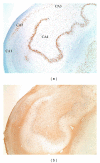
Figure 1
Figure 1
Hippocampal sclerosis. Neuronal loss in areas CA1, CA3, and CA4, with gliosis, (a) NeuN immunoreactivity. (b) glial fibrillary acidic protein immunoreactivity. Original magnification ×20.
Figure 2
Figure 2
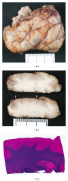
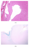
A cortical resection specimen from a child with hemimegalencephaly shows polymicrogyria. (a) external surface. (b) coronal sections, excessive convolution of cortical ribbon with shallow fused sulci (area between arrows). (c) micrograph (luxol fast blue-hematoxylin and eosin stain), several small wisps of subcortical white matter extending up to the cortical ribbon.
Figure 3
Figure 3
Focal cortical dysplasia (FCD). (a) FCD type Ia shows microcolumnar arrangements of cortical neurons with a preservation of cortical layering. NeuN immunoreactivity. Original magnification ×20. (b) FCD type IIa, cortical laminar disorganization and dysmorphic neurons (arrowhead), distributed throughout the entire cortical thickness (sparing the molecular layer) and subjacent white matter. Hematoxylin-eosin stain. Original magnification ×40.
Figure 4
Figure 4
Dysmorphic neurons. (a) hematoxylin-eosin stain. Original magnification ×400. (b) Bielschowsky stain. Original magnification ×400.
Figure 5
Figure 5
Balloon cells. Hematoxylin-eosin stain. Original magnification ×200.
Figure 6
Figure 6
Arteriovenous malformation comprises of mixture of arteries, veins, and abnormal vessels of variable wall thickness and caliber (arterialized veins) with intervening gliotic brain tissue. Extensive perilesional gliosis, calcification, and microhemorrhages, with hemosiderin deposition (not shown) are common features in vascular malformation. Movat stain. Original magnification ×100.
Figure 7
Figure 7
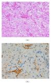
Ganglioglioma. (a) mixed population of neoplastic glial and ganglion (neuronal) cells. Hematoxylin-eosin stain. Original magnification ×200. (b) examples of neoplastic, binucleated neurons immunoreactive for synaptophysin. Original magnification ×400.
Figure 8
Figure 8
Dysembryoplastic  neuroepithelial  tumor. Oligodendroglia-like cells arranged in columns with occasional mature neurons and myxoid background. Hematoxylin-eosin stain. Original magnification ×200.
Figure 9
Figure 9
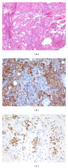
(a) pseudopapillae structures composed of hyalinized blood vessels and surrounded by glioneuronal cells. Papillary glioneuronal tumor may mimic vascular malformation. Hematoxylin-eosin stain. Original magnification ×100. (b) the glial component around blood vessels. Glial fibrillary acidic protein (GFAP). Original magnification ×400. (c) neuronal component in the interpapillary area. NeuN immunostain. Original magnification ×200.
Figure 10
Figure 10
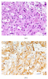
(a) pleomorphic xanthoastrocytoma with large, pleomorphic, and lipidized glial cells. Hematoxylin-eosin stain. (b) glial fibrillary acidic protein immunoreactivity (astrocyte marker). Original magnification ×600.
Figure 11
Figure 11
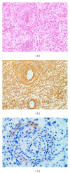
Angiocentric glioma. (a) angiocentric growth pattern of monomorphous bipolar cells. Hematoxylin-eosin stain. (b) corresponding section stained for glial fibrillary acidic protein shows cell processes wrapped around blood vessels. Original magnification ×200. (c) epithelial membrane antigen shows a dot-like immunoreactivity (arrow). Original magnification ×400.
Figure 12
Figure 12
Old trauma. (a) cortical cystic lesion with hemosiderin pigments. Hematoxylin-eosin stain. Original magnification ×40. (b) superficial iron deposit within the cortical tissue. Perl Prussian blue stain. Original magnification ×20.
Figure 13
Figure 13
Neurocysticercosis characterized by the presence of undulating, laminated, and membranous wall of a cysticercus. Hematoxylin-eosin stain. Original magnification ×40.
Figure 14
Figure 14
(a) perivascular lymphohistiocytic infiltrate in meningoencephalitis from a patient with temporal lobe epilepsy. Hematoxylin-eosin stain. (b) diffuse microglial infiltration with the formation of nodules. Immunostaining for CD68. Original magnification ×100.

References

    1. Wiebe S. Epidemiology of temporal lobe epilepsy. Canadian Journal of Neurological Sciences. 2000;27(1):S6–S10. - PubMed
    1. Tassi L, Meroni A, Deleo F, et al. Temporal lobe epilepsy: neuropathological and clinical correlations in 243 surgically treated patients. Epileptic Disorders. 2009;11(4):281–292. - PubMed
    1. Blümcke I, Thom M, Wiestler OD. Ammon’s horn sclerosis: a maldevelopmental disorder associated with temporal lobe epilepsy. Brain Pathology. 2002;12(2):199–211. - PMC - PubMed
    1. Lee MC, Kim GM, Woo YJ, et al. Pathogenic significance of neuronal migration disorders in temporal lobe epilepsy. Human Pathology. 2001;32(6):643–648. - PubMed
    1. Maher J, McLachlan RS. Febrile convulsions: is seizure duration the most important predictor of temporal lobe epilepsy? Brain. 1995;118(6):1521–1528. - PubMed

LinkOut - more resources