. 2012:2012:623190.
doi: 10.1155/2012/623190. Epub 2012 Aug 16.

The application of a three-step serum proteome analysis for the discovery and identification of novel biomarkers of hepatocellular carcinoma

Affiliations

The application of a three-step serum proteome analysis for the discovery and identification of novel biomarkers of hepatocellular carcinoma

Asako Kimura et al. Int J Proteomics. 2012.

Abstract

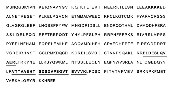
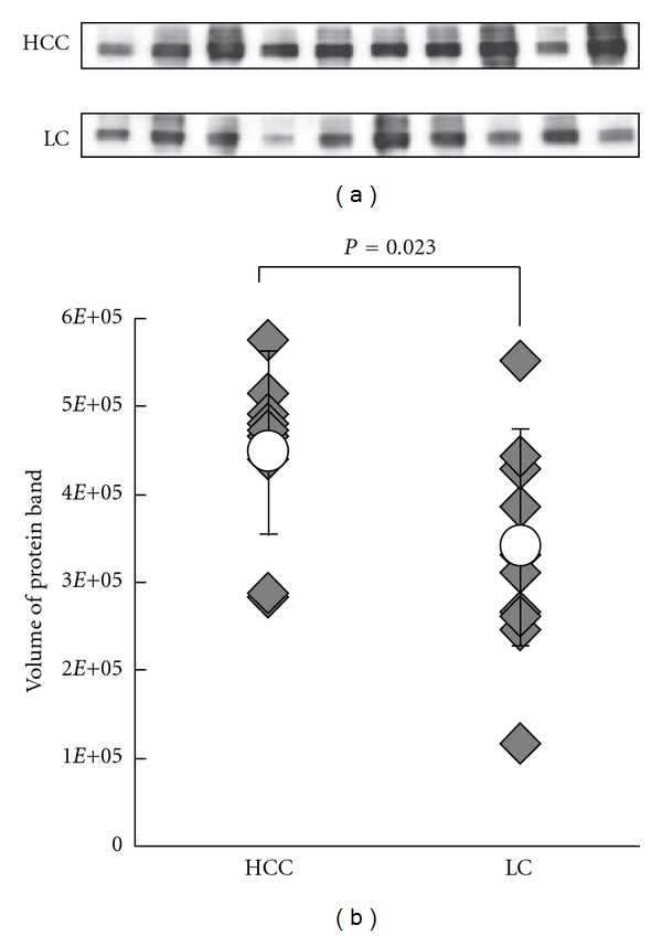
The representative tumor markers for HCC, AFP, and PIVKA-II are not satisfactory in terms of sensitivity and specificity in the early diagnosis of HCC. In search for novel markers for HCC, three-step proteome analyses were carried out in serum samples obtained from 12 patients with HCC and 10 with LC. As a first step, serum samples were subjected to antibody-based immunoaffinity column system that simultaneously removes twelve of abundant serum proteins. The concentrated flow-through was then fractionated using reversed-phase HPLC. Proteins obtained in each fraction were separated by SDS-PAGE. Serum samples obtained from patient with HCC and with LC were analyzed in parallel and their protein expression patterns were compared. A total of 83 protein bands were found to be upregulated in HCC serum. All the protein bands, the intensity of which was different between HCC and LC groups, were identified. Among them, clusterin was most significantly overexpressed (P = 0.023). The overexpression of serum clusterin was confirmed by ELISA using another validation set of HCC samples. Furthermore, serum clusterin was elevated in 40% of HCC cases in which both AFP and PIVKA-II were within their cut-off values. These results suggested that clusterin is a potential novel serum marker for HCC.


Figures

Figure 1
Figure 1
Schematic diagram of the experimental protocol.
Figure 2
Figure 2
Representative chromatogram during removal of highly abundant serum proteins by an immunoaffinity column. 100 μL of serum (diluted fivefold) was injected on the immunoaffinity column and was eluted (0.5 mL/min) as described in the experimental procedures. Flow-through fractions (12.1–20.0 min) were collected, and then a total of 4.0 mL fractions were concentrated to a volume of 80 μL using Vivaspin 2 for reversed-phase HPLC fractionation.
Figure 3
Figure 3
Representative chromatogram during fractionation by RP-HPLC of serum samples in which highly abundant proteins were immunodepleted. Concentrated immunodepleted samples were directly loaded onto the RP column and 40 fractions were collected every 0.5 min from 19.1 to 39.1 min ((a) arrow) as described in the experimental procedures. Among them, a total of 22 fractions were processed for SDS-PAGE analysis (b).
Figure 4
Figure 4
Representative SDS-PAGE pattern of immunodepleted serum sample after RP-HPLC fractionation (fraction number 13). 100 μL of serum samples from seven HCC patients and five LC patients was immunodepleted and injected onto the column. Forty fractions were collected and dried, and among them 22 fractions were separated using 10–20% SDS-PAGE. Each dried fraction was dissolved in 15 μL of sample buffer and loaded onto the gel as described in the experimental procedures. Following electrophoresis, proteins were visualized by silver staining (a). For protein identification, 300 μL of serum samples was prepared again and visualized by CBB staining (b). The intensities of 14 bands were increased in all the seven HCC patients. The representative examples are indicated by arrow heads.
Figure 5
Figure 5
Identification of clusterin by LC-MS/MS. The amino acid sequence of clusterin is shown. Matched peptide sequences are printed in bold and underlined.
Figure 6
Figure 6
Western blot analysis of clusterin in sera from HCC and LC groups. (a) Immunoedpleted sera of the 5 HCC and 5 LC SDS-PAGE cases and additional 5 HCC and 5 LC cases were separated by 10.0–12.0% SDS-PAGE and probed with anticlusterin. The expression levels are relatively higher in the HCC groups than LCs. (b) Differences in expression were analyzed by the Student's t-test. The expression levels of these proteins were upregulated significantly in HCC samples (P = 0.023). Rhombuses represent volumes of individual samples. Line indicates the range with the open circles indicating the mean values.
Figure 7
Figure 7
Concentration of clusterin in 64 patients with HCC, 60 patients with LC and 60 healthy individuals. Clusterin levels, quantified using ELISA, were significantly greater in HCC patients compared with LC, (P < 0.01) and normal subjects (P < 0.001).

References

    1. Ogunbiyi JO. Hepatocellular carcinoma in the developing world. Seminars in Oncology. 2001;28(2):179–187. - PubMed
    1. Lopez LJ, Marrero JA. Hepatocellular carcinoma. Current Opinion in Gastroenterology. 2004;20(3):248–253. - PubMed
    1. Fujiyama S, Tanaka M, Maeda S, Ashihara H, Hirata R, Tomita K. Tumor markers in early diagnosis, follow-up and management of patients with hepatocellular carcinoma. Oncology. 2002;62(1):57–63. - PubMed
    1. Szklaruk J, Silverman PM, Charnsangavej C. Imaging in the diagnosis, staging, treatment, and surveillance of hepatocellular carcinoma. American Journal of Roentgenology. 2003;180(2):441–454. - PubMed
    1. Johnson PJ. The role of serum alpha-fetoprotein estimation in the diagnosis and management of hepatocellular carcinoma. Clinics in Liver Disease. 2001;5(1):145–159. - PubMed