

Acute fibrinous and organizing pneumonia and undifferentiated connective tissue disease: a case report
- PMID: 22957292
- PMCID: PMC3420729
- DOI: 10.1155/2012/549298
Acute fibrinous and organizing pneumonia and undifferentiated connective tissue disease: a case report
Abstract
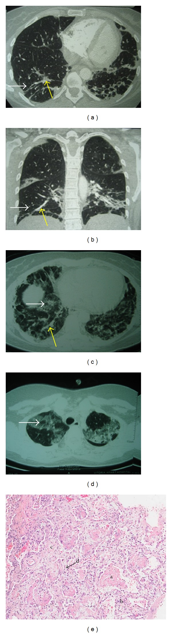
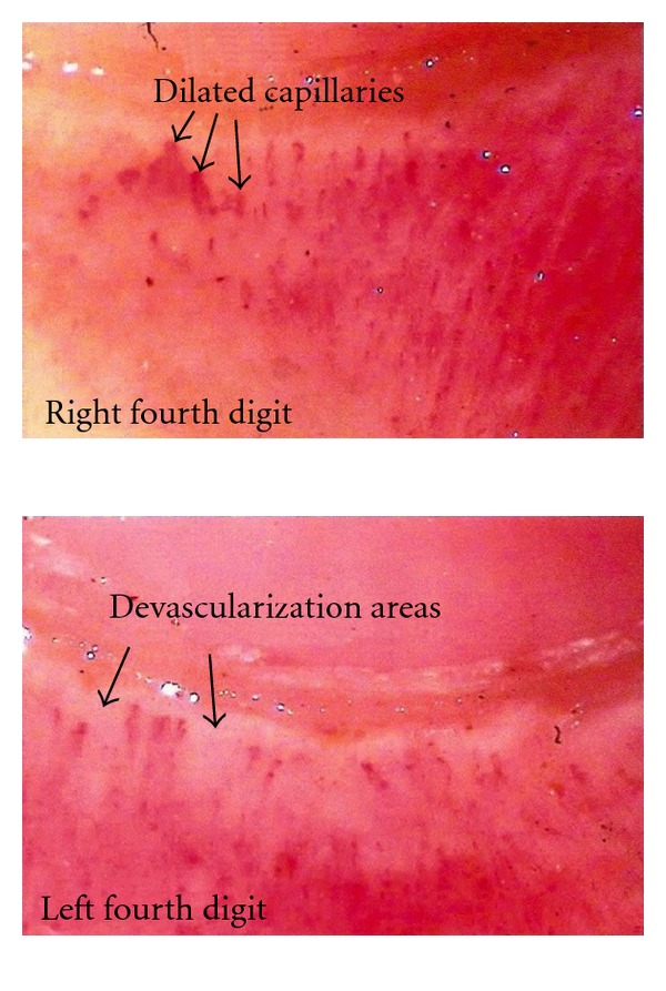
Acute fibrinous and organizing pneumonia (AFOP), recently described, is a histologic pattern characterized by the presence of fibrin "balls" within alveolar spaces. The term undifferentiated connective tissue disease (UCTD) is used to identify autoimmune systemic diseases that do not fulfill the criteria to be classified as a definitive connective tissue disease. The AFOP has never been reported in association with UCTD. The present reported case is a 39-year-old Caucasian, female with dry cough and progressive dyspnea. Eight months later, she was diagnosed with "organizing pneumonia" based on clinical history and radiologic images. She manifested Raynaud's Phenomenon, sicca syndrome, boot and gloves neuropathic pain, and previous hypothyroidism. Antinuclear antibody, rheumatoid factor, and specific autoantibodies were negative. Salivary gland biopsy and electroneuromyiography were normal. The capillaroscopy showed a "scleroderma" pattern with capillary deletion and ectasia. She experienced clinical and radiologic worsening. Despite being submitted to cyclophosphamide pulse, she developed hemorrhage and then died. Thoracotomy pulmonary specimen showed histological pattern of AFOP. This paper shows a rare association of AFOP with UCTD.
Figures
References
-
- Beasley MB, Franks TJ, Galvin JR, Gochuico B, Travis WD. Acute fibrinous and organizing pneumonia: a histologic pattern of lung injury and possible variant of diffuse alveolar damage. Archives of Pathology and Laboratory Medicine. 2002;126(9):1064–1070. - PubMed
-
- Beasley MB. The pathologist’s approach to acute lung injury. Archives of Pathology and Laboratory Medicine. 2010;134(5):719–727. - PubMed
-
- Cordeiro CR. Airway involvement in interstitial lung disease. Current Opinion in Pulmonary Medicine. 2006;12(5):337–341. - PubMed
Publication types
LinkOut - more resources
Full Text Sources
