. 2012 Oct 23;6(10):8824-36.
doi: 10.1021/nn302687n. Epub 2012 Sep 14.

Acute and chronic shear stress differently regulate endothelial internalization of nanocarriers targeted to platelet-endothelial cell adhesion molecule-1

Affiliations

Acute and chronic shear stress differently regulate endothelial internalization of nanocarriers targeted to platelet-endothelial cell adhesion molecule-1

Jingyan Han et al. ACS Nano. .

Abstract

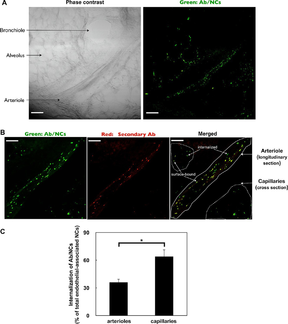
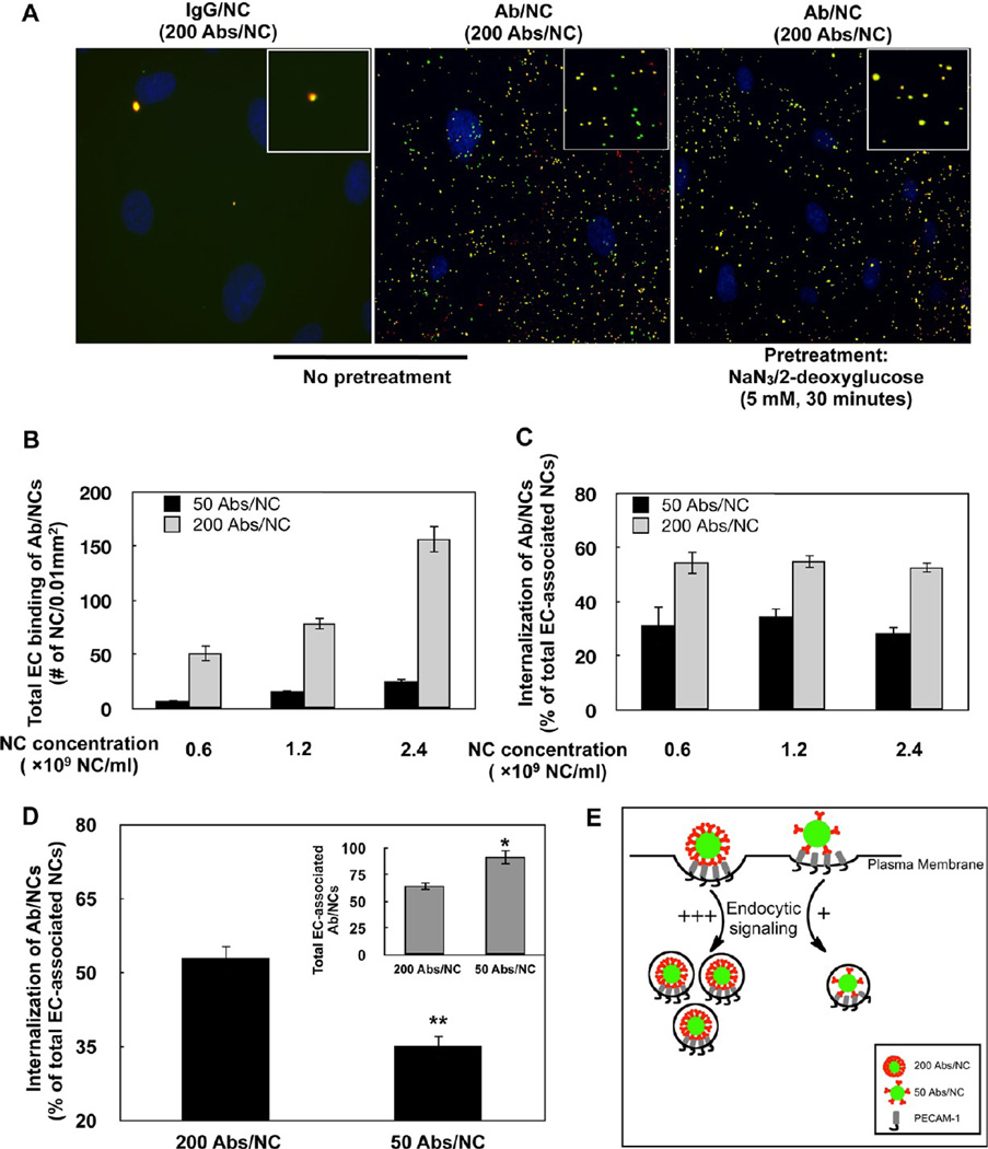
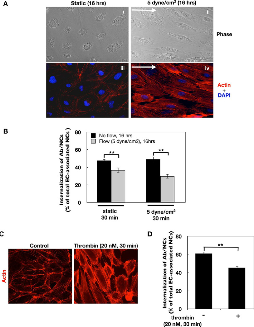
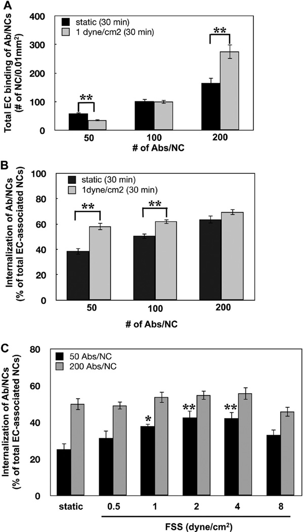
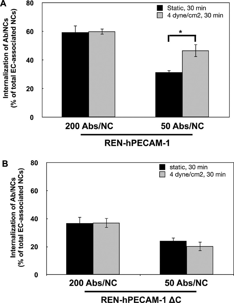
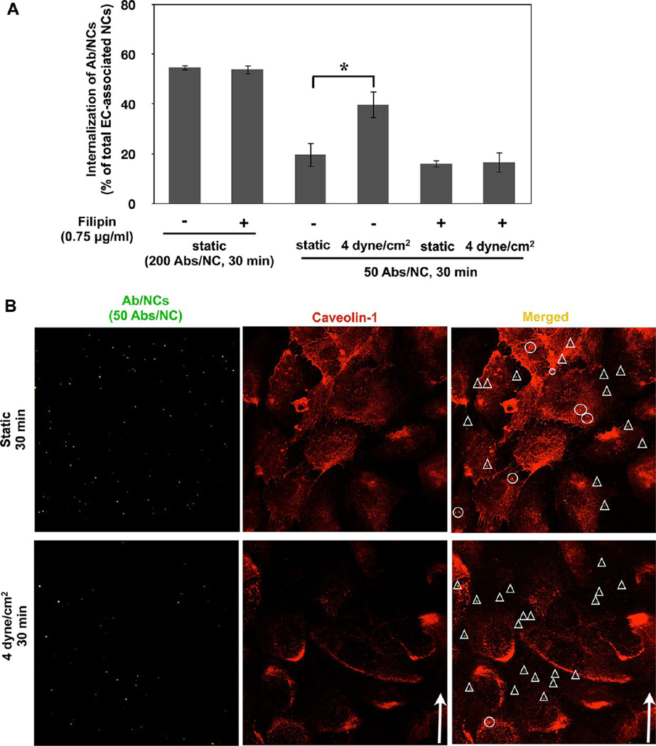
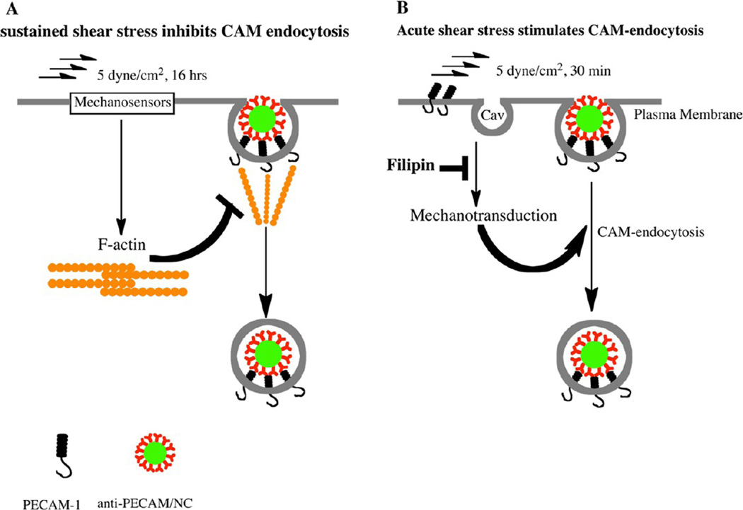
Intracellular delivery of nanocarriers (NC) is controlled by their design and target cell phenotype, microenvironment, and functional status. Endothelial cells (EC) lining the vascular lumen represent an important target for drug delivery. Endothelium in vivo is constantly or intermittently (as, for example, during ischemia-reperfusion) exposed to blood flow, which influences NC-EC interactions by changing NC transport properties, and by direct mechanical effects upon EC mechanisms involved in NC binding and uptake. EC do not internalize antibodies to marker glycoprotein PECAM(CD31), yet internalize multivalent NC coated with PECAM antibodies (anti-PECAM/NC) via a noncanonical endocytic pathway distantly related to macropinocytosis. Here we studied the effects of flow on EC uptake of anti-PECAM/NC spheres (~180 nm diameter). EC adaptation to chronic flow, manifested by cellular alignment with flow direction and formation of actin stress fibers, inhibited anti-PECAM/NC endocytosis consistent with lower rates of anti-PECAM/NC endocytosis in vivo in arterial compared to capillary vessels. Acute induction of actin stress fibers by thrombin also inhibited anti-PECAM/NC endocytosis, demonstrating that formation of actin stress fibers impedes EC endocytic machinery. In contrast, acute flow without stress fiber formation, stimulated anti-PECAM/NC endocytosis. Anti-PECAM/NC endocytosis did not correlate with the number of cell-bound particles under flow or static conditions. PECAM cytosolic tail deletion and disruption of cholesterol-rich plasmalemma domains abrogated anti-PECAM/NC endocytosis stimulation by acute flow, suggesting complex regulation of a flow-sensitive endocytic pathway in EC. The studies demonstrate the importance of the local flow microenvironment for NC uptake by the endothelium and suggest that cell culture models of nanoparticle uptake should reflect the microenvironment and phenotype of the target cells.


Conflict of interest statement

Conflict of Interest: The authors declare no competing financial interest.

Figures

Figure 1
Figure 1
Endocytosis of anti-PECAM/NCs targeted to pulmonary vasculature in vivo. (A) Visualization of pulmonary uptake of green fluorescence-labeled anti-PECAM/NC in vivo. Phase contrast (left) and fluorescence (right) images of a tissue section of the lung obtained from a mouse 30 min after intravenous injection of anti-PECAM/NC (Ab/NC below). At post-mortem, lung was perfused through pulmonary artery with RPMI medium for 5 min to remove unbound intravascular materials prior to fixation and slicing. Scale bars, 100 µm. (B) Endocytosis of Ab/NC in the pulmonary vasculature. A total of 30 min after intravenous injection of Ab/NC, lungs were perfused with buffer, as in panel A, and perfused further for 15 min with buffer containing Alexa Fluor 594 goat antirat IgG to counterstain surface-bound Ab/NC, followed by buffer perfusion to eliminate nonbound intravascular materials prior to fixation and slicing. Lung sections were analyzed with confocal fluorescence microscopy. Merged images show internalized Ab/NC as single-color labeled green particles and surface-bound Ab/NC as double-color labeled yellow particles. Scale bars, 40 µm. (C) The internalization was expressed as percentage of total amount of endothelial-associated NCs internalized. Data were collected from 6 images for each group. Values are expressed as mean ± SE (n = 6), *p < 0.05.
Figure 2
Figure 2
Endocytosis of anti-PECAM/NC in endothelial cell culture. (A) Static endothelial cells (EC) were incubated for 30 min at 37 °C with IgG/NC (left) of Ab/NC (middle and right), washed, and counterstained with secondary red anti-IgG. Merged fluorescence images show internalized (green) and surface-bound (yellow) Ab/NC in EC (middle). Endocytosis of Ab/NCs was blocked in ATP-depleted EC (right). (B) Endothelial binding (30 min incubation followed by washing) of Ab/NCs proportionally correlates with antibody surface density on particles and concentration of Ab/NC in incubation medium. Data were collected from eight images and presented as mean ± SE (n = 8). (C) Level of endocytosis of low and high avidity Ab/NC (50 and 200 Ab per particle) incubated with EC at indicated doses, as described in (B). (D) Internalization of low versus high avidity of Ab/NC incubated with EC at doses of 3 and 1 × 10⁹ NCs/mL, respectively, providing larger amount of cell-bound low-avidity Ab/NC versus high avidity Ab/NC (inset). In (C) and (D), the internalization was calculated as percentage of total amount of endothelial-bound Ab/NC, mean ± SE (n = 8); *p < 0.05 and **p < 0.01 in comparison with 200 Abs/NC group. (E) A schema represents the summary of results depicted in (B–D): endothelial endocytosis depends on the strength of the signal ignited by individual cell-bound Ab/NC (controlled by Ab surface density on a particle), not on the “collective” signaling by cell-bound Ab/NC (controlled by total number of cell-bound particles).
Figure 3
Figure 3
Endocytosis of anti-PECAM/NCs is inhibited in flow adapted EC. (A) Sustained exposure to flow induces EC alignment in the direction of flow and cytoskeletal remodeling. Confluent EC were exposed to static condition (i, iii)or5dyn/ cm2 laminar fluid shear stress (ii, iv) for 16 h. Cells were then fixed and stained for F-actin using Alexa-Fluor594-phalloidin. Images were taken using fluorescence microscope with a Plan Apo ×40 oil objective. Arrows show direction of flow. (B) In flow adapted EC, internalization of Ab/NC is inhibited. Cells were incubated or perfused (at 5 dyn/cm2) with HBSS medium containing anti-PECAM/NCs (200 Abs/NC, 2 × 10⁹ NCs/mL) for 30 min at 37 °C. Internalization was calculated and presented as mean ± SE (n = 8, **p < 0.01). (C) Thrombin-induced cytoskeletal remodeling in EC. Confluent EC were incubated without or with thrombin (20 nM) for 30 min, followed by immunostaining for F-actin. (D) Internalization of Ab/NC is inhibited in thrombin-treated EC. Mean ± SE (n = 8, **p < 0.01).
Figure 4
Figure 4
Acute exposure to flow stimulates endocytosis of anti-PECAM/NCs in EC. (A, B) Effect of flow (1 dyn/cm2, 30 min) on endothelial binding (A) and internalization (B) of Ab/NC carrying 50, 100, and 200 Ab molecules per NC. (C) Acute exposure to flow regulates endocytosis of Ab/NC (50 Abs/NC) in a flow rate-dependent manner. Data are presented as mean ( SE (n = 8, *p < 0.05, **p < 0.01).
Figure 5
Figure 5
Cytoplasmic domain of PECAM-1 mediates stimulation of endocytosis of anti-PECAM/NCs by flow. (A) Flow stimulates endocytosis of low-avidity Ab/NC in REN cells stably transfected to express human PECAM-1. (B) Effect of flow-induced stimulation of endocytosis of Ab/NC is abolished in REN cells transfected with human PECAM-1 lacking the cytoplasmic domain. Data expressed as mean ± SE (n = 6, *p < 0.05).
Figure 6
Figure 6
Cholesterol-rich membrane domains in endothelial cells are involved in stimulation of anti-PECAM/NC endocytosis by acute flow. (A) Disruption of cholesterol-enriched plasmalemma domains by filipin abolishes flow induced stimulation of endocytosis of low-avidity Ab/NC; mean ± SE (n = 8, *p < 0.05). (B) Ab/NC are not colocalized with caveolin-1 in endothelial cells either under static and flow conditions. Confluent EC were incubated or perfused with Ab/NC for 30 min at 37 °C, fixed, and immunostained for caveolin-1. Arrows indicate the direction of flow. Ab/NC in circles or triangles are colocalized (yellow) or noncolocalized (green) with caveolin-1, respectively.
Figure 7
Figure 7
Chronic and acute flow differently regulate endocytosis of anti-PECAM/NC. (A) Sustained exposure of EC to flow induces formation of actin stress fibers involved in cellular alignment, which impairs recruitment of actin in the fibers needed for endocytosis of Ab/NC. (B) Acute exposure of EC to flow stimulates endocytosis of Ab/NC likely via mechanisms involving cholesterol-rich domains of plasmalemma such as caveoli (cav).

References

    1. Champion JA, Mitragotri S. Role of Target Geometry in Phagocytosis. Proc. Natl. Acad. Sci. U.S.A. 2006;103:4930–4934. - PMC - PubMed
    1. Gratton SE, Ropp PA, Pohlhaus PD, Luft JC, Madden VJ, Napier ME, DeSimone JM. The Effect of Particle Design on Cellular Internalization Pathways. Proc. Natl. Acad. Sci. U.S.A. 2008;105:11613–11618. - PMC - PubMed
    1. Reilly MJ, Larsen JD, Sullivan MO. Polyplexes Traffic through Caveolae to the Golgi and Endoplasmic Reticulum en Route to the Nucleus. Mol. Pharm. 2012;9:1280–1290. - PubMed
    1. Bonner DK, Leung C, Chen-Liang J, Chingozha L, Langer R, Hammond PT. Intracellular Trafficking of Polyamidoamine-Poly(ethylene glycol) Block Copolymers in DNA Delivery. Bioconjugate Chem. 2011;22:1519–1525. - PMC - PubMed
    1. Evans CW, Fitzgerald M, Clemons TD, House MJ, Padman BS, Shaw JA, Saunders M, Harvey AR, Zdyrko B, Luzinov I, et al. Multimodal Analysis of PEI-Mediated Endocytosis of Nanoparticles in Neural Cells. ACS Nano. 2011;5:8640–8648. - PubMed

Publication types

Substances