

Surface sensing and lateral subcellular localization of WspA, the receptor in a chemosensory-like system leading to c-di-GMP production
- PMID: 22957788
- PMCID: PMC3501340
- DOI: 10.1111/mmi.12013
Surface sensing and lateral subcellular localization of WspA, the receptor in a chemosensory-like system leading to c-di-GMP production
Abstract
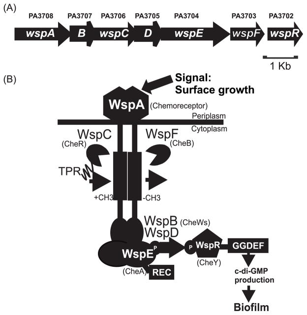
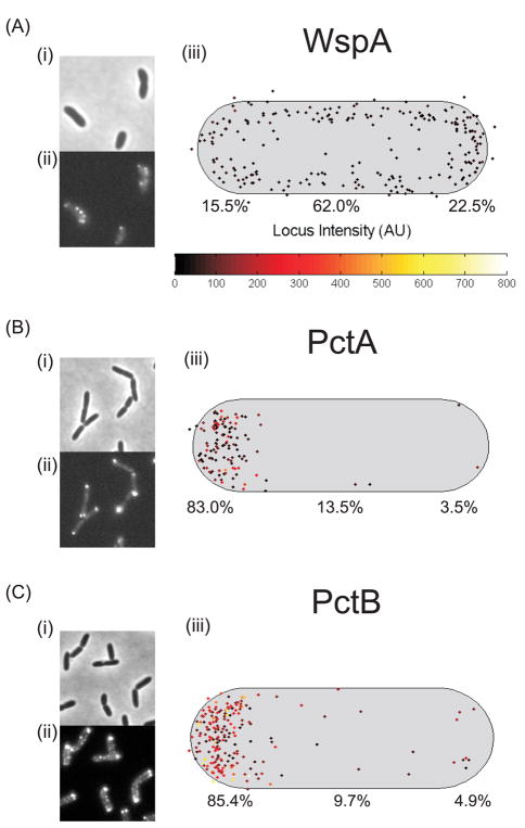
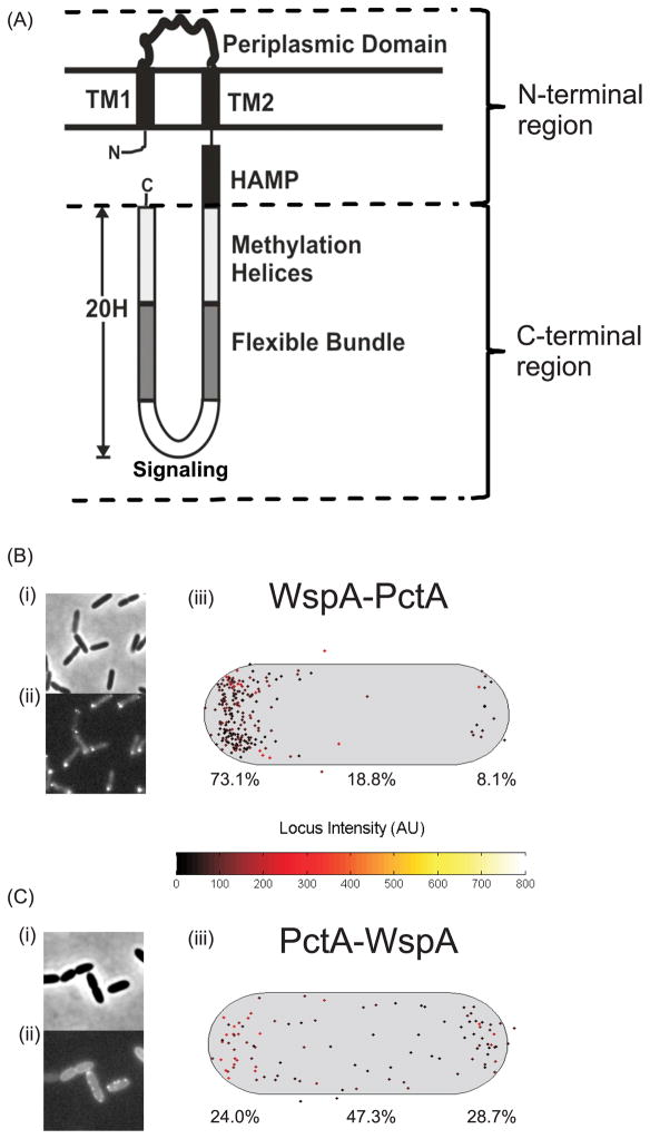
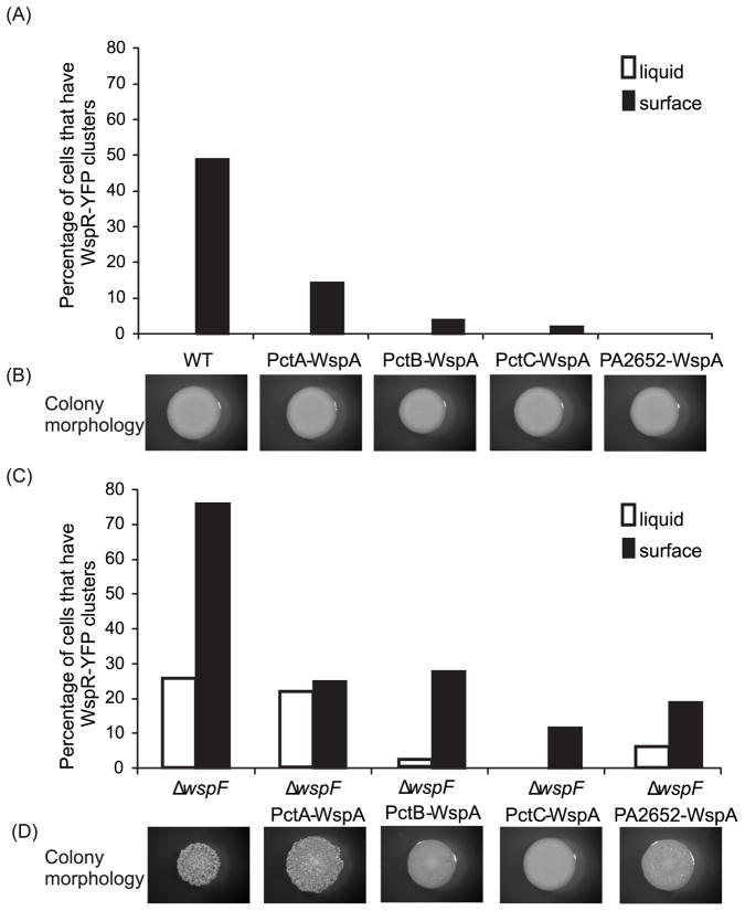
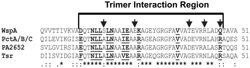
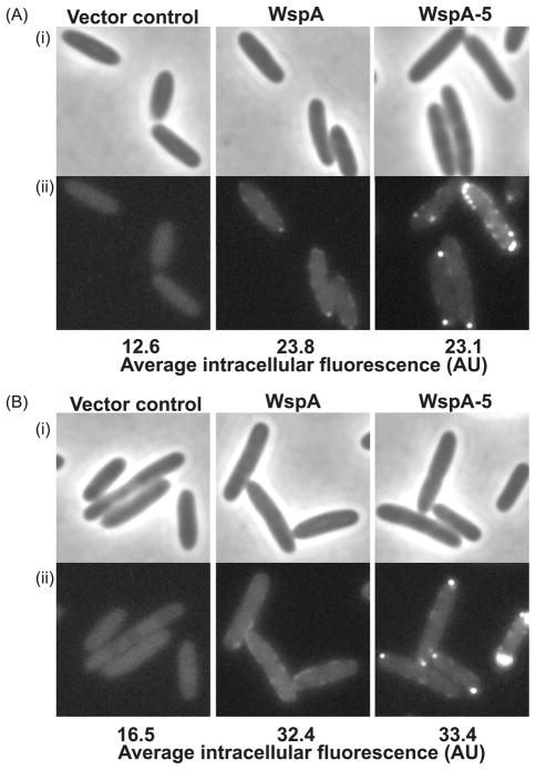
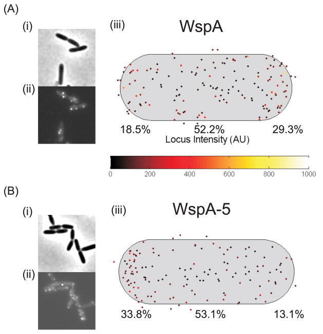
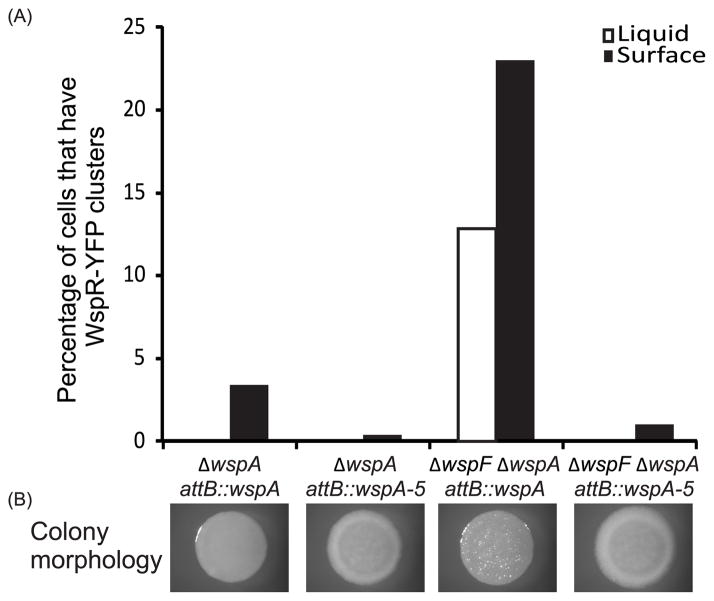
Pseudomonas aeruginosa responds to growth on agar surfaces to produce cyclic-di-GMP, which stimulates biofilm formation. This is mediated by an alternative cellular function chemotaxis-like system called Wsp. The receptor protein WspA, is bioinformatically indistinguishable from methyl-accepting chemotaxis proteins. However, unlike standard chemoreceptors, WspA does not form stable clusters at cell poles. Rather, it forms dynamic clusters at both polar and lateral subcellular locations. To begin to study the mechanism of Wsp signal transduction in response to surfaces, we carried out a structure-function study of WspA and found that its C-terminus is important for its lateral subcellular localization and function. When this region was replaced with that of a chemoreceptor for amino acids, WspA became polarly localized. In addition, introduction of mutations in the C-terminal region of WspA that rendered this protein able to form more stable receptor-receptor interactions, also resulted in a WspA protein that was less capable of activating signal transduction. Receptor chimeras with a WspA C-terminus and N-terminal periplasmic domains from chemoreceptors that sense amino acids or malate responded to surfaces to produce c-di-GMP. Thus, the amino acid sequence of the WspA periplasmic region did not need to be conserved for the Wsp system to respond to surfaces.
© 2012 Blackwell Publishing Ltd.
Figures

Similar articles
-
Subcellular location characteristics of the Pseudomonas aeruginosa GGDEF protein, WspR, indicate that it produces cyclic-di-GMP in response to growth on surfaces.Mol Microbiol. 2007 Dec;66(6):1459-73. doi: 10.1111/j.1365-2958.2007.06008.x. Epub 2007 Nov 19. Mol Microbiol. 2007. PMID: 18028314 Free PMC article.
-
Mutations in surface-sensing receptor WspA lock the Wsp signal transduction system into a constitutively active state.Environ Microbiol. 2022 Mar;24(3):1150-1165. doi: 10.1111/1462-2920.15763. Epub 2021 Sep 20. Environ Microbiol. 2022. PMID: 34499799
-
Activation Mechanism and Cellular Localization of Membrane-Anchored Alginate Polymerase in Pseudomonas aeruginosa.Appl Environ Microbiol. 2017 Apr 17;83(9):e03499-16. doi: 10.1128/AEM.03499-16. Print 2017 May 1. Appl Environ Microbiol. 2017. PMID: 28258142 Free PMC article.
-
PilZ Domain Protein FlgZ Mediates Cyclic Di-GMP-Dependent Swarming Motility Control in Pseudomonas aeruginosa.J Bacteriol. 2016 Jun 13;198(13):1837-46. doi: 10.1128/JB.00196-16. Print 2016 Jul 1. J Bacteriol. 2016. PMID: 27114465 Free PMC article.
-
Constitutive production of c-di-GMP is associated with mutations in a variant of Pseudomonas aeruginosa with altered membrane composition.Sci Signal. 2015 Apr 14;8(372):ra36. doi: 10.1126/scisignal.2005943. Sci Signal. 2015. PMID: 25872871
Cited by
-
Bacterial cyclic diguanylate signaling networks sense temperature.Nat Commun. 2021 Mar 31;12(1):1986. doi: 10.1038/s41467-021-22176-2. Nat Commun. 2021. PMID: 33790266 Free PMC article.
-
Concentration Dependent Effect of Plant Root Exudates on the Chemosensory Systems of Pseudomonas putida KT2440.Front Microbiol. 2019 Jan 30;10:78. doi: 10.3389/fmicb.2019.00078. eCollection 2019. Front Microbiol. 2019. PMID: 30761113 Free PMC article.
-
Sensory Perception in Bacterial Cyclic Diguanylate Signal Transduction.J Bacteriol. 2022 Feb 15;204(2):e0043321. doi: 10.1128/JB.00433-21. Epub 2021 Oct 4. J Bacteriol. 2022. PMID: 34606374 Free PMC article. Review.
-
Sensory Repertoire of Bacterial Chemoreceptors.Microbiol Mol Biol Rev. 2017 Oct 25;81(4):e00033-17. doi: 10.1128/MMBR.00033-17. Print 2017 Dec. Microbiol Mol Biol Rev. 2017. PMID: 29070658 Free PMC article. Review.
-
Methyl-accepting chemotaxis proteins: a core sensing element in prokaryotes and archaea.Cell Mol Life Sci. 2017 Sep;74(18):3293-3303. doi: 10.1007/s00018-017-2514-0. Epub 2017 Apr 13. Cell Mol Life Sci. 2017. PMID: 28409190 Free PMC article. Review.
References
-
- Alley MR. The highly conserved domain of the Caulobacter McpA chemoreceptor is required for its polar localization. Mol Microbiol. 2001;40:1335–1343. - PubMed
Publication types
MeSH terms
Substances
Grants and funding
LinkOut - more resources
Full Text Sources
