. 2012 Sep 7:9:212.
doi: 10.1186/1742-2094-9-212.

Neuroinflammation and oxidative stress in rostral ventrolateral medulla contribute to neurogenic hypertension induced by systemic inflammation

Affiliations

Neuroinflammation and oxidative stress in rostral ventrolateral medulla contribute to neurogenic hypertension induced by systemic inflammation

Kay L H Wu et al. J Neuroinflammation. .

Abstract

Background: In addition to systemic inflammation, neuroinflammation in the brain, which enhances sympathetic drive, plays a significant role in cardiovascular diseases, including hypertension. Oxidative stress in rostral ventrolateral medulla (RVLM) that augments sympathetic outflow to blood vessels is involved in neural mechanism of hypertension. We investigated whether neuroinflammation and oxidative stress in RVLM contribute to hypertension following chronic systemic inflammation.

Methods: In normotensive Sprague-Dawley rats, systemic inflammation was induced by infusion of Escherichia coli lipopolysaccharide (LPS) into the peritoneal cavity via an osmotic minipump. Systemic arterial pressure and heart rate were measured under conscious conditions by the non-invasive tail-cuff method. The level of the inflammatory markers in plasma or RVLM was analyzed by ELISA. Protein expression was evaluated by Western blot or immunohistochemistry. Tissue level of superoxide anion (O(2)(-)) in RVLM was determined using the oxidation-sensitive fluorescent probe dihydroethidium. Pharmacological agents were delivered either via infusion into the cisterna magna with an osmotic minipump or microinjection bilaterally into RVLM.

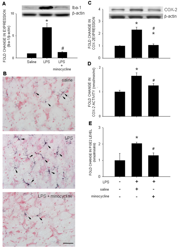
Results: Intraperitoneal infusion of LPS (1.2 mg/kg/day) for 14 days promoted sustained hypertension and induced a significant increase in plasma level of C-reactive protein, tumor necrosis factor-α (TNF-α), or interleukin-1β (IL-1β). This LPS-induced systemic inflammation was accompanied by activation of microglia, augmentation of IL-1β, IL-6, or TNF-α protein expression, and O(2)(-) production in RVLM, all of which were blunted by intracisternal infusion of a cycloxygenase-2 (COX-2) inhibitor, NS398; an inhibitor of microglial activation, minocycline; or a cytokine synthesis inhibitor, pentoxifylline. Neuroinflammation in RVLM was also associated with a COX-2-dependent downregulation of endothelial nitric oxide synthase and an upregulation of intercellular adhesion molecule-1. Finally, the LPS-promoted long-term pressor response and the reduction in expression of voltage-gated potassium channel, Kv4.3 in RVLM were antagonized by minocycline, NS398, pentoxifylline, or a superoxide dismutase mimetic, tempol, either infused into cisterna magna or microinjected bilaterally into RVLM. The same treatments, on the other hand, were ineffective against LPS-induced systemic inflammation.

Conclusion: These results suggest that systemic inflammation activates microglia in RVLM to induce COX-2-dependent neuroinflammation that leads to an increase in O(2)(-) production. The resultant oxidative stress in RVLM in turn mediates neurogenic hypertension.


Figures

Figure 1
Figure 1
Infusion of LPS into the peritoneal cavity elevates blood pressure and plasma level of pro-inflammatory cytokines. Time-course changes in mean SAP (MSAP) (A), HR (B), or plasma level of CRP (C), TNF-α (D), and IL-1β (E), measured on days 7 or 14 after intraperitoneal infusion of saline or LPS (1.2 mg/kg/day) via an osmotic minipump for 14 days, alone or with additional intracisternal infusion of NS398 (1.5 nmol/μL/h). Values are mean ± SEM (n=8 to 10 animals in each experimental group). *P <0.05 vs. saline-treatment group at corresponding time-intervals or sham-control group; #P <0.05 vs. LPS-treatment group at corresponding time-intervals in the post hoc Scheffé multiple-range test. Arrow indicates the time point during which the osmotic minipump was implanted into the peritoneal cavity.
Figure 2
Figure 2
Intraperitoneal infusion of LPS also elevates tissue level of pro-inflammatory cytokines in RVLM. Left panels: Time-course changes in tissue level of TNF-α, IL-1β, or IL-6 in RVLM after intraperitoneal infusion of saline or LPS (1.2 mg/kg/day), alone or with additional intracisternal infusion of NS398 (1.5 nmol/μL/h). Right panels: Tissue level of the proinflammatory cytokines in RVLM measured on day 7 after intraperitoneal LPS infusion, with additional intracisternal infusion of tempol (1 μmol/μL/h) or aCSF. Values are mean ± SEM (n=8 to 10 animals in each experimental group). *P <0.05 vs. corresponding saline-treatment group; #P <0.05 vs. corresponding LPS-treatment group in the post hoc Scheffé multiple-range test.
Figure 3
Figure 3
Systemic inflammation activates microglia and enhances COX-2 activity in RVLM. Gels (inset) and densitometric analysis of results from Western blot showing changes in expression of Iba-1 (A) or COX-2 (C), or representative photomicrographs showing immunoreactivity to Iba-1 (black color) (B), COX-2 activity (D), or tissue level of PGE2 (E) in RVLM, examined on day 7 after intraperitoneal infusion of saline or LPS (1.2 mg/kg/day), alone or with additional intracisternal infusion of minocycline (9 nmol/μL/). Values are mean ± SEM (n=8 to 10 animals in each experimental group). *P <0.05 vs. saline-treatment group; #P <0.05 vs. LPS-treatment group in the post hoc Scheffé multiple-range test. Scale bar in (A): 25 μm.
Figure 4
Figure 4
Intraperitoneal infusion of LPS induces endothelial dysfunction in RVLM. Representative gels (inset) and densitometric analysis of results from Western blot showing changes in expression of eNOS (A), iNOS (B), nNOS (C), or ICAM-1 (D), as well as photomicrographs showing distribution of vWF- (red fluorescence) and ICAM-1-immunoreactivity (green fluorescence) in RVLM, examined on day 7 after intraperitoneal infusion of saline or LPS (1.2 mg/kg/day), alone or with additional intracisternal infusion of NS398 (1.5 nmol/μL/h). Values are mean ± SEM (n=8 to 10 animals in each experimental group). *P <0.05 vs. saline-treatment group; #P <0.05 vs. LPS-treatment group in the post hoc Scheffé multiple-range test. Note that colocalization of vWF- and ICAM-1-immunoreactivity is shown in yellow color in (D). Scale bar in (D): 10 μm.
Figure 5
Figure 5
Intraperitoneal infusion of LPS induces neuroinflammation-associated oxidative stress in RVLM. Representative photomicrographs showing the distribution of dihydroethidium (red fluorescence) (A), or changes in tissue level of superoxide (B) in RVLM examined on day 7 after intraperitoneal infusion of saline or LPS (1.2 mg/kg/day), alone or with additional intracisternal infusion of minocycline (9 nmol/μL/h), pentoxifylline (PTX, 30 nmol/μL/h), NS398 (1.5 nmol/μL/h), or tempol (1 μmol/μL/h). Values are mean ± SEM (n=8 to 10 animals in each experimental group). *P <0.05 vs. saline-treatment group; #P <0.05 vs. LPS-treatment group in the post hoc Scheffé multiple-range test. A schematic drawing of rostral medulla oblongata is included in (A) to illustrate the location of RVLM from where the photomicrographs were taken. NA, nucleus ambiguous; NTS, nucleus tractus solitarii; V, nucleus spinalis trigemini. Scale bar in (A): 100 μm.
Figure 6
Figure 6
Intraperitoneal infusion of LPS upregulates NADPH oxidase subunits and antioxidants in RVLM. Representative gels (inset) and densitometric analysis of results from Western blot showing changes in expression of gp91phox (A), p47phox (B), Cu/ZnSOD (C), MnSOD (D), ecSOD (E), catalase (F), or glutathione peroxide (GPx) (G) in RVLM, determined on day 7 after intraperitoneal infusion of saline or LPS (1.2 mg/kg/day), alone or with additional intracisternal infusion of NS398 (1.5 nmol/μL/h) or minocycline (9 nmol/μL/h). Values are mean ± SEM (n=8 to 10 animals in each experimental group). *P <0.05 vs. saline-treatment group; #P <0.05 vs. LPS-treatment group in the post hoc Scheffé multiple-range test.
Figure 7
Figure 7
Oxidative stress in RVLM underpins hypertension induced by intraperitoneal infusion of LPS. Changes in MSAP (A,B) or plasma level of CRP (C) determined on day 7 after intraperitoneal infusion of saline or LPS (1.2 mg/kg/day), alone or with additional intracisternal infusion of aCSF or tempol (1μmol/μL/h) (A,C), or microinjection bilaterally into RVLM of tempol (100 pmol) (B). Values are mean ± SEM (n=8 to 10 animals in each experimental group). *P <0.05 vs. saline-treatment or sham-control group; #P <0.05 vs. LPS-treatment group in the post hoc Scheffé multiple-range test.
Figure 8
Figure 8
Intraperitoneal infusion of LPS downregulates Kv4.3 potassium channel in RVLM and increase the sympathetic neurogenic vasomotor activity. Representative gels (inset) and densitometric analysis of results from Western blot showing changes in expression of Kv4.3 channel protein in RVLM (A) or changes in the power density of the low frequency component of SAP spectrum (B), measured on day 7 after intraperitoneal infusion of saline or LPS (1.2 mg/kg/day), alone or with additional intracisternal infusion of minocycline (9 nmol/μL/h), NS398 (1.5 nmol/μL/h), PTX (30 nmol/μL/h), or tempol (1 μmol/μL/h). Values are mean ± SEM (n=8 to 10 animals in each experimental group). *P <0.05 vs. saline-treatment group; #P <0.05 vs. LPS-treatment group in the post hoc Scheffé multiple-range test

Similar articles

Cited by

References

    1. Libby P, Ridker PM, Hansson GK. Leducq Transatlantic Network on Atherothrombosis. Inflammation in atherosclerosis: from pathophysiology to practice. J Am Coll Cardiol. 2009;54:2129–2138. doi: 10.1016/j.jacc.2009.09.009. - DOI - PMC - PubMed
    1. Felder RB. Mineralocorticoid receptors, inflammation and sympathetic drive in a rat model of systolic heart failure. Exp Physiol. 2010;95:19–25. doi: 10.1113/expphysiol.2008.045948. - DOI - PMC - PubMed
    1. Straznicky NE, Eikelis N, Lambert EA, Esler MD. Mediators of sympathetic activation in metabolic syndrome obesity. Curr Hypertens Rep. 2008;10:440–447. doi: 10.1007/s11906-008-0083-1. - DOI - PubMed
    1. Paton JF, Waki H. Is neurogenic hypertension related to vascular inflammation of the brainstem? Neurosci Biobehav Rev. 2009;33:89–94. doi: 10.1016/j.neubiorev.2008.05.020. - DOI - PubMed
    1. Sprague AH, Khalil RA. Inflammatory cytokines in vascular dysfunction and vascular disease. Biochem Pharmacol. 2009;78:539–552. doi: 10.1016/j.bcp.2009.04.029. - DOI - PMC - PubMed

Publication types

MeSH terms