. 2010 Feb 28;5(1):22-30.
doi: 10.1186/2049-6958-5-1-22.

Regulatory T cells in induced sputum of asthmatic children: association with inflammatory cytokines

Affiliations

Regulatory T cells in induced sputum of asthmatic children: association with inflammatory cytokines

Agnès Hamzaoui et al. Multidiscip Respir Med. .

Abstract

Background and objective: CD4+CD25+ regulatory T (Treg) cells play an essential role in maintaining immune homeostasis. In this study, we investigated whether the induced sputum (IS) pool and the function of CD4+CD25+ Treg cells are altered in asthma pediatric patients.

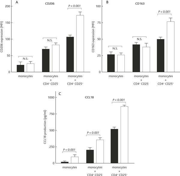
Methods: Treg activity was studied in the IS of 40 asthmatic children. CD3+ cells were analyzed for the expression of FoxP3 mRNA by real time reverse transcription-polymerase chain reaction (RT-PCR). IS cells from asthmatics and controls were stained for Treg markers and analyzed by flow cytometry. We also studied the ability of Treg cells to differentiate monocytes toward alternatively activated macrophages (AAM), and to suppress proinflammatory cytokines.

Results: (i) Mild and moderate asthmatics had significantly decreased expression of FoxP3/β-actin mRNA and decreased proportions of CD4+CD25highFoxP3+ cells compared to healthy children; (ii) patients with moderate asthma had even lower proportions of FoxP3 expression compared to mild asthmatic patients; (iii) monocytes cultured with Treg cells displayed typical features of AAM, including up-regulated expression of CD206 (macrophage mannose receptor) and CD163 (hemoglobin scavenger receptor), and an increased production of chemokine ligand 18 (CCL18). In addition, Treg cells from asthmatics have a reduced capacity to suppress LPS-proinflammatory cytokine production from monocytes/macrophages (IL-1, IL-6 and TNF-α).

Conclusion: Asthma pediatric patients display a decreased bronchial Treg population. The impaired bronchial Treg activity is associated with disease severity.


Figures

Figure 1
Figure 1
Antigen expression of t cells in healthy children and patients with mild and moderate bronchial asthma. Expression of forkhead box P3 (FoxP3) mRNA in T cells (A) and proportions of CD4+CD25highFoxP3+ cells in the CD4+ cell population (B), among healthy children and patients with mild and moderate bronchial asthma. Medians are indicated by a line inside each box, the 25th and 75th percentiles by the box limits, the lower and upper error bars represent the 10th and 90th percentiles, respectively. (C): CD4+CD25high and CD4+CD25high forkhead box P3 (FoxP3)+ cells in one representative moderate bronchial asthma children as measured by flow cytometry. Boxes (a) indicate cells in the CD25high population. Quadrants (b) indicate CD25highversus CD25low/intermediate cells and FoxP3+versus FoxP3- cells.
Figure 2
Figure 2
Treg cells induction of an alternative activated phenotype in monocytes/macrophages of moderate asthma children and controls. Monocytes were cultured without T cells (monocytes), with CD4+CD25- T cells, or with CD4+CD25+ T cells in the presence of anti-CD3 mAb (50 ng/ml). Moderate asthma = white bars; healthy controls = black bars. (A and B): The phenotype of monocytes was assessed after 40 h of culture by flow cytometry. The expression (average mean fluorescence intensity, MFI ± SEM) of CD206 and CD163 is shown for ten independent experiments, respectively. (C): The amount of CCL18 produced during the 40 h of co-culture was analyzed in the supernatant by ELISA. Significant differences were observed between moderate asthmatic children and control healthy children.
Figure 3
Figure 3
Failure to suppress the lps-induced proinflammatory response of monocytes/macrophages by treg cells in moderate bronchial asthma. Monocytes were cultured as described in the legend of Figure 2. After 40 h of co-culture, LPS (50 ng/ml) was added and 24 h later cytokine production was measured by ELISA (IL-6, TNF-α) or Luminex (IL-1β). The average production of ten independent experiments ± SD of proinflammatory cytokines is shown for the three culture conditions. Significant differences are seen in the graphs between moderate asthmatic children and healthy children. Moderate asthma = white bars; healthy controls = black bars.
Figure 4
Figure 4
Changes in proportions of cd4+cd25highfoxp3+ cells in the cd4+ cell population from eight patients with moderate asthma, before and after treatment. Eight patients received treatment with inhaled corticosteroid and were revisited after 6 weeks. Proportions of CD4+CD25highFoxP3+ cells in the CD4+ cell population were slightly raised, and the difference between before (mean ± SD: 0.85 ± 0.43) and after (1.32 ± 0.63) treatment with optimal inhaled corticosteroid reached a statistically significant level (p = 0.0035).

Similar articles

Cited by

References

    1. Saglani S, Payne DN, Zhu J, Wang Z, Nicholson AG, Bush A, Jeffery PK. Early detection of airway wall remodeling and eosinophilic inflammation in preschool wheezers. Am J Respir Crit Care Med. 2007;176:858–864. doi: 10.1164/rccm.200702-212OC. - DOI - PubMed
    1. Barbato A, Turato G, Baraldo S, Bazzan E, Calabrese F, Panizzolo C, Zanin ME, Zuin R, Maestrelli P, Fabbri LM, Saetta M. Epithelial damage and angiogenesis in the airways of children with asthma. Am J Respir Crit Care Med. 2006;174:975–981. doi: 10.1164/rccm.200602-189OC. - DOI - PubMed
    1. Pohunek P, Warner JO, Turzíková J, Kudrmann J, Roche WR. Markers of eosinophilic inflammation and tissue re-modelling in children before clinically diagnosed bronchial asthma. Pediatr Allergy Immunol. 2005;16:43–51. doi: 10.1111/j.1399-3038.2005.00239.x. - DOI - PubMed
    1. Afshar R, Medoff BD, Luster AD. Allergic asthma: a tale of many T cells. Clin Exp Allergy. 2008;38:1847–1857. doi: 10.1111/j.1365-2222.2008.03119.x. - DOI - PubMed
    1. Akdis M, Verhagen J, Taylor A, Karamloo F, Karagiannidis C, Crameri R, Thunberg S, Deniz G, Valenta R, Fiebig H, Kegel C, Disch R, Schmidt-Weber CB, Blaser K, Akdis CA. Immune responses in healthy and allergic individuals are characterized by a fine balance between allergen-specific T regulatory 1 and T helper 2 cells. J Exp Med. 2004;199:1567–1575. doi: 10.1084/jem.20032058. - DOI - PMC - PubMed