

Automated texture-based quantification of centrilobular nodularity and centrilobular emphysema in chest CT images
- PMID: 22958719
- PMCID: PMC3679917
- DOI: 10.1016/j.acra.2012.04.020
Automated texture-based quantification of centrilobular nodularity and centrilobular emphysema in chest CT images
Abstract
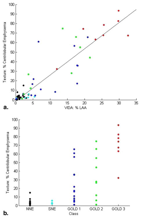
Rationale and objectives: Characterization of smoking-related lung disease typically consists of visual assessment of chest computed tomographic (CT) images for the presence and extent of emphysema and centrilobular nodularity (CN). Quantitative analysis of emphysema and CN may improve the accuracy, reproducibility, and efficiency of chest CT scoring. The purpose of this study was to develop a fully automated texture-based system for the detection and quantification of centrilobular emphysema (CLE) and CN in chest CT images.
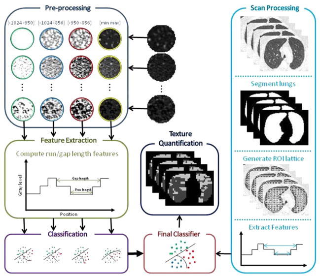
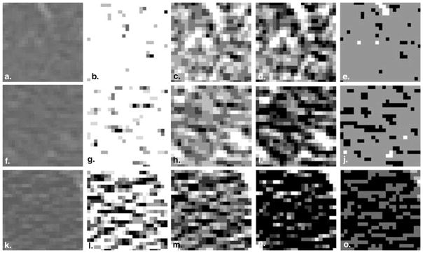
Materials and methods: A novel approach was used to prepare regions of interest (ROIs) within the lung parenchyma for representation by texture features associated with the gray-level run-length and gray-level gap-length methods. These texture features were used to train a multiple logistic regression classifier to discriminate between normal lung tissue, CN or "smoker's lung," and CLE. This classifier was trained and evaluated on 24 and 71 chest CT scans, respectively.
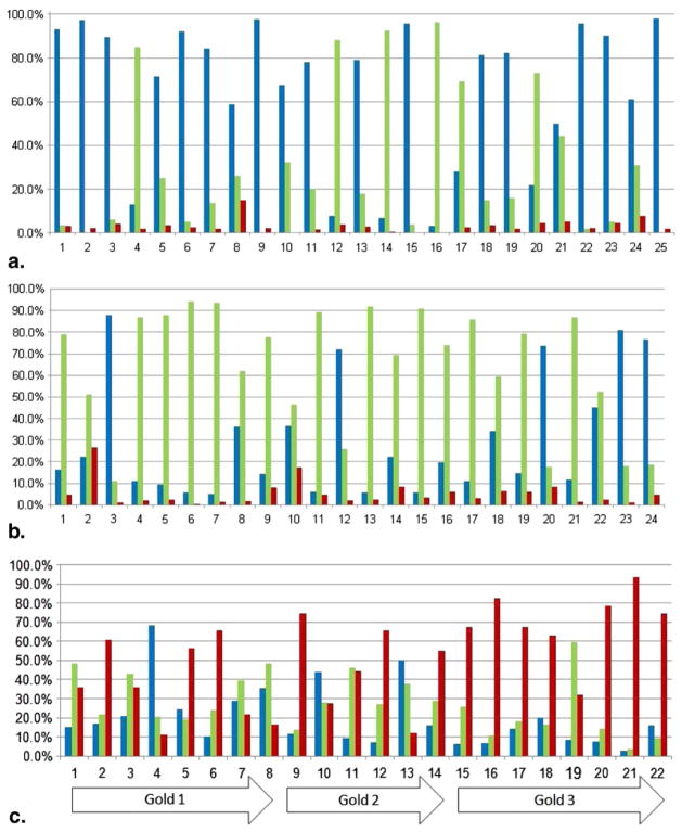
Results: During training, the classifier correctly classified 89% of ROIs depicting normal lung tissue, 74% of ROIs depicting CN, and 95% of ROIs manifesting CLE. When the performance of the classifier in quantifying extent of CN and CLE was evaluated on 71 chest CT scans, 65% of ROIs in smokers without CLE were classified as CN, compared to 31% in nonsmokers (P < .001) and 28% in smokers with CLE (P < .001).
Conclusions: The texture-based framework described herein facilitates successful discrimination among normal lung tissue, CN, and CLE and can be used for the automated quantification of smoking-related lung disease.
Copyright © 2012 AUR. Published by Elsevier Inc. All rights reserved.
Figures
References
-
- Kochanek KD, Xu J, Murphy SL, et al. Deaths: preliminary data for 2009. Natl Vit Stat Rep. 2001;59:1–51. - PubMed
-
- Hansell DM, Bankier AA, MacMahon H, et al. Fleischner society: glossary of terms for thoracic imaging. Radiology. 2008;246:697–722. - PubMed
-
- Remy-Jardin M, Edme JL, Boulenguez C, et al. Longitudinal follow-up study of smoker’s lung with thin-section CT in correlation with pulmonary function tests. Radiology. 2002;222:261–270. - PubMed
-
- Heyneman LE, Ward S, Lynch DA, et al. Respiratory bronchiolitis, respiratory bronchiolitis–associated interstitial lung disease, and desquamative interstitial pneumonia: different entities or part of the spectrum of the same disease process. AJR Am J Roentgenol. 1999;173:1617–1622. - PubMed
-
- Hersh CP, Washko GR, Jacobson FL, et al. Interobserver variability in the determination of upper lobe-predominant emphysema. Chest. 2007;131:424–431. - PubMed
MeSH terms
Grants and funding
LinkOut - more resources
Full Text Sources
Medical
Miscellaneous
