. 2012 Jul 6;4(1):19.
doi: 10.1186/1866-1955-4-19.

Reward circuitry dysfunction in psychiatric and neurodevelopmental disorders and genetic syndromes: animal models and clinical findings

Affiliations

Reward circuitry dysfunction in psychiatric and neurodevelopmental disorders and genetic syndromes: animal models and clinical findings

Gabriel S Dichter et al. J Neurodev Disord. .

Abstract

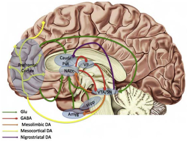
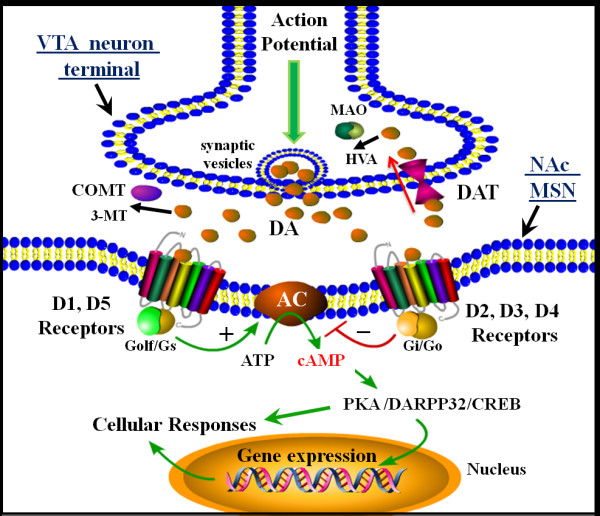
This review summarizes evidence of dysregulated reward circuitry function in a range of neurodevelopmental and psychiatric disorders and genetic syndromes. First, the contribution of identifying a core mechanistic process across disparate disorders to disease classification is discussed, followed by a review of the neurobiology of reward circuitry. We next consider preclinical animal models and clinical evidence of reward-pathway dysfunction in a range of disorders, including psychiatric disorders (i.e., substance-use disorders, affective disorders, eating disorders, and obsessive compulsive disorders), neurodevelopmental disorders (i.e., schizophrenia, attention-deficit/hyperactivity disorder, autism spectrum disorders, Tourette's syndrome, conduct disorder/oppositional defiant disorder), and genetic syndromes (i.e., Fragile X syndrome, Prader-Willi syndrome, Williams syndrome, Angelman syndrome, and Rett syndrome). We also provide brief overviews of effective psychopharmacologic agents that have an effect on the dopamine system in these disorders. This review concludes with methodological considerations for future research designed to more clearly probe reward-circuitry dysfunction, with the ultimate goal of improved intervention strategies.


Figures

Figure 1
Figure 1
Schematic illustration of the DA pathways and circuitry that regulate dopamine (DA) release in the human brain. The DA-containing neurons in the ventral tegmental area (VTA)/substantia nigra (SN) project to the nucleus accumbens (mesolimbic pathway; orange), to the cortex (mesocortical pathway; yellow) and caudate putamen (nigrostriatal pathway; purple). DA neuron firing rates are maintained at tonic levels in part due to steady-state inhibitory firing from the ventral pallidum. Excitatory glutamatergic fibers (green) project from the prefrontal cortex, amygdala, and hippocampus, that synapse on striatal targets, including the nucleus accumbens (NAc). The NAc sends GABAergic projections (red) to the ventral pallidum that suppress ventral pallidum inhibition of the VTA, thereby facilitating phasic burst firing of ventral tegmental area DA neurons. Note: Placement of structures is only approximate. Amyg, amygdala; Caud, caudate; DA, dopamine; GABA, GABAergic projections; Glu, glutamatergic projections; Hipp, hippocampus; Put, putamen; VP, ventral pallidum. (Figure and legend adapted with permission from Treadway and Zald [19].)
Figure 2
Figure 2
Schematic illustration of cellular mechanisms of neurotransmission in the mesolimbic dopamine (DA) reward pathway. Shown is a synapse between a ventral tegmental area DA neuron axon terminal and a medium spiny neuron (MSN) in the nucleus accumbens (NAc) in the ventral striatum. Transmission begins with an action potential that arrives to the terminal, inducing synaptic vesicle fusion and release of DA. The release of DA into the NAc stimulates various populations of MSNs, whose response to the transmitter depends on the types of DA receptors they express. DA stimulation of neurons containing D1 or D5 receptors (so-called D1-like receptors) results in activation of heterotrimeric Golf/Gs proteins, which activate the enzyme adenylyl cyclase, resulting in the synthesis of the second messenger cAMP. In contrast to this mechanism, DA stimulation of MSNs that express D2, D3 or D4 (or D2-like receptors) activate sheterotrimeric Gi/Go proteins, which inhibit adenylyl cyclase activity to decrease cAMP. The level of intracellular cAMP controls the activation of protein kinase A, which regulates additional signaling molecules including dopamine- and cAMP-regulated neuronal phosphoprotein of 32 kDa (DARPP-32) and the transcription factor cAMP response element binding (CREB) protein, both of which can modulate gene expression and additional cellular responses. The response to DA is generally terminated when DA is removed from the synapse by reuptake via the DA transporter (DAT). After reuptake, the transmitter can be repackaged into synaptic vesicles or may be degraded by the enzyme monoamine oxidase, resulting in the DA metabolite homovanillic acid. In addition, the enzyme catechol-o-methyltransferase (COMT) may also control DA levels by breaking down DA to 3-methoxytyramine (3-MT), AC, adenylyl cyclase; ATP; adensosine triphosphate; cAMP; cyclic adenosine monophosphate; HVA, homovanillic acid; MAO, monoamine oxidase; VTA, ventral tegmental area.

Similar articles

Cited by

References

    1. American Psychiatric Association. Diagnostic and statistical manual of mental disorders. 4. DSM-IV, Washington; 1994.
    1. Aupperle RL, Paulus MP. Neural systems underlying approach and avoidance in anxiety disorders. Dialogues Clin Neurosci. 2010;12(4):517–531. - PMC - PubMed
    1. Murray EA, Wise SP, Drevets WC. Localization of dysfunction in major depressive disorder: prefrontal cortex and amygdala. Biol Psychiatr. 2011;69(12):e43. - PMC - PubMed
    1. Kerns JG, Cohen JD, MacDonald AW, Johnson MK, Stenger VA, Aizenstein H, Carter CS. Decreased conflict- and error-related activity in the anterior cingulate cortex in subjects with Schizophrenia. Am J Psychiatr. 2005;162(10):1833–1839. - PubMed
    1. Pannu Hayes J, Labar KS, Petty CM, McCarthy G, Morey RA. Alterations in the neural circuitry for emotion and attention associated with posttraumatic stress symptomatology. Psychiatr Res. 2009;172(1):7–15. - PMC - PubMed