

Uterine biology in pigs and sheep
- PMID: 22958877
- PMCID: PMC3436697
- DOI: 10.1186/2049-1891-3-23
Uterine biology in pigs and sheep
Abstract
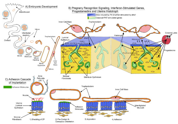
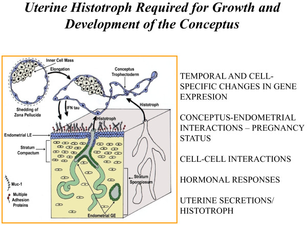
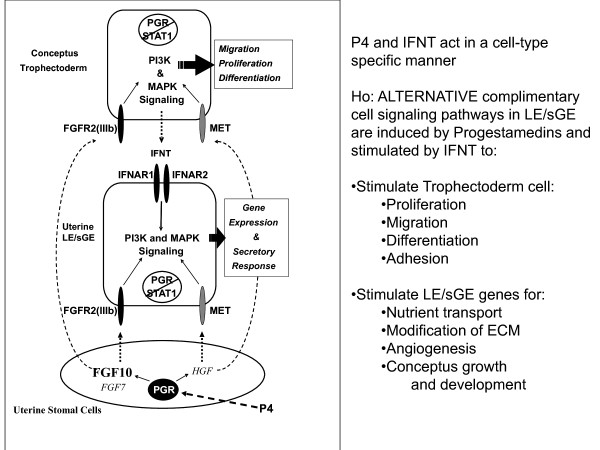
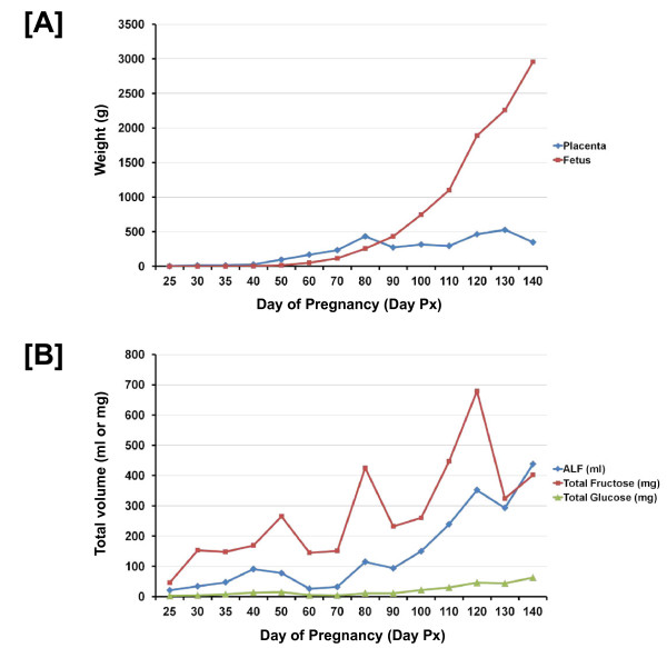
There is a dialogue between the developing conceptus (embryo-fetus and associated placental membranes) and maternal uterus which must be established during the peri-implantation period for pregnancy recognition signaling, implantation, regulation of gene expression by uterine epithelial and stromal cells, placentation and exchange of nutrients and gases. The uterus provide a microenvironment in which molecules secreted by uterine epithelia or transported into the uterine lumen represent histotroph required for growth and development of the conceptus and receptivity of the uterus to implantation. Pregnancy recognition signaling mechanisms sustain the functional lifespan of the corpora lutea (CL) which produce progesterone, the hormone of pregnancy essential for uterine functions that support implantation and placentation required for a successful outcome of pregnancy. It is within the peri-implantation period that most embryonic deaths occur due to deficiencies attributed to uterine functions or failure of the conceptus to develop appropriately, signal pregnancy recognition and/or undergo implantation and placentation. With proper placentation, the fetal fluids and fetal membranes each have unique functions to ensure hematotrophic and histotrophic nutrition in support of growth and development of the fetus. The endocrine status of the pregnant female and her nutritional status are critical for successful establishment and maintenance of pregnancy. This review addresses the complexity of key mechanisms that are characteristic of successful reproduction in sheep and pigs and gaps in knowledge that must be the subject of research in order to enhance fertility and reproductive health of livestock species.
Figures
Similar articles
-
Roles of conceptus secretory proteins in establishment and maintenance of pregnancy in ruminants.Asian-Australas J Anim Sci. 2012 Jan;25(1):1-16. doi: 10.5713/ajas.2011.r.08. Asian-Australas J Anim Sci. 2012. PMID: 25049471 Free PMC article.
-
Uterine receptivity to implantation of blastocysts in mammals.Front Biosci (Schol Ed). 2011 Jan 1;3(2):745-67. doi: 10.2741/s184. Front Biosci (Schol Ed). 2011. PMID: 21196409 Review.
-
Insights into the Regulation of Implantation and Placentation in Humans, Rodents, Sheep, and Pigs.Adv Exp Med Biol. 2022;1354:25-48. doi: 10.1007/978-3-030-85686-1_2. Adv Exp Med Biol. 2022. PMID: 34807435 Review.
-
Amino acids and conceptus development during the peri-implantation period of pregnancy.Adv Exp Med Biol. 2015;843:23-52. doi: 10.1007/978-1-4939-2480-6_2. Adv Exp Med Biol. 2015. PMID: 25956294 Review.
-
Epidermal growth factor: Porcine uterine luminal epithelial cell migratory signal during the peri-implantation period of pregnancy.Mol Cell Endocrinol. 2016 Jan 15;420:66-74. doi: 10.1016/j.mce.2015.11.023. Epub 2015 Nov 24. Mol Cell Endocrinol. 2016. PMID: 26620571
Cited by
-
The Male Fetal Biomarker INSL3 Reveals Substantial Hormone Exchange between Fetuses in Early Pig Gestation.PLoS One. 2016 Mar 31;11(3):e0152689. doi: 10.1371/journal.pone.0152689. eCollection 2016. PLoS One. 2016. PMID: 27031644 Free PMC article.
-
Within- and between-Breed Selection Signatures in the Original and Improved Valachian Sheep.Animals (Basel). 2022 May 25;12(11):1346. doi: 10.3390/ani12111346. Animals (Basel). 2022. PMID: 35681809 Free PMC article.
-
Dietary requirements of synthesizable amino acids by animals: a paradigm shift in protein nutrition.J Anim Sci Biotechnol. 2014 Jun 14;5(1):34. doi: 10.1186/2049-1891-5-34. eCollection 2014. J Anim Sci Biotechnol. 2014. PMID: 24999386 Free PMC article. Review.
-
Evolutionary perspectives into placental biology and disease.Appl Transl Genom. 2013 Sep 18;2:64-69. doi: 10.1016/j.atg.2013.07.001. eCollection 2013 Dec 1. Appl Transl Genom. 2013. PMID: 27896057 Free PMC article. Review.
-
Dietary supplementation with N-acetyl-L-cysteine ameliorates hyperactivated ERK signaling in the endometrium that is linked to poor pregnancy outcomes following ovarian stimulation in pigs.J Anim Sci Biotechnol. 2024 Nov 6;15(1):148. doi: 10.1186/s40104-024-01109-1. J Anim Sci Biotechnol. 2024. PMID: 39501409 Free PMC article.
References
-
- Guillomot M. Cellular interactions during implantation in domestic ruminants. J Reprod Fertil. 1995;49:39–51. - PubMed
-
- Bazer FW, First NL. Pregnancy and parturition. J Anim Sci. 1983;57(Suppl 2):425–460. - PubMed
-
- Gray CA, Bazer FW, Spencer TE. Uterine glands: developmental biology and function during pregnancy. Ann Rev Biomed Sci. 2001;3:85–126.
-
- Bazer FW, Burghardt RC, Johnson GA, Spencer TE, Wu G. Interferons and progesterone for establishment and maintenance of pregnancy: Interactions among novel cell signaling pathways. Reprod Biol. 2008;8(3):179–211. - PubMed
LinkOut - more resources
Full Text Sources
