Smad6 and Smad7 are co-regulated with hepcidin in mouse models of iron overload
- PMID: 22960056
- PMCID: PMC7731909
- DOI: 10.1016/j.bbadis.2012.08.013
Smad6 and Smad7 are co-regulated with hepcidin in mouse models of iron overload
Abstract
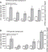
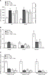
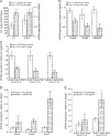
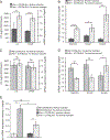
The inhibitory Smad7 acts as a critical suppressor of hepcidin, the major regulator of systemic iron homeostasis. In this study we define the mRNA expression of the two functionally related Smad proteins, Smad6 and Smad7, within pathways known to regulate hepcidin levels. Using mouse models for hereditary hemochromatosis (Hfe-, TfR2-, Hfe/TfR2-, Hjv- and hepcidin1-deficient mice) we show that hepcidin, Smad6 and Smad7 mRNA expression is coordinated in such a way that it correlates with the activity of the Bmp/Smad signaling pathway rather than with liver iron levels. This regulatory circuitry is disconnected by iron treatment of Hfe-/- and Hfe/TfR2 mice that significantly increases hepatic iron levels as well as hepcidin, Smad6 and Smad7 mRNA expression but fails to augment pSmad1/5/8 levels. This suggests that additional pathways contribute to the regulation of hepcidin, Smad6 and Smad7 under these conditions which do not require Hfe.
Copyright © 2012 Elsevier B.V. All rights reserved.
Figures


Similar articles
-
Defective bone morphogenic protein signaling underlies hepcidin deficiency in HFE hereditary hemochromatosis.Hepatology. 2010 Oct;52(4):1266-73. doi: 10.1002/hep.23814. Hepatology. 2010. PMID: 20658468
-
Iron regulation of hepcidin despite attenuated Smad1,5,8 signaling in mice without transferrin receptor 2 or Hfe.Gastroenterology. 2011 Nov;141(5):1907-14. doi: 10.1053/j.gastro.2011.06.077. Epub 2011 Jul 13. Gastroenterology. 2011. PMID: 21745449 Free PMC article.
-
Altered hepatic BMP signaling pathway in human HFE hemochromatosis.Blood Cells Mol Dis. 2010 Dec 15;45(4):308-12. doi: 10.1016/j.bcmd.2010.08.010. Epub 2010 Sep 21. Blood Cells Mol Dis. 2010. PMID: 20863724 Free PMC article.
-
[Pathophysiology and genetics of classic HFE (type 1) hemochromatosis].Presse Med. 2007 Sep;36(9 Pt 2):1271-7. doi: 10.1016/j.lpm.2007.03.038. Epub 2007 May 22. Presse Med. 2007. PMID: 17521857 Review. French.
-
Non-HFE hemochromatosis.Semin Liver Dis. 2005 Nov;25(4):450-60. doi: 10.1055/s-2005-923316. Semin Liver Dis. 2005. PMID: 16315138 Review.
Cited by
-
Bone morphogenic proteins in iron homeostasis.Bone. 2020 Sep;138:115495. doi: 10.1016/j.bone.2020.115495. Epub 2020 Jun 23. Bone. 2020. PMID: 32585319 Free PMC article. Review.
-
MyD88 Regulates the Expression of SMAD4 and the Iron Regulatory Hormone Hepcidin.Front Cell Dev Biol. 2018 Aug 31;6:105. doi: 10.3389/fcell.2018.00105. eCollection 2018. Front Cell Dev Biol. 2018. PMID: 30234111 Free PMC article.
-
Scavenging Reactive Oxygen Species Production Normalizes Ferroportin Expression and Ameliorates Cellular and Systemic Iron Disbalances in Hemolytic Mouse Model.Antioxid Redox Signal. 2018 Aug 10;29(5):484-499. doi: 10.1089/ars.2017.7089. Epub 2018 Mar 2. Antioxid Redox Signal. 2018. PMID: 29212341 Free PMC article.
-
Atorvastatin Inhibits Ferroptosis of H9C2 Cells by regulatingSMAD7/Hepcidin Expression to Improve Ischemia-Reperfusion Injury.Cardiol Res Pract. 2022 Nov 8;2022:3972829. doi: 10.1155/2022/3972829. eCollection 2022. Cardiol Res Pract. 2022. PMID: 36398315 Free PMC article.
-
Regulation of the Iron Homeostatic Hormone Hepcidin.Adv Nutr. 2017 Jan 17;8(1):126-136. doi: 10.3945/an.116.013961. Print 2017 Jan. Adv Nutr. 2017. PMID: 28096133 Free PMC article. Review.
References
-
- Andrews NC, Iron homeostasis: insights from genetics and animal models, Nat. Rev. Genet. 1 (2000) 208–217. - PubMed
-
- Lesbordes-Brion JC, Viatte L, Bennoun M, Lou DQ, Ramey G, Houbron C, Hamard G, Kahn A, Vaulont S, Targeted disruption of the hepcidin 1 gene results in severe hemochromatosis, Blood 108 (2006) 1402–1405. - PubMed
-
- Feder JN, Gnirke A, Thomas W, Tsuchihashi Z, Ruddy DA, Basava A, Dormishian F, Domingo R Jr., Ellis MC, Fullan A, Hinton LM, Jones NL, Kimmel BE, Kronmal GS, Lauer P, Lee VK, Loeb DB, Mapa FA, McClelland E, Meyer NC, Mintier GA, Moeller N, Moore T, Morikang E, Prass CE, Quintana L, Starnes SM, Schatzman RC, Brunke KJ, Drayna DT, Risch NJ, Bacon BR, Wolff RK, A novel MHC class I-like gene is mutated in patients with hereditary haemochromatosis, Nat. Genet. 13 (1996) 399–408. - PubMed
-
- Bridle KR, Frazer DM, Wilkins SJ, Dixon JL, Purdie DM, Crawford DH, Subramaniam VN, Powell LW, Anderson GJ, Ramm GA, Disrupted hepcidin regulation in HFE-associated haemochromatosis and the liver as a regulator of body iron homoeostasis, Lancet 361 (2003) 669–673. - PubMed
-
- Wallace DF, Summerville L, Crampton EM, Frazer DM, Anderson GJ, Subramaniam VN, Combined deletion of Hfe and transferrin receptor 2 in mice leads to marked dysregulation of hepcidin and iron overload, Hepatology 50 (2009) 1992–2000. - PubMed
Publication types
MeSH terms
Substances
Grants and funding
LinkOut - more resources
Full Text Sources
Other Literature Sources
Medical
Molecular Biology Databases

